Abstract
So far, the antibacterial activity of some organic and inorganic compounds has been studied. Barium zirconate titanate [Ba(ZrxTi1-x)O3] (x = 0.05) nanoparticle is an example of inorganic materials. In vitro studies have provided evidence for the antibacterial activity of this nanoparticle. In the current study, the nano-powder was synthesized by sol-gel method. X-ray diffraction showed that the powder was single-phase and had a perovskite structure at the calcination temperature of 1000 °C. Antibacterial activity of the desired nanoparticle was assessed on two gram-positive (Staphylococcus aureus PTCC1431 and Micrococcus luteus PTCC1625) and two gram-negative (Escherichia coli HP101BA 7601c and clinically isolated Klebsiella pneumoniae) bacteria according to Radial Diffusion Assay (RDA). The results showed that the antibacterial activity of BZT nano-powder on both gram-positive and gram-negative bacteria was acceptable. The minimum inhibitory concentration of this nano-powder was determined. The results showed that MIC values for E. coli, K. pneumoniae, M. luteus and S. aureus were about 2.3 μg/mL, 7.3 μg/mL, 3 μg/mL and 12 μg/mL, respectively. Minimum bactericidal concentration (MBC) was also evaluated and showed that the growth of E. coli, K. pneumoniae, M. luteus and S. aureus could be decreased at 2.3, 14, 3 and 18 μg/mL of BZT. Average log reduction in viable bacteria count in time-kill assay ranged between 6 Log10 cfu/mL to zero after 24 h of incubation with BZT nanoparticle.
Keywords: nanoparticles, antibiotics, barium zirconate titanate, ceramics, electron microscopy
Introduction
Nowadays, nano-science is going to affect all aspects of life. It has been shown that chemically synthesized nanoparticles (NPs) have antibacterial effects on gram-positive and gram-negative bacteria (Ruparelia et al., 2008; Valodkar et al., 2012; Sreelakshmi et al., 2011; Wang et al., 2011; Allahverdiyev et al., 2011; Mishra et al., 2011; Musarrat et al., 2010; Damm et al., 2008; Yoksan and Chirachanchai 2009; Ramyadevi et al., 2012; Prasad et al., 2011). Some nanoparticles even show inhibitory effect on the bacterial growth when they are mixed with other compounds and nano-powders (Li et al., 2006). Researches have shown the antibacterial properties of some polymers which are made by nanoparticles for use in the surface area of medical instruments (Monteiro et al., 2009; Singh and Nalwa 2011). These nanoparticles seem to be useful in gene therapy studies, medical studies and drug delivery systems (DD systems) in the near future (Pinto-Alphandary et al., 2000; Pagonis et al., 2010; Prow et al., 2011). Ceramic nanoparticles are inorganic systems with porous characteristics which were recently developed as drug vehicles (Sekhon and Kamboj 2010; Fontana et al., 1998). Some studies even showed their non-toxic effects on human cells (Sharma et al., 2011; Martinez-Gutierrez et al., 2010). Recently, an organic nanoparticle has been produced which is completely non-toxic, biodegradable and nimble in the way it uses light and heat to treat cancer and deliver drugs (Vollmer et al., 2012; Hung et al., 2010). Currently, researchers are able to encapsulate drugs in nanoparticles with the size of viruses. Nanoparticles are effective in drug delivery due to the fact that these nanoparticles, in combination with organic compounds like lipids and glycoproteins, could precisely detect the damaged cells and deliver the drugs (Lovell et al., 2011; Sim and Wallis 2011). Designing carbohydrate nanoparticles for prolonged efficacy of antibacterial peptide is now under investigation (Bi et al., 2011). Syntheses of nanoparticles are highly cost-effective. Some of the nanoparticles such as gold, copper and silver nano-powders with strong germicidal properties have been synthesized, but these metals are expensive and their high production cost does not make them potential candidate for use as antibacterial agents. Therefore, producing less expensive nano-powders with acceptable antibacterial properties would be of great interests in nano and medical science era. Such inexpensive, germicidal and easy producible nanoparticles would have great role in pharmacology and medical science as well as drug discovery for designing new antibacterial agents and nano scale drug carriers. In this study, the aim was to produce a less expensive nano-material with antibacterial properties. Therefore, the barium zirconate titanate [Ba(ZrxTi1-x)O3] (x = 0.05) nanoparticle was synthesized and tested on E. coli, K. pneumoniae, M. luteus and S. aureus as representative of gram-negative and gram-positive bacteria.
Experimental
Preparation
[Ba(ZrxTi1-x)O3] (x = 0.05) nanoparticle was prepared by a sol-gel process (Yu and Xia 2012). The raw materials in this experiment were barium nitrate [Ba(NO3)2], zirconium nitrate [ZrO(NO3)2] and titanium isopropoxide Ti[OCH(CH3)2]4. By dissolving barium nitrate and zirconium nitrate in distilled water, aqueous solution of each cations (Ba+2, Zr+4) was prepared. For preparation of Ti+4, titanium (IV) isopropoxide was dissolved in the mixture of nitric and citric acid (Ghasemifard et al., 2009b). The solutions of barium, titanium and zirconium were added to the aqueous solution of citric acid under continuous stirring at 55–60 °C, with the constant pH of 7.0. In order to keep the pH constant, ammonium hydroxide was added to the solution (Ghasemifard et al., 2009a). The sol form of BZT was heated to about 80 °C to evaporate all water and to obtain the gel. When excessive nitric acid was added, the gel temperature increased rapidly, this caused the final color of the powder to become black. After auto-combustion of the gels, the resultant powders were calcinated at 1000 °C to obtain the desired single-phase powders.
Antibacterial assay
Antibacterial activity of synthesized nanoparticles were tested on gram-positive and gram-negative bacteria according to the radial diffusion assay (RDA) for antibacterial agents (R.I. Lehrer 1991). Staphylococcus aureus PTCC1431 and Micrococcus luteus PTCC1625 as gram-positive and Escherichia coli HP101BA 7601c and a clinical isolate of Klebsiellae pneumoniae as gram-negative bacteria were prepared for antibacterial assay. In order to obtain mid-logarithmic phase microorganism, 100 μL of the culture was transferred to 100 mL of fresh TSB media culture and incubated for an additional 3 h at 37 °C, and therefore bacteria were used in their logarithmic phase for antibacterial assay. For this purpose, 4 × 10⁶ cfu (Colony Forming Units) was poured into five mL of 10 mM cold phosphate buffer and was mixed with 1% agarose (Sigma-Aldrich) in 0.03% tripticase soy broth (TSB) as an underlay culture, and was then poured into the plate. Subsequently, specific amount of BZT nanoparticles was dispersed and dissolved in the same buffer and was poured into the punched well in a plate. After 3 h incubation at 37 °C, overlay media culture containing pre-autoclaved 6% TSB and 1% agarose was gently poured into the plate and was kept at 37 °C for 12 h. For bactericidal efficiency, antibacterial activity of BZT was assessed for the duration of 24 h. For this purpose, specific amount of bacteria were cultured in 96 well plate and the absorbance at 600 nm was measured each 3 h and compared to controls (bacteria without antibacterial agent). The concentration of bacteria was defined as logarithm to the base 10.
MIC and MBC determination
Similar to other antibacterial agents, nanoparticles are subjected to minimum inhibitory concentration (MIC) and minimum bactericidal concentration (MBC) determination. In microbiology, MIC is defined as the lowest concentration of an antibacterial compound that inhibits the visible growth of a microorganism after an overnight incubation (Andrews 2001). Two gram-positive (Staphylococcus aureus PTCC1431 and Micrococcus luteus PTCC1625) and two gram-negative bacteria (Escherichia coli HP101BA 7601c and a clinical isolate of Klebsiella pneumoniae) were chosen for antibacterial tests and MIC and MBC assay. A specific amount of bacteria (4 × 10⁶ cfu) was prepared and after treating with serial dilution of BZT, was poured into the 96-well plates and was incubated at 37 °C for 24 h. Afterward, the absorbance was recorded at 600 nm for each well using an enzyme-linked immunosorbent assay (ELISA) reader and the results were compared to the control sample. This procedure was performed in triplicate.
MBC is defined as the lowest concentration of antimicrobial that will prevent the growth of an organism after subculture on to antibiotic free media. For MBC test, 20 μL of bacteria suspension was inoculated on to agar plate from 2 first well that showed no bacteria growth. The plate was then incubated for an additional 24 h at 37 °C.
Hemolysis assay
Hemolytic activity of BZT was determined according to Minn et al. method (Minn et al., 1998). For this purpose, 2 mL of human red blood cells (hRBCs) were washed several times with 5 mL of cold phosphate buffered saline (PBS) by centrifugation at 4,000 rpm (3600 g) for 10 min. Washed cells were diluted to a final volume of 40 mL of PBS. Hemolysis assay for the desired nanoparticle was determined at relatively high concentration of 20 μg/mL in which 20 μL of BZT were added to 180 μL of 5% diluted erythrocytes and the treated cells were kept at 37 °C for 30 min. 0.1% Triton X-100 was used as positive control with 100% hemolytic activity. After 30 min, the solution was centrifuged at 4,000 rpm for 5 min, and the supernatant was mildly diluted to 1 mL of PBS. Absorbance of the solution was measured at 567 nm.
Results and Discussion
X-ray diffraction and other physicochemical properties of BZT
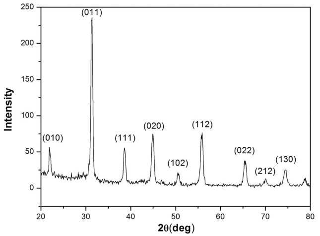
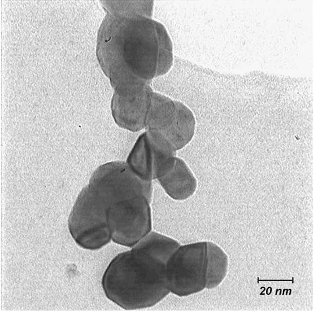
Ba(Zr0.1Ti0.9)O3 nanoparticles were prepared by a sol-gel process. The sizes and other physicochemical properties of the nanoparticles were determined by XRD and TEM image. The phase formation of BZT powder was investigated using X-ray diffraction analysis at room temperature (29 °C) in the range (20–80 degree) with CuKα radiation. Figure 1 shows the x-ray diffraction patterns of BZT powders calcinated at 1000 °C. It is evident that powders have a perovskite cubic structure without extra phases. Cubic structure with general formula of ABO3 is the most important characteristics of perovskites. The typical TEM image of the BZT powders is shown in Figure 2. The primary particle size of the BZT powder was found to be approximately 25 nm in diameter.
Figure 1.
XRD patterns of BZT nano-powders at room temperature.
Figure 2.
TEM image of the BZT nano-powder calcinated at temperatures of 1000 °C.
Antibacterial assay
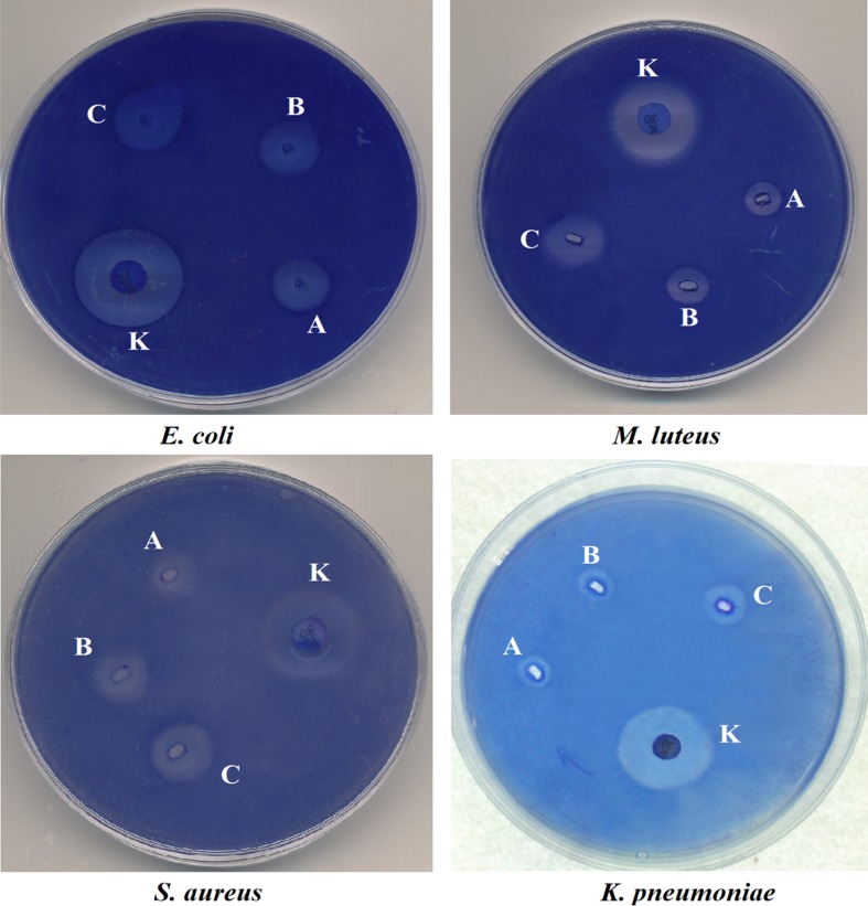
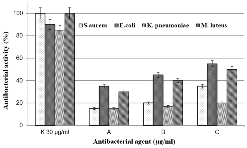
According to previously described methods for antibacterial and MIC assay, bacteria were cultured and the nano-powder with different concentrations was poured into the punched wells. After 12 h incubation at 37 °C, the growth inhibitory zone around the wells was obvious (Figure 3). Several independent experiments confirmed that these nano-powders have antibacterial activity on both tested gram-positive and gram-negative bacteria, but the mechanism of such antibacterial properties is not yet understood. For antibacterial assay of BZT nano-powders, each 1 mm diameter of an inhibition zone from the center of the halo, was expressed as Units (1 mm = 1 U) and was calculated after subtracting the diameter of the central well. Finally, the highest amount of antibacterial activity was defined as 100% activity and others were compared to it (Figure 4).
Figure 3.
Antibacterial activity of BZT on E. coli, M. luteus, K. pneumoniae and S. aureus. K is abbreviation for kanamycin 30 μg and A, B, and C show the concentrations of 2, 5, and 10 μg/mL of BZT nanoparticle, respectively.
Figure 4.
Antibacterial properties of BZT nanoparticle on E. coli, K. pneumoniae, M. luteus and S. aureus. (K is the abbreviation for standard 30 μg/mL kanamycin and A, B and C show BZT in the concentration of 2, 5 and 10 μg/mL respectively.)
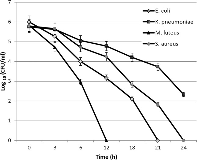
The reported antibacterial activity is in close competence with some bactericidal, synthetic nanoparticles such as silver and copper nanoparticles which inhibits the growth of bacteria; with the inhibition zone of 26 mm (Prasad et al., 2011; Ramyadevi et al., 2012). According to our data, the synthesized nano-powder has germicidal power on both gram-positive and gram-negative bacteria. The results for bactericidal efficiency and time kill assessment in a period of 24 h showed effective reduction of bacteria concentration (Figure 5).
Figure 5.
Reduction in initial bacterial concentration after 24 h of incubation with BZT at MIC values. Bacteria concentration is defined as Log10 (CFU/mL).
MIC and MBC determination
The overall MIC values for these nanoparticles were 2.3 μg/mL, 7.3 μg/mL, 3 μg/mL and 12 μg/mL for E. coli, K. pneumoniae, M. luteus and S. aureus, respectively. This value for E. coli (MTCC 443) is reported to be 40 μg/mL and 140 μg/mL for silver and copper nanoparticle, respectively (Ruparelia et al., 2008). According to the reported MIC values by Ruparelia et al, this value for Ag and Cu nanoparticles against S. aureus (NCIM 2079) is 120 μg/mL and 140 μg/mL, respectively. Minimum bactericidal concentration for E. coli, K. pneumoniae, M. luteus and S. aureus was reported to be 2.3, 14, 3 and 18 μg/mL (Table 1).
Table 1.
Minimum inhibitory (MIC) and bactericidal (MBC) concentrations of BZT nano-powders.
| Bacteria | MIC (μg/mL) | MBC (μg/mL) |
|---|---|---|
| E. coli (HP101BA 7601c) | 2.3 | 2.3 |
| K. pneumoniae | 7.3 | 14 |
| M. luteus (PTCC1625) | 3 | 3 |
| S. aureus (PTCC1431) | 12 | 18 |
Hemolysis assay
Hemolysis assay is a standard biological method to investigate cytotoxicity of an agent on red blood cells. For BZT nano-powders, 6.5% hemolytic activity was observed at 20 μg/mL in comparison with Triton X-100 as positive control with 100% hemolysis. Low hemolytic activity makes them potential candidates for further studies in drug delivery and microbiology. But more studies on the cytotoxicity of this nanoparticle are desired to verify their non-toxic effects on human cells.
Conclusions
In the present study, barium zirconate titanate nanoparticle has been synthesized and tested for antibacterial activity. Results showed that the desired nano-powders had satisfactory antibacterial properties with slightly hemolytic activity which probably make them a candidate as potential antibacterial agents in DD systems. In the recent decade, some nanoparticles have been introduced that showed antibacterial and anti-cancer properties and consequently studied for their potential as antibacterial agents (Selvaraj et al., 2010; Fontana et al., 1998). Studies show that some nanoparticles and nanostructures, especially carbon nanotubes and nanoceramics, are widely used in medicine and medical instruments due to their unique chemical and physical structures (Ercan et al., 2011; Zhou et al., 2010). Gelain et al., in 2011 reported that some of these nanostructures can be useful in the development of cell and tissue engineering procedures and they could increase the drug efficiency (Gelain et al., 2011). They also have role in food industry, agriculture and human and veterinary medicine (Wolska et al., 2012). The enhanced antibiotic efficacy of these nano-powders in combination with conventional antibacterials on HIV-1 virus and other pathological infections has also been confirmed by several independent researches (Wolska et al., 2012; Mahajan et al., 2012; Dar et al., 2013; Mirzajani et al., 2011). Due to their nano size and biocompatibility with cells and because these nanoparticles have exhibited potential as drug delivery system, nanoceramics have attracted many attentions for further studies in pharmacology and nanomedicine (Roy et al., 2003). Due to ceramic nature of BZT nanoparticle, it is suggested to evaluate the potential of BZT nanoparticle as coatings in variety of medical or surgical instruments. Using nanostructures and nanoceramics may provide millimeter-scale precision at a much lower cost compared to current technologies in medicine, drug delivery and pharmaceutical sciences (Kaufman et al., 2013). But, much more studies are required to prove the suggested applications of nanostructures.
Acknowledgments
In this study, the desired nano-powder was synthesized at the department of Physics, at Ferdowsi University of Mashhad, Mashhad, Iran. And, we would like to thank the staffs at the Electro-ceramic and Nano science research group for their kind assistance.
Footnotes
Declaration of interests:
The authors report no declarations of interest.
References
- Allahverdiyev AM, Abamor ES, Bagirova M, Rafailovich M. Antimicrobial effects of TiO(2) and Ag(2)O nanoparticles against drug-resistant bacteria and leishmania parasites. Future Microbiol. 2011;6:933–940. doi: 10.2217/fmb.11.78. [DOI] [PubMed] [Google Scholar]; Allahverdiyev AM, Abamor ES, Bagirova M, Rafailovich M (2011) Antimicrobial effects of TiO(2) and Ag(2)O nanoparticles against drug-resistant bacteria and leishmania parasites. Future Microbiol 6:933–940. [DOI] [PubMed]
- Andrews JM. Determination of minimum inhibitory concentrations. J Antimicrob Chemother. 2001;48(Suppl 1):5–16. doi: 10.1093/jac/48.suppl_1.5. [DOI] [PubMed] [Google Scholar]; Andrews JM (2001) Determination of minimum inhibitory concentrations. J Antimicrob Chemother 48 Suppl 1:5–16. [DOI] [PubMed]
- Bi L, Yang L, Narsimhan G, Bhunia AK, Yao Y. Designing carbohydrate nanoparticles for prolonged efficacy of antimicrobial peptide. J Controlled Release. 2011;150:150–156. doi: 10.1016/j.jconrel.2010.11.024. [DOI] [PubMed] [Google Scholar]; Bi L, Yang L, Narsimhan G, Bhunia AK, Yao Y (2011) Designing carbohydrate nanoparticles for prolonged efficacy of antimicrobial peptide. J Controlled Release 150:150–156. [DOI] [PubMed]
- Damm C, Münstedt H, Rösch A. The antimicrobial efficacy of polyamide 6/silver-nano- and microcomposites. Mater Chem Phys. 2008;108:61–66. [Google Scholar]; Damm C, Münstedt H, Rösch A (2008) The antimicrobial efficacy of polyamide 6/silver-nano- and microcomposites. Mater Chem Phys 108:61–66.
- Dar MA, Ingle A, Rai M. Enhanced antimicrobial activity of silver nanoparticles synthesized by Cryphonectria sp. evaluated singly and in combination with antibiotics. Nanomedicine. 2013;9:105–110. doi: 10.1016/j.nano.2012.04.007. [DOI] [PubMed] [Google Scholar]; Dar MA, Ingle A, Rai M (2013) Enhanced antimicrobial activity of silver nanoparticles synthesized by Cryphonectria sp. evaluated singly and in combination with antibiotics. Nanomedicine 9:105–110. [DOI] [PubMed]
- Ercan B, Taylor E, Alpaslan E, Webster TJ. Diameter of titanium nanotubes influences anti-bacterial efficacy. Nanotechnology. 2011;22:295102. doi: 10.1088/0957-4484/22/29/295102. [DOI] [PubMed] [Google Scholar]; Ercan B, Taylor E, Alpaslan E, Webster TJ (2011) Diameter of titanium nanotubes influences anti-bacterial efficacy. Nanotechnology 22:295102. [DOI] [PubMed]
- Fontana G, Pitarresi G, Tomarchio V, Carlisi B, San Biagio PL. Preparation, characterization and in vitro antimicrobial activity of ampicillin-loaded polyethylcyanoacrylate nanoparticles. Biomaterials. 1998;19:1009–1017. doi: 10.1016/s0142-9612(97)00246-9. [DOI] [PubMed] [Google Scholar]; Fontana G, Pitarresi G, Tomarchio V, Carlisi B, San Biagio PL (1998) Preparation, characterization and in vitro antimicrobial activity of ampicillin-loaded polyethylcyanoacrylate nanoparticles. Biomaterials 19:1009–1017. [DOI] [PubMed]
- Gelain F, Silva D, Caprini A, Taraballi F, Natalello A, Villa O, Nam KT, Zuckermann RN, Doglia SM, Vescovi A. BMHP1-derived self-assembling peptides: hierarchically assembled structures with self-healing propensity and potential for tissue engineering applications. ACS Nano. 2011;5:1845–1859. doi: 10.1021/nn102663a. [DOI] [PubMed] [Google Scholar]; Gelain F, Silva D, Caprini A, Taraballi F, Natalello A, Villa O, Nam KT, Zuckermann RN, Doglia SM, Vescovi A (2011) BMHP1-derived self-assembling peptides: hierarchically assembled structures with self-healing propensity and potential for tissue engineering applications. ACS Nano 5:1845–1859. [DOI] [PubMed]
- Ghasemifard M, Hosseini S, Bagheri-Mohagheghi M, Shahtahmasbi N. Structure comparison of PMN-PT and PMN-PZT nanocrystals prepared by gel-combustion method at optimized temperatures. Physica E. 2009a;41:1701–1706. [Google Scholar]; Ghasemifard M, Hosseini S, Bagheri-Mohagheghi M, Shahtahmasbi N (2009a) Structure comparison of PMN-PT and PMN-PZT nanocrystals prepared by gel-combustion method at optimized temperatures. Physica E 41:1701–1706.
- Ghasemifard M, Hosseini S, Khorrami G. Synthesis and structure of PMN-PT ceramic nanopowder free from pyrochlore phase. Ceram Int. 2009b;35:2899–2905. [Google Scholar]; Ghasemifard M, Hosseini S, Khorrami G (2009b) Synthesis and structure of PMN-PT ceramic nanopowder free from pyrochlore phase. Ceram Int 35:2899–2905.
- Hung LH, Teh SY, Jester J, Lee AP. PLGA micro/nanosphere synthesis by droplet microfluidic solvent evaporation and extraction approaches. Lab chip. 2010;10:1820–1825. doi: 10.1039/c002866e. [DOI] [PubMed] [Google Scholar]; Hung LH, Teh SY, Jester J, Lee AP (2010) PLGA micro/nanosphere synthesis by droplet microfluidic solvent evaporation and extraction approaches. Lab chip 10:1820–1825. [DOI] [PubMed]
- Kaufman JJ, Ottman R, Tao G, Shabahang S, Banaei EH, Liang X, Johnson SG, Fink Y, Chakrabarti R, Abouraddy AF. In-fiber production of polymeric particles for biosensing and encapsulation. Proc Nat Acad Sci USA. 2013;110:15549–15554. doi: 10.1073/pnas.1310214110. [DOI] [PMC free article] [PubMed] [Google Scholar]; Kaufman JJ, Ottman R, Tao G, Shabahang S, Banaei EH, Liang X, Johnson SG, Fink Y, Chakrabarti R, Abouraddy AF (2013) In-fiber production of polymeric particles for biosensing and encapsulation. Proc Nat Acad Sci USA 110:15549–15554. [DOI] [PMC free article] [PubMed]
- Li Y, Leung P, Yao L, Song QW, Newton E. Antimicrobial effect of surgical masks coated with nanoparticles. J Hosp Infect. 2006;62:58–63. doi: 10.1016/j.jhin.2005.04.015. [DOI] [PubMed] [Google Scholar]; Li Y, Leung P, Yao L, Song QW, Newton E (2006) Antimicrobial effect of surgical masks coated with nanoparticles. J Hosp Infect 62:58–63. [DOI] [PubMed]
- Lovell JF, Jin CS, Huynh E, Jin H, Kim C, Rubinstein JL, Chan WCW, Cao W, Wang LV, Zheng G. Porphysome nanovesicles generated by porphyrin bilayers for use as multimodal biophotonic contrast agents. Nat Mater. 2011;10:324–332. doi: 10.1038/nmat2986. [DOI] [PubMed] [Google Scholar]; Lovell JF, Jin CS, Huynh E, Jin H, Kim C, Rubinstein JL, Chan WCW, Cao W, Wang LV, Zheng G (2011) Porphysome nanovesicles generated by porphyrin bilayers for use as multimodal biophotonic contrast agents. Nat Mater 10:324–332. [DOI] [PubMed]
- Mahajan SD, Aalinkeel R, Law WC, Reynolds JL, Nair BB, Sykes DE, Yong KT, Roy I, Prasad PN, Schwartz SA. Anti-HIV-1 nanotherapeutics: promises and challenges for the future. Int J Nanomedicine. 2012;7:5301–5314. doi: 10.2147/IJN.S25871. [DOI] [PMC free article] [PubMed] [Google Scholar]; Mahajan SD, Aalinkeel R, Law WC, Reynolds JL, Nair BB, Sykes DE, Yong KT, Roy I, Prasad PN, Schwartz SA (2012) Anti-HIV-1 nanotherapeutics: promises and challenges for the future. Int J Nanomedicine 7:5301–5314. [DOI] [PMC free article] [PubMed]
- Martinez-Gutierrez F, Olive PL, Banuelos A, Orrantia E, Nino N, Sanchez EM, Ruiz F, Bach H, Av-Gay Y. Synthesis, characterization, and evaluation of antimicrobial and cytotoxic effect of silver and titanium nanoparticles. Nanomedicine. 2010;6:681–688. doi: 10.1016/j.nano.2010.02.001. [DOI] [PubMed] [Google Scholar]; Martinez-Gutierrez F, Olive PL, Banuelos A, Orrantia E, Nino N, Sanchez EM, Ruiz F, Bach H, Av-Gay Y (2010) Synthesis, characterization, and evaluation of antimicrobial and cytotoxic effect of silver and titanium nanoparticles. Nanomedicine 6:681–688. [DOI] [PubMed]
- Minn I, Kim HS, Kim SC. Antimicrobial peptides derived from pepsinogens in the stomach of the bullfrog, Rana catesbeiana. Biochim biophys acta. 1998;1407:31–39. doi: 10.1016/s0925-4439(98)00023-4. [DOI] [PubMed] [Google Scholar]; Minn I, Kim HS, Kim SC (1998) Antimicrobial peptides derived from pepsinogens in the stomach of the bullfrog, Rana catesbeiana. Biochim biophys acta 1407:31–39. [DOI] [PubMed]
- Mirzajani F, Ghassempour A, Aliahmadi A, Esmaeili MA. Antibacterial effect of silver nanoparticles on Staphylococcus aureus. Res Microbiol. 2011;162:542–549. doi: 10.1016/j.resmic.2011.04.009. [DOI] [PubMed] [Google Scholar]; Mirzajani F, Ghassempour A, Aliahmadi A, Esmaeili MA (2011) Antibacterial effect of silver nanoparticles on Staphylococcus aureus. Res Microbiol 162:542–549. [DOI] [PubMed]
- Mishra A, Tripathy SK, Yun SI. Bio-synthesis of gold and silver nanoparticles from Candida guilliermondii and their antimicrobial effect against pathogenic bacteria. J Nanosci Nanotechnol. 2011;11:243–248. doi: 10.1166/jnn.2011.3265. [DOI] [PubMed] [Google Scholar]; Mishra A, Tripathy SK, Yun SI (2011) Bio-synthesis of gold and silver nanoparticles from Candida guilliermondii and their antimicrobial effect against pathogenic bacteria. J Nanosci Nanotechnol 11:243–248. [DOI] [PubMed]
- Monteiro DR, Gorup LF, Takamiya AS, Ruvollo-Filho AC, Camargo ERd, Barbosa DB. The growing importance of materials that prevent microbial adhesion: antimicrobial effect of medical devices containing silver. Int J Antimicrob Ag. 2009;34:103–110. doi: 10.1016/j.ijantimicag.2009.01.017. [DOI] [PubMed] [Google Scholar]; Monteiro DR, Gorup LF, Takamiya AS, Ruvollo-Filho AC, Camargo ERd, Barbosa DB (2009) The growing importance of materials that prevent microbial adhesion: antimicrobial effect of medical devices containing silver. Int J Antimicrob Ag 34:103–110. [DOI] [PubMed]
- Musarrat J, Dwivedi S, Singh BR, Al-Khedhairy AA, Azam A, Naqvi A. Production of antimicrobial silver nanoparticles in water extracts of the fungus Amylomyces rouxii strain KSU-09. Bioresour Technol. 2010;101:8772–8776. doi: 10.1016/j.biortech.2010.06.065. [DOI] [PubMed] [Google Scholar]; Musarrat J, Dwivedi S, Singh BR, Al-Khedhairy AA, Azam A, Naqvi A (2010) Production of antimicrobial silver nanoparticles in water extracts of the fungus Amylomyces rouxii strain KSU-09. Bioresour Technol 101:8772–8776. [DOI] [PubMed]
- Pagonis TC, Chen J, Fontana CR, Devalapally H, Ruggiero K, Song X, Foschi F, Dunham J, Skobe Z, Yamazaki H, Kent R, Tanner ACR, Amiji MM, Soukos NS. Nanoparticle-based Endodontic Antimicrobial Photodynamic Therapy. J Endodont. 2010;36:322–328. doi: 10.1016/j.joen.2009.10.011. [DOI] [PMC free article] [PubMed] [Google Scholar]; Pagonis TC, Chen J, Fontana CR, Devalapally H, Ruggiero K, Song X, Foschi F, Dunham J, Skobe Z, Yamazaki H, Kent R, Tanner ACR, Amiji MM, Soukos NS (2010) Nanoparticle-based Endodontic Antimicrobial Photodynamic Therapy. J Endodont 36:322–328. [DOI] [PMC free article] [PubMed]
- Pinto-Alphandary H, Andremont A, Couvreur P. Targeted delivery of antibiotics using liposomes and nanoparticles: research and applications. Int J Antimicrob Ag. 2000;13:155–168. doi: 10.1016/s0924-8579(99)00121-1. [DOI] [PubMed] [Google Scholar]; Pinto-Alphandary H, Andremont A, Couvreur P (2000) Targeted delivery of antibiotics using liposomes and nanoparticles: research and applications. Int J Antimicrob Ag 13:155–168. [DOI] [PubMed]
- Prasad T, Elumalai EK, Khateeja S. Evaluation of the antimicrobial efficacy of phytogenic silver nanoparticles. Asian Pac J Trop Biomed. 2011;1(1, Supplement):S82–S85. [Google Scholar]; Prasad T, Elumalai EK, Khateeja S (2011) Evaluation of the antimicrobial efficacy of phytogenic silver nanoparticles. Asian Pac J Trop Biomed 1 (1, Supplement):S82–S85.
- Prow TW, Grice JE, Lin LL, Faye R, Butler M, Becker W, Wurm EM, Yoong C, Robertson TA, Soyer HP, Roberts MS. Nanoparticles and microparticles for skin drug delivery. Adv Drug Deliv Rev. 2011;63:470–491. doi: 10.1016/j.addr.2011.01.012. [DOI] [PubMed] [Google Scholar]; Prow TW, Grice JE, Lin LL, Faye R, Butler M, Becker W, Wurm EM, Yoong C, Robertson TA, Soyer HP, Roberts MS (2011) Nanoparticles and microparticles for skin drug delivery. Adv Drug Deliv Rev 63:470–491. [DOI] [PubMed]
- Lehrer RI, Rosenman M, Harwig SS, Jackson R, Eisenhauer P. Ulterasensitive assay for endogenous antimicrobial polypeptides. J Immunol Methods. 1991;137:167–173. doi: 10.1016/0022-1759(91)90021-7. [DOI] [PubMed] [Google Scholar]; Lehrer RI, Rosenman M, Harwig SS, Jackson R, Eisenhauer P (1991) Ulterasensitive assay for endogenous antimicrobial polypeptides. J Immunol Methods 137:167–173 [DOI] [PubMed]
- Ramyadevi J, Jeyasubramanian K, Marikani A, Rajakumar G, Rahuman AA. Synthesis and antimicrobial activity of copper nanoparticles. Mater Lett. 2012;71:114–116. [Google Scholar]; Ramyadevi J, Jeyasubramanian K, Marikani A, Rajakumar G, Rahuman AA (2012) Synthesis and antimicrobial activity of copper nanoparticles. Mater Lett 71:114–116.
- Roy I, Ohulchanskyy TY, Pudavar HE, Bergey EJ, Oseroff AR, Morgan J, Dougherty TJ, Prasad PN. Ceramic-based nanoparticles entrapping water-insoluble photosensitizing anticancer drugs: a novel drug-carrier system for photodynamic therapy. J Am Chem Soc. 2003;125:7860–7865. doi: 10.1021/ja0343095. [DOI] [PubMed] [Google Scholar]; Roy I, Ohulchanskyy TY, Pudavar HE, Bergey EJ, Oseroff AR, Morgan J, Dougherty TJ, Prasad PN (2003) Ceramic-based nanoparticles entrapping water-insoluble photosensitizing anticancer drugs: a novel drug-carrier system for photodynamic therapy. J Am Chem Soc 125:7860–7865. [DOI] [PubMed]
- Ruparelia JP, Chatterjee AK, Duttagupta SP, Mukherji S. Strain specificity in antimicrobial activity of silver and copper nanoparticles. Acta Biomaterialia. 2008;4:707–716. doi: 10.1016/j.actbio.2007.11.006. [DOI] [PubMed] [Google Scholar]; Ruparelia JP, Chatterjee AK, Duttagupta SP, Mukherji S (2008) Strain specificity in antimicrobial activity of silver and copper nanoparticles. Acta Biomaterialia 4:707–716. [DOI] [PubMed]
- Sekhon BS, Kamboj SR. Inorganic nanomedicine-Part 2. Nanomed-Nanotechnol. 2010;6:612–618. doi: 10.1016/j.nano.2010.04.003. [DOI] [PubMed] [Google Scholar]; Sekhon BS, Kamboj SR (2010) Inorganic nanomedicine-Part 2. Nanomed-Nanotechnol 6:612–618. [DOI] [PubMed]
- Selvaraj V, Grace AN, Alagar M, Hamerton I. Antimicrobial and anticancer efficacy of antineoplastic agent capped gold nanoparticles. J Biomed Nanotechnol. 2010;6:129–137. doi: 10.1166/jbn.2010.1115. [DOI] [PubMed] [Google Scholar]; Selvaraj V, Grace AN, Alagar M, Hamerton I (2010) Antimicrobial and anticancer efficacy of antineoplastic agent capped gold nanoparticles. J Biomed Nanotechnol 6:129–137. [DOI] [PubMed]
- Sharma A, Tandon A, Tovey JC, Gupta R, Robertson JD, Fortune JA, Klibanov AM, Cowden JW, Rieger FG, Mohan RR. Polyethylenimine-conjugated gold nanoparticles: Gene transfer potential and low toxicity in the cornea. Nanomedicine. 2011;7:505–513. doi: 10.1016/j.nano.2011.01.006. [DOI] [PMC free article] [PubMed] [Google Scholar]; Sharma A, Tandon A, Tovey JC, Gupta R, Robertson JD, Fortune JA, Klibanov AM, Cowden JW, Rieger FG, Mohan RR (2011) Polyethylenimine-conjugated gold nanoparticles: Gene transfer potential and low toxicity in the cornea. Nanomedicine 7:505–513. [DOI] [PMC free article] [PubMed]
- Sim RB, Wallis R. Surface properties: Immune attack on nanoparticles. Nat Nano. 2011;6:80–81. doi: 10.1038/nnano.2011.4. [DOI] [PubMed] [Google Scholar]; Sim RB, Wallis R (2011) Surface properties: Immune attack on nanoparticles. Nat Nano 6:80–81. [DOI] [PubMed]
- Singh R, Nalwa HS. Medical applications of nanoparticles in biological imaging, cell labeling, antimicrobial agents, and anticancer nanodrugs. J Biomed Nanotechnol. 2011;7:489–503. doi: 10.1166/jbn.2011.1324. [DOI] [PubMed] [Google Scholar]; Singh R, Nalwa HS (2011) Medical applications of nanoparticles in biological imaging, cell labeling, antimicrobial agents, and anticancer nanodrugs. J Biomed Nanotechnol 7:489–503. [DOI] [PubMed]
- Sreelakshmi C, Datta KK, Yadav JS, Reddy BV. Honey derivatized Au and Ag nanoparticles and evaluation of its antimicrobial activity. J Nanosci Nanotechnol. 2011;11:6995–7000. doi: 10.1166/jnn.2011.4240. [DOI] [PubMed] [Google Scholar]; Sreelakshmi C, Datta KK, Yadav JS, Reddy BV (2011) Honey derivatized Au and Ag nanoparticles and evaluation of its antimicrobial activity. J Nanosci Nanotechnol 11:6995–7000. [DOI] [PubMed]
- Valodkar M, Rathore PS, Jadeja RN, Thounaojam M, Devkar RV, Thakore S. Cytotoxicity evaluation and antimicrobial studies of starch capped water soluble copper nanoparticles. J Hazard Mater. 2012;201–202:244–249. doi: 10.1016/j.jhazmat.2011.11.077. [DOI] [PubMed] [Google Scholar]; Valodkar M, Rathore PS, Jadeja RN, Thounaojam M, Devkar RV, Thakore S (2012) Cytotoxicity evaluation and antimicrobial studies of starch capped water soluble copper nanoparticles. J Hazard Mater 201–202:244–249. [DOI] [PubMed]
- Vollmer C, Thomann R, Janiak C. Organic carbonates as stabilizing solvents for transition-metal nanoparticles. Dalton Trans. 2012;41:9722–9727. doi: 10.1039/c2dt30668a. [DOI] [PubMed] [Google Scholar]; Vollmer C, Thomann R, Janiak C (2012) Organic carbonates as stabilizing solvents for transition-metal nanoparticles. Dalton Trans 41:9722–9727. [DOI] [PubMed]
- Wang L, Luo J, Shan S, Crew E, Yin J, Zhong CJ, Wallek B, Wong SS. Bacterial inactivation using silver-coated magnetic nanoparticles as functional antimicrobial agents. Anal Chem. 2011;83:8688–8695. doi: 10.1021/ac202164p. [DOI] [PMC free article] [PubMed] [Google Scholar]; Wang L, Luo J, Shan S, Crew E, Yin J, Zhong CJ, Wallek B, Wong SS (2011) Bacterial inactivation using silver-coated magnetic nanoparticles as functional antimicrobial agents. Anal Chem 83:8688–8695. [DOI] [PMC free article] [PubMed]
- Wolska KI, Grzes K, Kurek A. Synergy between novel antimicrobials and conventional antibiotics or bacteriocins. Pol J Microbiol. 2012;61:95–104. [PubMed] [Google Scholar]; Wolska KI, Grzes K, Kurek A (2012) Synergy between novel antimicrobials and conventional antibiotics or bacteriocins. Pol J Microbiol 61:95–104 [PubMed]
- Yoksan R, Chirachanchai S. Silver nanoparticles dispersing in chitosan solution: Preparation by γ-ray irradiation and their antimicrobial activities. Mater Chem Phys. 2009;115:296–302. [Google Scholar]; Yoksan R, Chirachanchai S (2009) Silver nanoparticles dispersing in chitosan solution: Preparation by γ-ray irradiation and their antimicrobial activities. Mater Chem Phys 115:296–302.
- Yu Y-H, Xia M. Preparation and characterization of ZnTiO3 powders by sol-gel process. Mater Lett. 2012;77:10–12. [Google Scholar]; Yu Y-H, Xia M (2012) Preparation and characterization of ZnTiO3 powders by sol-gel process. Mater Lett 77:10–12.
- Zhou Y, Huang W, Liu J, Zhu X, Yan D. Self-assembly of hyperbranched polymers and its biomedical applications. Adv Mater. 2010;22:4567–4590. doi: 10.1002/adma.201000369. [DOI] [PubMed] [Google Scholar]; Zhou Y, Huang W, Liu J, Zhu X, Yan D (2010) Self-assembly of hyperbranched polymers and its biomedical applications. Adv Mater 22:4567–4590. [DOI] [PubMed]


