

Abstract
Schwann cells (SCs) hold promise for spinal cord injury (SCI) repair; however, there are limitations for use as a lone treatment. We showed acute inhibition of the phosphatase and tensin homologue (PTEN) by bisperoxovanadium (bpV) was neuroprotective and enhanced function following cervical hemicontusion SCI. We hypothesized that combining acute bpV therapy and delayed SC engraftment would further improve neuroprotection and recovery after cervical SCI. Adult female Sprague Dawley (SD) rats were randomly sorted into 5 groups: sham, vehicle, bpV, SC transplantation, and bpV + SC transplantation. SCs were isolated from adult green fluorescent protein (GFP)-expressing SD rats (GFP-SCs). 200 g/kg bpV(pic) was administered intraperitoneally (i.p.) twice daily for 7 days post-SCI in bpV-treated groups. GFP-SCs (1 × 10⁶ in 5 μl medium) were transplanted into the lesion epicenter at the 8th day post-SCI. Forelimb function was tested for 10 weeks and histology was assessed. bpV alone significantly reduced lesion (by 40%, p<0.05) and cavitation (by 65%, p<0.05) and improved functional recovery (p <0.05) compared to injury alone. The combination promoted similar neuroprotection (p<0.01 vs. injury); however, GFP-SCs alone did not. Both SC-transplanted groups exhibited remarkable long-term SC survival, SMI-31+ axon ingrowth and RECA-1+ vasculature presence in the SC graft; however, bpV + SCs promoted an 89% greater axon-to-lesion ratio than SCs only. We concluded that bpV likely contributed largely to the neuroprotective and functional benefits while SCs facilitated considerable host-tissue interaction and modification. The combination of the two shows promise as an attractive strategy to enhance recovery after SCI.
Keywords: spinal cord injury, Schwann cell transplantation, bpV, PTEN, neuroprotection
Introduction
Schwann cells (SCs) are dynamic participants in peripheral nerve function and repair, and have been widely studied as a therapy for central nervous system (CNS) injury. In spinal cord injury (SCI) research, SCs have been studied in various contexts ranging from peripheral nerve transplantation (David and Aguayo, 1981; David and Aguayo, 1985; Houle, 1991; Houle and Cote, 2013; Tom et al., 2013; Tom et al., 2009), SC-seeded channel engraftment (Deng et al., 2013; Deng et al., 2011; Iannotti et al., 2003; Xu et al., 1997; Xu et al., 1995a; Xu et al., 1995b; Xu et al., 1999) to cell suspension injection into or adjacent to the site of injury (Golden et al., 2007; Hu et al., 2013; Pearse et al., 2004a; Pearse et al., 2004b; Pearse et al., 2007; Schaal et al., 2007; Siriphorn et al., 2010; Takami et al., 2002). Many recent studies have investigated co-transplanting SCs with other cell types including olfactory ensheathing cells (OECs) (Pearse et al, 2007; Takami et al, 2002) and glial restricted precursors (GRPs) (Hu et al, 2013). Hormonal therapy combined with SC transplantation has also been reported (Siriphorn et al, 2010).
The potential benefits of SCs in the injured spinal cord are many (Oudega and Xu, 2006); nonetheless, key limitations to SC transplantation as a single therapy for SCI persist. Approximately 80% of transplanted cells die by necrosis and apoptosis by the end of the first week after transplantation (Hill et al., 2007; Hill et al., 2006). Although SCs can support considerable growth of propriospinal (Iannotti et al., 2004), reticulospinal (Schaal et al, 2007), raphaespinal (Novikova et al., 2008) and axons of other origin, these fibers fail to exit the graft into caudal host tissue due to the inhibitory glial scar that encapsulates the contusive lesion. Delaying SC transplantation 7–10 days after injury avoids cytotoxicity of the acute injury and allows for cystic cavity formation for cell engraftment (Hill et al., 2007; Hill et al., 2006; Martin, 1996). Adding a neuroprotective therapy prior to SC transplantation may enhance tissue sparing, repair, and influence the SC graft following transplantation. Combination therapies may be necessary for advancing SC transplantation research and its potential as a viable clinical treatment for SCI (Bunge, 2008).
Approximately 60% of human SCI occurs at the cervical level, yet strikingly few studies investigated SC transplantation following cervical SCI (Schaal et al., 2007; Novikova et al., 2008; Siriphorn et al., 2010). A better understanding of effects and benefits of cervical SC transplantation and new therapeutic combinations in cervical SCI models are necessary. Combining a pharmacological approach with SC transplantation is clinically practical due to ease and rapidity of administration in acutely injured patients. In addition, autologous transplants require several weeks to isolate, purify, and expand a sufficient number of a patient’s own SCs. A neuroprotective small-molecule therapy prior to SC transplantation may reduce secondary damage and promote a more robust spinal response to the delayed SC graft.
Bisperoxovanadium (bpV) compounds are dual protein-lipid phosphatases best known for their potent and specific inhibition of the phosphatase and tensin homolog deleted on chromosome ten (PTEN) (Schmid et al., 2004), an important regulator of cell survival and protein synthesis among other functions. PTEN antagonizes phosphatidylinositol-3-kinase (PI3K) activity, limiting survival signaling through Akt and its effectors such as mammalian target of rapamycin (mTOR) (Nave et al., 1999), bcl-2-associated death promoter (BAD) (Datta et al., 1997), and glycogen synthase kinase 3β (GSK3β) (Cross et al., 1995). Neuroprotective and reparative effects of bpVs have been widely demonstrated in neurological disease and injury models (Mao et al., 2013; Song et al., 2010; Sury et al., 2011; Yang et al., 2007; Zhang et al., 2007; Zhao et al., 2013) including SCI (Nakashima et al., 2008; Walker et al., 2012).
Our studies showed that bpV(pic) promoted significant neuroprotection and recovery when given during the first week following cervical hemicontusion SCI in rats (Walker et al., 2012; Walker and Xu, 2014). Combining acute bpV(pic) and sub-acute SC transplantation may promote additional benefits by compensating for individual treatment limitations and complementing therapeutic strengths. We hypothesized that administering bpV(pic) acutely followed by subacute SC transplantation would provide benefits over either single treatment alone. To test this hypothesis, we employed systemic bpV(pic) treatment and transplanted green-fluorescent protein (GFP) transgenic SCs (GFP-SCs) for optimal histological identification. Using a novel forelimb functional assessment and histological analyses, our findings shed light on potential benefits of this combination therapy in a clinically-relevant cervical hemicontusive SCI model.
Materials and Methods
Schwann cell culture
Schwann cells (SCs) were purified and expanded as previously described (Morrissey et al., 1991; Xu et al., 1995b; Xu et al., 1999). In brief, SCs were harvested from the sciatic nerves of transgenic homozygote green fluorescent protein (GFP)-Rosa expressing adult female Sprague-Dawley (SD) rats (George Smith, Temple University), followed by purification and expansion in culture. Cultures were > 98% pure SCs upon collection for transplantation (Xu et al, 1997; Xu et al, 1995b). Purified cells at passage 3 or 4 were collected for transplantation into the spinal lesion cavity.
Animal groups and surgical procedures
Adult female Sprague-Dawley rats (200–250 g, Harlan) (n = 34) were housed in an environmentally-controlled facility on a 12:12 light:dark cycle with ad libitum water and food access. The overall experimental design is illustrated in Fig. 1. Prior to surgery, animals were randomly assigned to five groups: 1) Sham (n = 6), 2) Saline-treated only (Veh, n = 6), 3) bpV(pic) treated (n = 6), 4) GFP-SCs (n = 7), 5) bpV(pic) + GFP-SCs (n = 9). The experimental groups were established based on findings reported in previous publications testing the effects of bpV and SCs alone (Walker et al., 2012; Wang and Xu, 2014). In these experiments, the injury only groups and the injury plus saline group showed no difference. Therefore, we used the injury plus saline group as the non-treatment control. This helped to reduce the number of animals necessary for the present study, and allow for an emphasis on the comparison of the three treatment groups, bpV, SCs, and bpV + SCs to each other and compared to injury and sham animals. Due to respiratory complications from surgery (n = 2) or inadequate contusion injury as recognized by rapid functional recovery (n =1), the final animal number for the study was 31 (Sham = 6, Veh = 5, bpV = 5, SCs = 7, bpV + SCs = 8). Final impactor displacement of the spinal cord was 2.02 ± 0.21 mm and impact velocity was 0.47 ± 0.02 m/s, indicating consistent contusive SCI among experimental animals.
Figure 1. Experimental design and treatments for the bpV(pic)/GFP-SC combination study.

(A) Time course and milestones of the experiment. (B) The structure of bpV(pic). (C) A photomicrograph of confluent pure GFP-SCs used for transplantation. Scale bar = 200 μm.
For surgical procedures, animals were anaesthetized intraperitoneally (IP) with a ketamine/xylazine cocktail ([87 mg/kg]/[12 mg/kg]), and received laminectomy only (sham operation) or unilateral cervical SCI performed as previously published (Walker et al, 2012). Briefly, the 5th cervical vertebra was stabilized with a customized device (Zhang et al., 2004) and a partial unilateral laminectomy was performed to expose the right side of the cord. A moderate unilateral injury was produced using the NYU/MASCIS Impactor (Gruner, 1992) (2.5 mm tip, 10g weight, 12.5 mm height) using previously published methods (Gensel et al., 2006; Walker et al., 2012). Sham animals underwent surgery but did not receive injury. All animals received subcutaneous injection of 5 mL 0.9% saline for hydration and were monitored for 24 hours in housing with controlled temperature. Animals were returned to the campus animal housing facility under veterinarian-guided observation and care upon recovery. A laboratory animal technician assisted in animal care. All procedures and surgeries were approved under the Guide for the Care and Use of Laboratory Animals (National Research Council) and the Guidelines of the Indiana University School of Medicine Institutional Animal Care and Use Committee.
Intraspinal GFP-SC transplantation
Transplantation of GFP-SCs was slightly modified from previously published methods (Hu et al., 2013). Seven to eight days post-injury, GFP-SCs were harvested from culture flasks using 0.05% Trypsin-EDTA, washed and suspended in DMEM + 10% FBS on ice for cell transplantation. In preparation for transplantation, injured and sham rats were anesthetized using ketamine/xylazine and the surgical site was re-opened. Two groups of animals, SCs only and bpV + SCs, were designated for SC transplantation into the lesion cavity epicenter. The spine was stabilized as described, and cell suspension (1 × 10⁶ GFP-SCs in 5 μl DMEM medium) was stereotaxically injected into to lesion epicenter at a depth of 1.2 mm through a glass micropipette with an outer diameter of 50–70 μm and beveled sharpened tip at a rate of ~1 μl/min. After injection, the pipette was left in place for 2 min to prevent cell leakage. Animals not receiving SC injection were surgically opened and the sham laminectomy or injury site exposed. Following surgery, animals were allowed to recover under conditions described for the initial surgical procedures.
bpV(pic) administration
Groups were randomly designated to receive intraperitoneal (IP) injections of bpV(pic) or 0.9% saline vehicle according to methods previously described (Walker et al, 2012). Briefly, 400 μg/kg/day bpV(pic) (Enzo Life Sciences, Inc., Farmingdale, NY) was injected IP beginning immediately after injury, 2 hours post-injury, and twice daily for 7 days following SCI. Ultimately, two groups received bpV(pic) and the rest, including sham animals, received IP injections of saline according to the described treatment schedule.
Behavioral testing
For assessment of forelimb sensorimotor functional recovery, rats were provided flavored cereal rings in their home cage prior to and once per week for 11 weeks following injury (10 weeks after transplantation surgery) and tested as previously described (Walker et al. 2012). Briefly, the rats were scored on a 0 to 8 point scale, with 8 being the maximal score, according to their ability to support, grasp, and manipulate treats with the injured and non-injured forelimb while eating the treats. Coordinated treat manipulation was defined as obvious consistent coordinated handling of the treat by both forelimbs during eating. Three trials (rings) were scored and the average score was presented for each rat during a testing session. Two individuals blinded to the animals’ conditions performed the test.
Lesion and spared tissue assessment
Twelve weeks post-injury (11 weeks post-transplantation), all rats were given an overdose of ketamine/xylazine and transcardially perfused with 4% paraformaldehyde in 0.1 M PBS. Spinal tissue was dissected and processed as previously described (Liu et al., 2006; Walker et al., 2012). In brief, a 1 cm segment of cervical cord including the injury epicenter was isolated, cryopreserved, and sectioned using a cryostat (Leica) at 20 μm thickness in the transverse or longitudinal horizontal planes on Superfrost Plus slides (Fisher Scientific). Tissue was kept at −20°C until utilized. Cresyl violet acetate staining with eosin counterstaining (CVE) of sectioned tissue was used to aid in identification of the injury epicenter. Adjacent sections were processed as previously described for immunofluorescence labeling. Briefly, tissues were blocked for non-specific binding of antibody and incubated overnight at 4°C with primary antibody against glial fibrillary acidic protein (GFAP), a marker of reactive astrocytes and the glial scar (1:200, Sigma). To use a clearly defined lesion border, GFAP immunolabeling demarcated the interior and exterior of the lesion by the labeling of the glial scar interface. This interface served as the boundaries of the lesion, and the area interior to this interface was assessed as a measure of lesion area at the epicenter. As the inverse of this value, spared tissue was calculated as all area of the ipsilateral cord outside the internal glial scar interface.
Lesion cavity and ventral horn neuron quantification
CVE staining was used to quantify the epicenter cavity area (Fig. 4) and number of ventral horn neurons at and 2 mm rostral and caudal to the injury epicenter (Fig. 5). Open cavities were traced using contour mapping in Neurolucida software (Microbrightfield, Inc., Williston, VT) to obtain cavity area. To quantify spared ventral horn neurons, a horizontal line was drawn across the transverse section passing through the central canal to establish a standard anatomical region for analysis. All identifiable ipsilateral neurons ventral to this line, exhibiting dark, evenly distributed cresyl violet staining, were manually quantified using Neurolucida software.
Figure 4. bpV, SCs, used alone or in combination, reduced lesion cavity.

(A–D) Cresyl violet/eosin (CVE) staining shows cavities at the lesion epicenter in different groups. All three bpV and/or SC treatment groups (B–D) showed marked reduction of lesion cavities as compared to the Vehicle-treated control (A). (E) The three treatment groups showed statistically significant reduction of lesion cavitation as compared to the Vehicle group, although no difference was found between these treatment groups. *, p< 0.05; **, p< 0.01. Scale bar = 1 mm.
Figure 5. bpV and bpV + SCs increased the number of ventral horn neurons adjacent to the epicenter of injury.

(A) Cresyl violet/eosin-stained cross sections cut at 2mm rostral, epicenter, and 2 mm caudal to the injury in groups received different treatments. Ventral horn neurons were clearly seen (right column). (B) Only bpV and bpV + SCs groups significantly increased the number of ventral horn neurons at 2 mm caudal to the lesion. *, p< 0.05. Scale bar: 2 mm rostral & epicenter = 1mm; 2mm caudal = 250 μm.
Quantification of the GFP-SC graft
To provide an estimation of the SC graft relation to the lesion, both GFP-labeling and CVE staining of transverse sections were used to identify the graft at the epicenter of injury, which corresponded to region of maximal graft area. At the epicenter, SC grafts, if present, were traced using contour mapping in Neurolucida for each transplanted animal in the SCs group and bpV + SCs group. The average SC graft area per group was determined, and was presented as mean absolute graft area at the lesion epicenter. The total area of the graft for each animal was also divided by the epicenter section lesion area, which provided a % SC graft size of lesion area.
Immunofluorescence labeling
Immunofluorescence double labeling was performed on sections prepared as described above and incubated with the following primary antibodies simultaneously with rabbit anti-GFAP (1:200): mouse anti-SMI31, a marker for axons (1:1000; EMD Millipore, Billerica, MA), and mouse anti-rat endothelial cell antigen-1 (RECA-1), a marker for rat vascular endothelium (1:200) (ABD Serotec, Inc., Raleigh, NC). The next day, sections were incubated with rhodamine-, fluoroisothiocyanate-, or AMCA-conjugated goat anti-rabbit or anti-mouse antibodies (1:100, Jackson ImmunoResearch Lab, West Grove, PA). Coverslips were mounted on the slides with Fluoromount G (Southern Biotech, Birmingham, AL). Pre-immune serum was used as a control to confirm the specificity of the antibody. Images were obtained with an Olympus BX60 epifluorescent microscope and Neurolucida software (Microbrightfield, Inc.).
Statistical Analysis
A two-tailed unpaired Student’s t-test was used to determine statistical significance between two groups. Statistical significance between multiple groups was determined using a one-way ANOVA with post-hoc analysis if significance was established. For functional assessment and comparison between groups over time, a repeated-measures ANOVA was performed followed by Tukey’s post-hoc analysis. All statistics were calculated using GraphPad Prism 5.0 software (GraphPad, Inc., La Jolla, CA), with a p value < 0.05 considered statistically significant.
Results
bpV and bpV + SCs significantly improved forelimb function
To test the recovery of sensorimotor function, a cereal ring treat-handling test was performed for 10 weeks post-transplantation. Throughout testing, all groups performed dependably with an overall trend in improvement (Fig. 2). A repeated-measures ANOVA showed significant treatment effects on scoring throughout the study (F [4, 26] = 21, p< 0.0001). At Week 10, only animals in the bpV (p < 0.05) and bpV + SCs (p < 0.01) treatment groups scored significantly higher than saline-treated (Vehicle) controls. No statistical significance was observed between bpV and SCs, bpV and bpV + SCs, or SCs and bpV + SCs treatment groups at the end of testing; however, at least once during the testing period all groups exhibited significantly improved scores over vehicle-only treated animals (Fig. 2).
Figure 2. Forelimb sensorimotor assessment scores.

Rats were tested on coordinated forelimb movement and manipulation of cereal rings during eating. Post-SCI behavior was performed 1 week after SCI and just before the 2nd surgery. At the end of the study bpV + SCs and bpV-only group scores were significantly higher than the Vehicle group. bpV + SCs, *, p< 0.05 vs. Veh; ** p< 0.01 vs. Veh; *** p< 0.001 vs. Veh. bpV, #, p< 0.05 vs. Veh; ## p< 0.01 vs. Veh; ### p< 0.001 vs. Veh. SCs, $, p< 0.05 vs. Veh; $$, p< 0.01 vs. Veh; $$$ p< 0.001 vs. Veh.
bpV and bpV + SCs significantly reduced lesion and cavitation, and increased spared tissue and ventral horn neurons
As a measure of neuroprotection, lesion area, spared tissue, and cavity size were calculated. At the conclusion of the study, tissue from both bpV and bpV + SCs groups showed significantly reduced lesion area and increased spared tissue area (p < 0.01 & p < 0.05, respectively; Fig. 3F&G). The bpV treatment groups also showed significantly reduced lesion area and increased spared tissue over the SC transplantation group (p < 0.05; Fig. 3F&G). Decreased lesion area highly correlated with improved functional assessment scores (R2 = 0.93) (Fig. 3H). Lesion cavity area was significantly reduced in all treated animals (bpV and bpV + SCs, p < 0.01; SCs only, p < 0.05) compared to the Vehicle control animals (Fig. 4A–E); however, no significant difference was observed between the treatment groups. Similar to the trend observed for lesion and cavity reduction and overall tissue sparing, both bpV and bpV + SCs treatments promoted significant ventral horn neuron sparing compared to the Vehicle treatment (p < 0.05; Fig. 5A & B), though only 2.0 mm caudal to the injury epicenter. A similar trend was observed 2.0 mm rostral to the epicenter, but the difference was not statistically significant. Almost no neurons were observed at the injury epicenter in any group.
Figure 3. bpV and bpV + SCs reduced lesion and enhanced spared tissue.

(A) Schematic drawing illustrated how lesion and spared issue are defined. (B–E) GFAP immunostaining clearly defined astrocytic glial boarder around the lesion in all groups. Marked reduction of lesion was found in bpV and bpV + SCs treatment groups. (F and G) Significant reduction of percent lesion area (F) and increase in percent spared tissue (G) were found as compared to Vehicle-treated group. Animals treated with SCs alone exhibited a trend in neuroprotective measures, but showed no statistical significance compared to the Vehicle group. (H) Lesion reduction closely correlated with the mean functional scores of each group (R2 = 0.93). *, p < 0.05. Scale bar = 1 mm. Bars = mean ± SEM.
Influence of SC graft area on lesion reduction in SC and bpV + SC groups
SC graft area was outlined and quantified at the lesion epicenter. Results showed that total SC graft area is similar between SC only and the bpV + SCs treatment groups (Fig. 6E). However, as the lesion area affects and may be influenced by the graft, to best compare the graft size between both groups was to divide the cross-sectional area of the SC graft at the epicenter by the area of the lesion for that animal. This revealed a much different comparison, showing a greater graft-to-lesion ratio in the bpV + SCs group compared to SC transplantation only; however, this difference was not statistically significant (Fig. 6F). However, whether the SC transplantation enhanced lesion reduction or the lesion in which the graft was initially injected was smaller cannot be determined from these data. Nonetheless, functional assessment results suggest bpV may mediate significant neuroprotection and lesion reduction within the first week post-injury. Also, both bpV and bpV + SC groups show similar lesion reduction ability which suggests bpV may have already reduced the lesion cavity before SCs were transplanted, resulting in the observed trend in increased graft-to-lesion ratio.
Figure 6. Comparison of GFP-SC graft area between SCs and bpV + SCs groups.
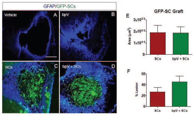
(A–D) SCs in both transplantation groups (C and D) but not in the Vehicle (A) or bpV (B) group showed GFP labeling in the lesion, indicating the survival of the grafted SCs. (E) Areas occupied by grafted GFP-SCs was similar between the SCs and bpV + SCs groups. (F) bpV + SCs exhibited greater GFP-SC/Lesion Area ratio than animals receiving SC transplantation alone although the difference was not statistically significant. Scale bar = 1 mm.
Axonal and vascular growth within the SC grafts
To determine whether GFP-SC grafts promoted ingrowth of axons and vasculature, immunofluorescent staining of SMI-31 and RECA-1, respectively, at the center of graft was examined (Fig. 7). Animals received no SC transplantation were served as controls. As shown in Fig. 7C, non-transplanted rats exhibited little to no SMI-31+ axons within the boundaries of GFAP. In contrast, SC transplantation only (Fig. 7G) and bpV + SCs (Fig. 7K) groups promoted remarkable axonal growth into the SC graft. Overall, total axonal area within the graft (Fig. 7M) and axonal area as a percentage of maximal graft area (Fig. 7N) was similar between the two transplantation groups. Nonetheless, as bpV + SCs promoted a higher graft-to-lesion ratio, a significant increase of 89 ± 20% of SMI-31+ axons over SCs only as a ratio of axons to lesion size (Fig. 7O). Likewise, lack of vascular presence in the lesion cavity was found in non-transplanted animals (Fig. 8A & A′). In contrast, evident of vascular ingrowth into the graft region at the lesion site was found in the SC only (Fig. 8B & B′) and bpV + SCs groups (Fig. 8C & C′). Like axonal presence in the graft, vasculature was similarly distributed between the two SC-transplanted groups.
Figure 7. SC transplantation promoted axonal growth within the lesion.

(A–L) High power cross-section view of the ipsilateral lesion epicenter in the Vehicle, SCs, and bpV-SCs groups. (A–D) In the Vehicle control group receiving no SC transplantation, no axonal growth into the lesion was found. (E–L) In groups that receiving SCs (E–H) or bpV-SC (I–L), considerable axonal growth, immunolabeled by SMI-31, into the lesion site was found (G, K) and they were closely associated with grafted GFP-SCs (F–J). (M–O) There was no statistically significance difference between the total SMI-31+ area (M), nor SMI-31+ as a fraction of SC-graft area (N). However, axonal growth into the graft as an index of lesion size was significantly greater in the bpV-SC treatment group as compared with the SCs only group (O). *, p < 0.05. Scale bar = 750 μm.
Figure 8. SCs promoted vascular growth into the graft.

(A–A′) In the Vehicle control group receiving no SC transplantation, no RECA-1+ vasculature was found within the lesion. (B–C′) RECA-1+ vascular growth into the GFP-SC grafts was clearly seen in groups receiving SCs (B–B′) and bpV + SCs (C–C′). White arrowheads indicate RECA-1+ blood vessels. Scale bar = 1 mm (A), 500 μm (B & B′), 50 μm (C & C′).
Discussion
bpV alone or combined with SC transplantation promoted significant forelimb function recovery and neuroprotection
In the present study, we show that bpV promotes neuroprotection and functional recovery, and that these benefits are similar to those provided by the combination of bpV and SCs. This evidence confirms bpV as a valuable therapy for improving functional recovery, which is likely closely tied to its neuroprotective effects. When transplanted alone, SCs did not significantly improve forelimb functional ability at the end of the study (Fig. 2), but showed remarkable survival throughout the nearly 3 month long study (Fig. 6). In a recent report, we showed that SCs isolated from this transgenic GFP-expressing SD rat strain showed proliferative and considerable survival potential as assessed through 1 year post-transplantation in a contusive SCI lesion produced in SD rats (Wang and Xu, 2014). As SD rats are outbred rats, this recent study involved long-term administration of the immunosuppressant cyclosporin A to repress potential rejection of the SC graft by the rats. As administering cyclosporin A could affect the pharmacologic effects of bpV utilized in our study, we did not use cyclosporin in any animals in the present study. Upon final histological assessment, we observed relatively healthy SC grafts in nearly all of the transplanted rats (7 of 9). These results suggest that the GFP-SCs survived for approximately three months in SD rats without the use of immunosuppressant therapy in the current study.
Although SCs were identifiable in animals transplanted with SCs or bpV + SCs, treatment with bpV, regardless of subsequent SC transplantation, promoted greater functional recovery over SCs alone. Even still, there was no distinct evidence or observation that bpV directly affected the SC graft or that it influenced host tissue interaction with the transplanted SCs. The SCs, however, play a unique role in providing permissive substrate for axonal growth and revascularization through the injury site. We cannot determine whether SCs definitively contributed to recovery of forelimb function based on our data presented here. As we only used one behavioral assessment for forelimb recovery, this test may not be sensitive for detection of recovery following SC transplantation. Alternatively, SCs alone may not be sufficient to meaningfully increase forelimb recovery as suggested in other studies. Thus, developing new repair strategies combining SC transplantation and other efficacious strategies such as the use of bpV acute neuroprotection may enhance functional recovery.
Based on functional assessment results, bpV’s effect on function recovery is maintained over the course of both 6 (Walker et al., 2012) and 11 weeks post-SCI. Further affirming these results, the combination therapy showed very similar neuroprotective effects, as well as sustained improvement in functional outcome over the course of this study. SC transplantation alone did not promote significant neuroprotection compared to the Vehicle group (Fig. 3). In addition, behavioral assessment scores inversely correlated with lesion area (Fig. 3H). Nevertheless, our findings suggest bpV may have played a more important role in neuroprotection and functional recovery than SC transplantation. Also, both bpV and bpV + SC groups showed similar lesion reduction ability which raises the possibility that bpV may have already reduced the lesion before SCs were transplanted. Despite not having cells transplanted into the lesion, bpV exhibited similar lesion cavity reduction to both the SCs and bpV + SCs treatment groups, further supporting this interpretation. In continuation of the beneficial influence of bpV on neuroprotection, only bpV and bpV + SCs groups showed significant ventral horn neuron sparing caudal to the injury, and a similar trend rostrally (Fig. 5).
Influence of bpV on SCs and host axon growth into the lesion
Although the total area of the SC graft at the epicenter was similar between groups (1.88 × 10⁵ ± 6.3 × 10⁴ vs. 1.87 × 10⁵ ± 5.4 × 104 μm2; Fig. 6E), the graft-to-lesion ratio was higher in the combination group compared to animals that received only SC transplantation (Fig. 6E & F). Reduced lesion area promoted by acute bpV treatment resulted in a somewhat greater graft-to-lesion ratio (Fig. 6F), which may have contributed to a significantly higher axon/lesion area value (Fig. 7D). However, no statistically significant differences were observed between the SC and bpV + SC treatment groups concerning graft/lesion ratio. When investigating axonal presence within the lesion in non-grafted animals (Fig. 7A), almost no axons were observed inside the GFAP boundaries of the lesion. Using SCs as a lone therapy, Schaal and colleagues transplanted cells into the contused cervical spinal cord and observed supraspinal axonal growth into the graft, as well as SC-mediated forelimb functional recovery (Schaal et al., 2007). In 2008, one group loaded SCs into poly-β-hydroxybutyrate (PBH) channels, implanted the graft into the hemisected gap, and identified serotonergic raphespinal and calcitonin gene-related peptide (CGRP) fibers, but not descending motor rubrospinal axons within the graft (Novikova et al., 2008). Although we observed axonal growth into the graft, our study did not assess the origin of these axons. As no significant difference in functional improvement was observed between bpV-treated and bpV + SCs treated animals, the significant axonal occupation within the lesion may have been incidental to the reduced lesion area concerning effects on functional recovery.
SCs promoted vascular ingrowth
A similar pattern was observed for RECA-1+ vasculature. In both transplant groups, SC transplantation promoted clear vascular growth at the epicenter of the graft (Fig. 8B–C′), while negligible vasculature was observed in the lesions of non-SC transplanted animals (Fig. 8A & A′). To our knowledge, this study is the first to report vascular growth into the SC graft in a cervical SCI model. The exact mechanism influencing vascular generation, or the influence these vessels imparted on the grafted SCs or axonal ingrowth in this model is unclear. Evidence suggests angiogenesis is beneficial following SCI (Han et al., 2010), and can promote axonal sprouting and growth (Dray et al., 2009; Loy et al., 2001), all findings that support the results observed in the present study. Although both SCs and bpV + SC transplantation groups exhibited vascular labeling within the SC graft, we do not have sufficient evidence to determine whether the combination strategy was better at promoting vascularization of the graft than SC transplantation alone. Further analysis of vascular growth in the graft will help identify whether these vessels are functional, and whether their effects contribute benefits to the grafted cells or surrounding host tissue.
Association between treatment and functional recovery
What potentially contributed to the functional improvement in the bpV and bpV + SCs groups based on our histological findings? This outcome could have resulted from both gray and white matter sparing in these groups as both treatments promoted significantly reduced lesion areas and increased overall spared tissue at the lesion epicenter, a measure that significantly correlates with lesion severity and behavioral analysis outcomes following contusive SCI (Bresnahan et al., 1987; Noble and Wrathall, 1985). However, as all injured animals failed to fully extend and grasp the treats with the digits suggests chronic corticospinal tract damage in the ipsilateral dorsal column. Therefore, further analysis of bpV’s effects on this tract could help shed light on mechanisms to target CST protection and repair to further improve the rats’ ability to perform the treat manipulation exercise.
In a thoracic contusive SCI, SCs have demonstrated the ability to enhance Basso, Beattie, and Bresnahan (BBB) (Basso et al., 1995) hindlimb locomotor scores (Barakat et al., 2005; Takami et al., 2002). Conversely, Pearse and colleagues showed no benefit in BBB scoring for animals transplanted only with SCs over injury alone, although SCs could significantly improve hindpaw rotation in footprint analysis (Pearse et al., 2004a; Pearse et al., 2007). Evidence suggests SCs are more effective for repair than other cell types for transplantation, such as olfactory ensheathing cells or glia (OECs or OEGs) for treating experimental contusive SCI (Takami et al., 2002); however, in most studies, combining SCs with other therapies or genetically-modifying SCs promotes more notable recovery in rat behavioral assessments than SCs alone even when SCs exhibit a benefit. In one recent study, injured animals grafted with SCs genetically modified to overexpress the neurotrophic factor GDNF showed improved locomotor recovery over SCs alone (Deng et al., 2013). These mixed results suggest that although SCs promote positive results as a therapy for SCI, their effects may not be sufficient for recovery unless other therapies are combined. Our recent research supports this notion, as SC transplantation did not improve BBB scores or footfall errors over control animals (Wang and Xu, 2014). Various objective measures of gait during locomotion also showed no significant difference compared to controls. We did observe incredible survival capability of the transplanted SCs, however, which should inspire continued optimism in the use of SC transplantation in combined therapies.
Biphasic versus overlapping combination therapy
Remarkably few studies have been reported on SC transplantation at the cervical level, however, those that were performed focused on combining SCs with an additional treatment or strategy to produce host and SC graft effects. Our study continues this trend with the biphasic combination treatment of acute bpV and subacute SC transplantation. The most comparable study of the cervical SC transplantation studies investigated a co-treatment of 17β-estradiol with transplantation of SCs into the hemi-contused cervical spinal cord (Siriphorn et al., 2010). In this study, a 17β-estradiol (E2) pellet (5 mg/pellet, 21-day continuous release) was implanted subcutaneously 30 min following injury. After a delay of 8 days post-SCI, 5-(and-6)-carboxyfluoreceindiacetatesuccinimidyl ester (CFSE)-labeled SCs were transplanted into the contusion site and animals were allowed to survive for 7 days following transplantation. The overlapping administration of 17β-estradiol and SC transplantation, aimed at testing E2 effects on SC survival, is interesting and yielded novel information. In our case, we examined a two-phase treatment regimen with bpV(pic) administration and SC transplantation to determine whether the biphasic treatments lead to synergistic effects. We are investigating potential effects of bpV on SCs in vitro to address this possibility, as well as other potential combination benefits of bpV and SCs for SCI.
Conclusion
Ultimately, the results of this study suggest bpV likely promoted most of the neuroprotective benefits, and played a larger role in functional recovery than SC transplantation when the two therapies were combined. Transplanted SCs elicited a dynamic host tissue response, promoting considerable axonal and vascular ingrowth over the course of the study. The impact of these responses on recovery, however, is indeterminable from the observed results. The vasculature observed in the graft, if functional, could have provided nutrients and growth components necessary for long-term SC survival and axon growth. The bpV component of our combination therapy possibly enhanced recovery through acute sparing of neurons and other cells from secondary damage following SCI. If combined with another treatment to degrade the glial scar or allow SC overexpression of trophic factors, transplantation of SCs may contribute more substantially to neuroprotection, axonal growth into caudal host tissue, and forelimb functional recovery. Future studies will address these and other issues concerning the potential of SCs in combination therapies following SCI.
Highlights.
Schwann cells (SCs) are a promising candidate for cell therapy after spinal cord injury
However, Schwann cells have limitations when used alone
Bisperoxovanadium (bpV) compound is a promising neuroprotective agent
Combining bpV with SC transplantation enhanced neuroprotection and recovery
Acknowledgments
This work was supported in part by National Institutes of Health (NIH/NINDS R01 NS059622, NS050243, NS073636), and DOD (CDMRP W81XWH-12-1-0562) to XMX, and NIH F31NS 071863 to CLW), the Indiana Spinal Cord and Brain Injury Research Funds, the Mari Hulman George Endowment Funds. We appreciate the use of the Core facility of the Spinal Cord and Brain Injury Research Group/Stark Neurosciences Research Institute at Indiana University. We also thank Drs. Theodore Cummins, Xiao-Ming Jin, and Feng Zhou for critical reading and constructive advice in the preparation of this manuscript.
Footnotes
Publisher's Disclaimer: This is a PDF file of an unedited manuscript that has been accepted for publication. As a service to our customers we are providing this early version of the manuscript. The manuscript will undergo copyediting, typesetting, and review of the resulting proof before it is published in its final citable form. Please note that during the production process errors may be discovered which could affect the content, and all legal disclaimers that apply to the journal pertain.
References
- Barakat DJ, Gaglani SM, Neravetla SR, Sanchez AR, Andrade CM, Pressman Y, Puzis R, Garg MS, Bunge MB, Pearse DD. Survival, integration, and axon growth support of glia transplanted into the chronically contused spinal cord. Cell transplantation. 2005;14:225–240. doi: 10.3727/000000005783983106. [DOI] [PubMed] [Google Scholar]
- Basso DM, Beattie MS, Bresnahan JC. A sensitive and reliable locomotor rating scale for open field testing in rats. Journal of neurotrauma. 1995;12:1–21. doi: 10.1089/neu.1995.12.1. [DOI] [PubMed] [Google Scholar]
- Bresnahan JC, Beattie MS, Todd FDI, Noyes DH. A behavioral and anatomical analysis of spinal cord injury produced by a feedback-controlled impaction device. Experimental neurology. 1987;95:548–570. doi: 10.1016/0014-4886(87)90299-8. [DOI] [PubMed] [Google Scholar]
- Bunge MB. Novel combination strategies to repair the injured mammalian spinal cord. The journal of spinal cord medicine. 2008;31:262–269. doi: 10.1080/10790268.2008.11760720. [DOI] [PMC free article] [PubMed] [Google Scholar]
- Cross DA, Alessi DR, Cohen P, Andjelkovich M, Hemmings BA. Inhibition of glycogen synthase kinase-3 by insulin mediated by protein kinase B. Nature. 1995;378:785–789. doi: 10.1038/378785a0. [DOI] [PubMed] [Google Scholar]
- Datta SR, Dudek H, Tao X, Masters S, Fu H, Gotoh Y, Greenberg ME. Akt phosphorylation of BAD couples survival signals to the cell-intrinsic death machinery. Cell. 1997;91:231–241. doi: 10.1016/s0092-8674(00)80405-5. [DOI] [PubMed] [Google Scholar]
- David S, Aguayo AJ. Axonal elongation into peripheral nervous system “bridges” after central nervous system injury in adult rats. Science. 1981;214:931–933. doi: 10.1126/science.6171034. [DOI] [PubMed] [Google Scholar]
- David S, Aguayo AJ. Axonal regeneration after crush injury of rat central nervous system fibres innervating peripheral nerve grafts. Journal of neurocytology. 1985;14:1–12. doi: 10.1007/BF01150259. [DOI] [PubMed] [Google Scholar]
- Deng LX, Deng P, Ruan Y, Xu ZC, Liu NK, Wen X, Smith GM, Xu XM. A Novel Growth-Promoting Pathway Formed by GDNF-Overexpressing Schwann Cells Promotes Propriospinal Axonal Regeneration, Synapse Formation, and Partial Recovery of Function after Spinal Cord Injury. J Neurosci. 2013;33:5655–5667. doi: 10.1523/JNEUROSCI.2973-12.2013. [DOI] [PMC free article] [PubMed] [Google Scholar]
- Deng LX, Hu J, Liu N, Wang X, Smith GM, Wen X, Xu XM. GDNF modifies reactive astrogliosis allowing robust axonal regeneration through Schwann cell-seeded guidance channels after spinal cord injury. Experimental neurology. 2011;229:238–250. doi: 10.1016/j.expneurol.2011.02.001. [DOI] [PMC free article] [PubMed] [Google Scholar]
- Dray C, Rougon G, Debarbieux F. Quantitative analysis by in vivo imaging of the dynamics of vascular and axonal networks in injured mouse spinal cord. Proc Natl Acad Sci U S A. 2009;106:9459–9464. doi: 10.1073/pnas.0900222106. [DOI] [PMC free article] [PubMed] [Google Scholar]
- Gensel JC, Tovar CA, Hamers FP, Deibert RJ, Beattie MS, Bresnahan JC. Behavioral and histological characterization of unilateral cervical spinal cord contusion injury in rats. Journal of neurotrauma. 2006;23:36–54. doi: 10.1089/neu.2006.23.36. [DOI] [PubMed] [Google Scholar]
- Golden KL, Pearse DD, Blits B, Garg MS, Oudega M, Wood PM, Bunge MB. Transduced Schwann cells promote axon growth and myelination after spinal cord injury. Experimental neurology. 2007;207:203–217. doi: 10.1016/j.expneurol.2007.06.023. [DOI] [PMC free article] [PubMed] [Google Scholar]
- Gruner JA. A monitored contusion model of spinal cord injury in the rat. Journal of neurotrauma. 1992;9:123–128. doi: 10.1089/neu.1992.9.123. [DOI] [PubMed] [Google Scholar]
- Han S, Arnold SA, Sithu SD, Mahoney ET, Geralds JT, Tran P, Benton RL, Maddie MA, D’Souza SE, Whittemore SR, Hagg T. Rescuing vasculature with intravenous angiopoietin-1 and alpha v beta 3 integrin peptide is protective after spinal cord injury. Brain: a journal of neurology. 2010;133:1026–1042. doi: 10.1093/brain/awq034. [DOI] [PMC free article] [PubMed] [Google Scholar]
- Hill CE, Hurtado A, Blits B, Bahr BA, Wood PM, Bartlett Bunge M, Oudega M. Early necrosis and apoptosis of Schwann cells transplanted into the injured rat spinal cord. Eur J Neurosci. 2007;26:1433–1445. doi: 10.1111/j.1460-9568.2007.05771.x. [DOI] [PubMed] [Google Scholar]
- Hill CE, Moon LD, Wood PM, Bunge MB. Labeled Schwann cell transplantation: cell loss, host Schwann cell replacement, and strategies to enhance survival. Glia. 2006;53:338–343. doi: 10.1002/glia.20287. [DOI] [PubMed] [Google Scholar]
- Houle JD. Demonstration of the potential for chronically injured neurons to regenerate axons into intraspinal peripheral nerve grafts. Experimental neurology. 1991;113:1–9. doi: 10.1016/0014-4886(91)90139-4. [DOI] [PubMed] [Google Scholar]
- Houle JD, Cote MP. Axon regeneration and exercise-dependent plasticity after spinal cord injury. Ann N Y Acad Sci. 2013;1279:154–163. doi: 10.1111/nyas.12052. [DOI] [PMC free article] [PubMed] [Google Scholar]
- Hu JG, Wang XF, Deng LX, Liu NK, Gao X, Chen JH, Zhou FC, Xu XM. Co-transplantation of glial restricted precursor cells and Schwann cells promotes functional recovery after spinal cord injury. Cell transplantation. 2013 doi: 10.3727/096368912X661373. [DOI] [PubMed] [Google Scholar]
- Iannotti C, Li H, Yan P, Lu X, Wirthlin L, Xu XM. Glial cell line-derived neurotrophic factor-enriched bridging transplants promote propriospinal axonal regeneration and enhance myelination after spinal cord injury. Experimental neurology. 2003;183:379–393. doi: 10.1016/s0014-4886(03)00188-2. [DOI] [PubMed] [Google Scholar]
- Iannotti C, Zhang YP, Shields CB, Han Y, Burke DA, Xu X-M. A novel neuroprotective role for glial cell line-derived neurotrophic factor following moderate spinal cord contusion injury. Experimental neurology. 2004 doi: 10.1016/j.expneurol.2004.05.033. (In press) [DOI] [PubMed] [Google Scholar]
- Liu NK, Zhang YP, Titsworth WL, Jiang X, Han S, Lu PH, Shields CB, Xu XM. A novel role of phospholipase A(2) in mediating spinal cord secondary injury. Ann Neurol. 2006;59:606–619. doi: 10.1002/ana.20798. [DOI] [PubMed] [Google Scholar]
- Loy DN, Crawford CH, Darnall JB, Burke DA, Whittemore SR. The temporal progression of angiogenesis and basal lamina deposition following contusive spinal cord injury n the adult rat. Experimental neurology. 2001 doi: 10.1002/cne.10168. [DOI] [PubMed] [Google Scholar]
- Mao L, Jia J, Zhou X, Xiao Y, Wang Y, Mao X, Zhen X, Guan Y, Alkayed NJ, Cheng J. Delayed administration of a PTEN inhibitor BPV improves functional recovery after experimental stroke. Neuroscience. 2013;231:272–281. doi: 10.1016/j.neuroscience.2012.11.050. [DOI] [PMC free article] [PubMed] [Google Scholar]
- Martin JH. Differential spinal projections from the forelimb areas of the rostral and caudal subregions of primary motor cortex in the cat. Experimental brain research Experimentelle Hirnforschung Experimentation cerebrale. 1996;108:191–205. doi: 10.1007/BF00228094. [DOI] [PubMed] [Google Scholar]
- Morrissey TK, Kleitman N, Bunge RP. Isolation and functional characterization of Schwann cells derived from adult peripheral nerve. J Neurosci. 1991;11:2433–2442. doi: 10.1523/JNEUROSCI.11-08-02433.1991. [DOI] [PMC free article] [PubMed] [Google Scholar]
- Nakashima S, Arnold SA, Mahoney ET, Sithu SD, Zhang YP, D’Souza SE, Shields CB, Hagg T. Small-molecule protein tyrosine phosphatase inhibition as a neuroprotective treatment after spinal cord injury in adult rats. J Neurosci. 2008;28:7293–7303. doi: 10.1523/JNEUROSCI.1826-08.2008. [DOI] [PMC free article] [PubMed] [Google Scholar]
- Nave BT, Ouwens M, Withers DJ, Alessi DR, Shepherd PR. Mammalian target of rapamycin is a direct target for protein kinase B: identification of a convergence point for opposing effects of insulin and amino-acid deficiency on protein translation. The Biochemical journal. 1999;344(Pt 2):427–431. [PMC free article] [PubMed] [Google Scholar]
- Noble LJ, Wrathall JR. Spinal cord contusion in the rat: Morphometric analyses of alterations in the spinal cord. Experimental neurology. 1985;88:135–149. doi: 10.1016/0014-4886(85)90119-0. [DOI] [PubMed] [Google Scholar]
- Novikova LN, Pettersson J, Brohlin M, Wiberg M, Novikov LN. Biodegradable poly-beta-hydroxybutyrate scaffold seeded with Schwann cells to promote spinal cord repair. Biomaterials. 2008;29:1198–1206. doi: 10.1016/j.biomaterials.2007.11.033. [DOI] [PubMed] [Google Scholar]
- Oudega M, Xu XM. Schwann cell transplantation for repair of the adult spinal cord. Journal of neurotrauma. 2006;23:453–467. doi: 10.1089/neu.2006.23.453. [DOI] [PubMed] [Google Scholar]
- Pearse DD, Marcillo AE, Oudega M, Lynch MP, Wood PM, Bunge MB. Transplantation of Schwann cells and olfactory ensheathing glia after spinal cord injury: does pretreatment with methylprednisolone and interleukin-10 enhance recovery? Journal of neurotrauma. 2004a;21:1223–1239. doi: 10.1089/neu.2004.21.1223. [DOI] [PubMed] [Google Scholar]
- Pearse DD, Pereira FC, Marcillo AE, Bates ML, Berrocal YA, Filbin MT, Bunge MB. cAMP and Schwann cells promote axonal growth and functional recovery after spinal cord injury. Nature medicine. 2004b;10:610–616. doi: 10.1038/nm1056. [DOI] [PubMed] [Google Scholar]
- Pearse DD, Sanchez AR, Pereira FC, Andrade CM, Puzis R, Pressman Y, Golden K, Kitay BM, Blits B, Wood PM, Bunge MB. Transplantation of Schwann cells and/or olfactory ensheathing glia into the contused spinal cord: Survival, migration, axon association, and functional recovery. Glia. 2007;55:976–1000. doi: 10.1002/glia.20490. [DOI] [PubMed] [Google Scholar]
- Schaal SM, Kitay BM, Cho KS, Lo TP, Jr, Barakat DJ, Marcillo AE, Sanchez AR, Andrade CM, Pearse DD. Schwann cell transplantation improves reticulospinal axon growth and forelimb strength after severe cervical spinal cord contusion. Cell transplantation. 2007;16:207–228. doi: 10.3727/000000007783464768. [DOI] [PubMed] [Google Scholar]
- Schmid AC, Byrne RD, Vilar R, Woscholski R. Bisperoxovanadium compounds are potent PTEN inhibitors. FEBS Lett. 2004;566:35–38. doi: 10.1016/j.febslet.2004.03.102. [DOI] [PubMed] [Google Scholar]
- Siriphorn A, Chompoopong S, Floyd CL. 17beta-Estradiol protects Schwann cells against H(2)O(2)-induced cytotoxicity and increases transplanted Schwann cell survival in a cervical hemicontusion spinal cord injury model. J Neurochem. 2010;115:864–872. doi: 10.1111/j.1471-4159.2010.06770.x. [DOI] [PubMed] [Google Scholar]
- Song W, Volosin M, Cragnolini AB, Hempstead BL, Friedman WJ. ProNGF induces PTEN via p75NTR to suppress Trk-mediated survival signaling in brain neurons. J Neurosci. 2010;30:15608–15615. doi: 10.1523/JNEUROSCI.2581-10.2010. [DOI] [PMC free article] [PubMed] [Google Scholar]
- Sury MD, Vorlet-Fawer L, Agarinis C, Yousefi S, Grandgirard D, Leib SL, Christen S. Restoration of Akt activity by the bisperoxovanadium compound bpV(pic) attenuates hippocampal apoptosis in experimental neonatal pneumococcal meningitis. Neurobiol Dis. 2011;41:201–208. doi: 10.1016/j.nbd.2010.09.007. [DOI] [PMC free article] [PubMed] [Google Scholar]
- Takami T, Oudega M, Bates ML, Wood PM, Kleitman N, Bunge MB. Schwann cell but not olfactory ensheathing glia transplants improve hindlimb locomotor performance in the moderately contused adult rat thoracic spinal cord. J Neurosci. 2002;22:6670–6681. doi: 10.1523/JNEUROSCI.22-15-06670.2002. [DOI] [PMC free article] [PubMed] [Google Scholar]
- Tom VJ, Sandrow-Feinberg HR, Miller K, Domitrovich C, Bouyer J, Zhukareva V, Klaw MC, Lemay MA, Houle JD. Exogenous BDNF enhances the integration of chronically injured axons that regenerate through a peripheral nerve grafted into a chondroitinase-treated spinal cord injury site. Experimental neurology. 2013;239:91–100. doi: 10.1016/j.expneurol.2012.09.011. [DOI] [PMC free article] [PubMed] [Google Scholar]
- Tom VJ, Sandrow-Feinberg HR, Miller K, Santi L, Connors T, Lemay MA, Houle JD. Combining peripheral nerve grafts and chondroitinase promotes functional axonal regeneration in the chronically injured spinal cord. J Neurosci. 2009;29:14881–14890. doi: 10.1523/JNEUROSCI.3641-09.2009. [DOI] [PMC free article] [PubMed] [Google Scholar]
- Walker CL, Walker MJ, Liu NK, Risberg EC, Gao X, Chen J, Xu XM. Systemic bisperoxovanadium activates Akt/mTOR, reduces autophagy, and enhances recovery following cervical spinal cord injury. PLoS One. 2012;7:e30012. doi: 10.1371/journal.pone.0030012. [DOI] [PMC free article] [PubMed] [Google Scholar]
- Walker CL, Xu XM. PTEN inhibitor bisperoxovanadium protects oligodendrocytes and myelin and prevents neuronal atrophy in adult rats following cervical hemicontusive spinal cord injury. Neuroscience letters. 2014 doi: 10.1016/j.neulet.2014.02.039. [DOI] [PMC free article] [PubMed] [Google Scholar]
- Wang X, Xu XM. Long-term survival, axonal growth-promotion, and myelination of Schwann cells grafted into contused spinal cord in adult rats. Experimental neurology. 2014;261:308–319. doi: 10.1016/j.expneurol.2014.05.022. [DOI] [PMC free article] [PubMed] [Google Scholar]
- Xu XM, Chen A, Guenard V, Kleitman N, Bunge MB. Bridging Schwann cell transplants promote axonal regeneration from both the rostral and caudal stumps of transected adult rat spinal cord. Journal of neurocytology. 1997;26:1–16. doi: 10.1023/a:1018557923309. [DOI] [PubMed] [Google Scholar]
- Xu XM, Guenard V, Kleitman N, Aebischer P, Bunge MB. A combination of BDNF and NT-3 promotes supraspinal axonal regeneration into Schwann cell grafts in adult rat thoracic spinal cord. Experimental neurology. 1995a;134:261–272. doi: 10.1006/exnr.1995.1056. [DOI] [PubMed] [Google Scholar]
- Xu XM, Guenard V, Kleitman N, Bunge MB. Axonal regeneration into Schwann cell-seeded guidance channels grafted into transected adult rat spinal cord. The Journal of comparative neurology. 1995b;351:145–160. doi: 10.1002/cne.903510113. [DOI] [PubMed] [Google Scholar]
- Xu XM, Zhang SX, Li H, Aebischer P, Bunge MB. Regrowth of axons into the distal spinal cord through a Schwann-cell-seeded mini-channel implanted into hemisected adult rat spinal cord. Eur J Neurosci. 1999;11:1723–1740. doi: 10.1046/j.1460-9568.1999.00591.x. [DOI] [PubMed] [Google Scholar]
- Yang P, Dankowski A, Hagg T. Protein tyrosine phosphatase inhibition reduces degeneration of dopaminergic substantia nigra neurons and projections in 6-OHDA treated adult rats. Eur J Neurosci. 2007;25:1332–1340. doi: 10.1111/j.1460-9568.2007.05384.x. [DOI] [PubMed] [Google Scholar]
- Zhang QG, Wu DN, Han D, Zhang GY. Critical role of PTEN in the coupling between PI3K/Akt and JNK1/2 signaling in ischemic brain injury. FEBS Lett. 2007;581:495–505. doi: 10.1016/j.febslet.2006.12.055. [DOI] [PubMed] [Google Scholar]
- Zhang YP, Iannotti C, Shields LB, Han Y, Burke DA, Xu XM, Shields CB. Dural closure, cord approximation, and clot removal: enhancement of tissue sparing in a novel laceration spinal cord injury model. Journal of neurosurgery. 2004;100:343–352. doi: 10.3171/spi.2004.100.4.0343. [DOI] [PubMed] [Google Scholar]
- Zhao J, Qu Y, Wu J, Cao M, Ferriero DM, Zhang L, Mu D. PTEN inhibition prevents rat cortical neuron injury after hypoxia-ischemia. Neuroscience. 2013 doi: 10.1016/j.neuroscience.2013.02.046. [DOI] [PubMed] [Google Scholar]