Abstract
Background & Purpose
Much attention has been solely paid to physical outcome measures for return to sport after injury in the past. However, current research shows that the psychological component of these injuries can be more predictive of return to sport than physical outcome measures. The purpose of this case report is to describe the successful return to sport following surgery of a complicated tibia and fibula fracture of a Division I collegiate women's soccer player with a low level of kinesiophobia.
Case Description
A 22‐year‐old female sustained a closed traumatic mid‐shaft fracture of her tibia and fibula. During a high velocity play she sustained a direct blow while colliding with an opposing player's cleats. As a result of the play, her distal tibia was displaced 908 to the rest of her leg. She underwent a closed reduction and tibial internal fixation with an intramedullary rod. Outcome scores were tracked using the IKDC and TSK‐11. The IKDC measures symptoms, function, and sport activity related to knee injuries. The TSK‐11 measures fear of movement and re‐injury, which was important to assess during this case due to the gruesome nature of the injury.
Outcomes
At 4 months, the subject became symptomatic over the fibula and was diagnosed with a fibular nonunion fracture. This was unexpected due to the low incidence of and usual asymptomatic nature of fibular nonunion fractures, which required an additional surgery. TSK‐11 scores ranged from 19‐20 throughout, signifying low levels of kinesiophobia. IKDC scores improved from 8.05 to 60.92. The subject ultimately signed a professional soccer contract.
Discussion
The rehabilitation of this subject was complex due to her low levels of kinesiophobia, self‐guided overtraining, and the potential role they may have had in her fibular nonunion fracture. This case study demonstrates a successful outcome despite a unique injury presentation, multiple surgeries, and low levels of kinesiophobia. While a low level of kinesiophobia can be detrimental to rehabilitation compliance, it may have benefited her in the long‐term.
Level of Evidence
5
Keywords: Fracture, kinesiophobia, soccer
Background and Purpose
There is a high prevalence of fractures in Division I college sports, especially in athletes participating in contact sports such as soccer.1 In fact, the majority of injuries that soccer players experience are high impact traumas, with slide tackles being the most common lower extremity fracture mechanism.2 While lower extremity fractures are common in soccer, research regarding prognosis and outcomes for high impact fractures is lacking.3-4 This is problematic due to the variables associated with making a safe return to play decision. Therefore, prognostic factors that may positively or negatively affect the rehabilitation process and ultimately return to sport must be recognized.
Previous attention has been paid to physical outcome measures for return to sport. However, current research shows that the psychological component of these injuries can be more predictive of return to play than physical outcome measures.5-10 Athletes that sustain traumatic injuries may never return to their sport due to fear of re‐injury.5-10 This fear of re‐injury is an example of how a negative psychological state can hinder an outcome. Inversely, a positive psychological response to injury and rehabilitation correlates with a more rapid return to sport within a year.5-10
Kinesiophobia has been defined as an “excessive, irrational, and debilitating fear of physical movement and activity resulting from a feeling of vulnerability to painful injury or re‐injury.” 11 The fear associated with kinesiophobia can be heightened in injuries due to trauma.11 Behavior that is guided by kinesiophobia has the potential to negatively impact outcomes for patients with pain.12-13 According to the fear avoidance model (FAM) of exaggerated pain perception proposed by Lethem et al14, pain perception involves both a sensory and an emotional reaction component. Fear of pain is an important feature of the emotional reaction in that it can bring out two different forms of coping responses: confrontation or avoidance. An individual motivated by fear typically avoids both painful experiences and activities. Fear avoidance behaviors can also be indicative of high psychological distress, which has further been associated with poor clinical outcomes.14
Evidence supports the assessment of pain‐related fear in patients across multiple musculoskeletal conditions ranging from sub‐acute to chronic conditions.5-8,12-13,15-26 Higher levels of pain have been shown to be predictive of higher levels of disability, and fear of pain has been associated with kinesiophobia. 5-8,12-13,15-26 In order to help produce the best clinical outcomes it is important to identify patients who are at risk for kinesiophobia. Outcome tools such as the Tampa Scale for Kinesiophobia (TSK) and its shortened‐version the TSK‐11, can be used to better understand the psychological impact of an injury. The TSK questionnaire involves items that incorporate fear of injury, fear of pain and a person's ability to perceive and report symptoms.11The combination of both physical and psychological outcome tools can help to facilitate a safe return to play.
The purpose of this case report is to describe the successful return to sport following surgery of a complicated tibia and fibula fracture of a Division I collegiate women's soccer player with a low level of kinesiophobia.
Case Description: Patient History and Systems Review
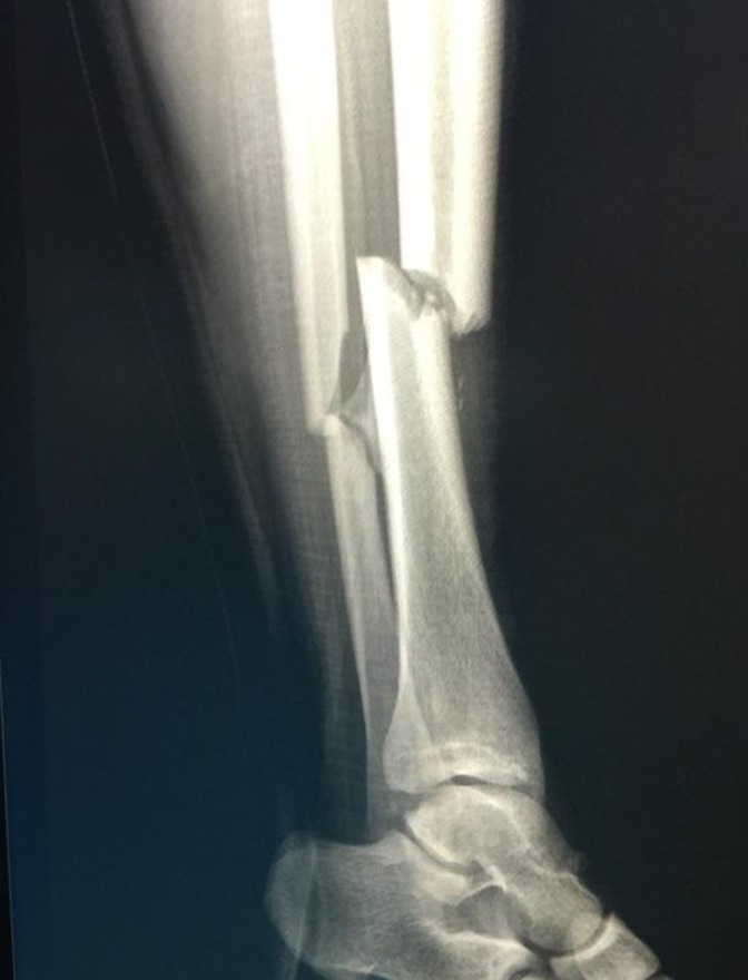
A 22 year‐old female Division I collegiate soccer player with no previous significant medical history or history of injury sustained a displaced, closed mid‐shaft right tibia and fibula fracture while playing in a soccer game. During a high‐velocity play, she received a direct impact to her anteromedial tibia by an opponent in an attempt to win a 50/50 ball. Despite the use of shin guards, the force of the impact was so great that they did not prevent this injury from occurring. As a result, her distal tibia was medially displaced 90° to the rest of her leg. She was immediately transported to the local emergency department.
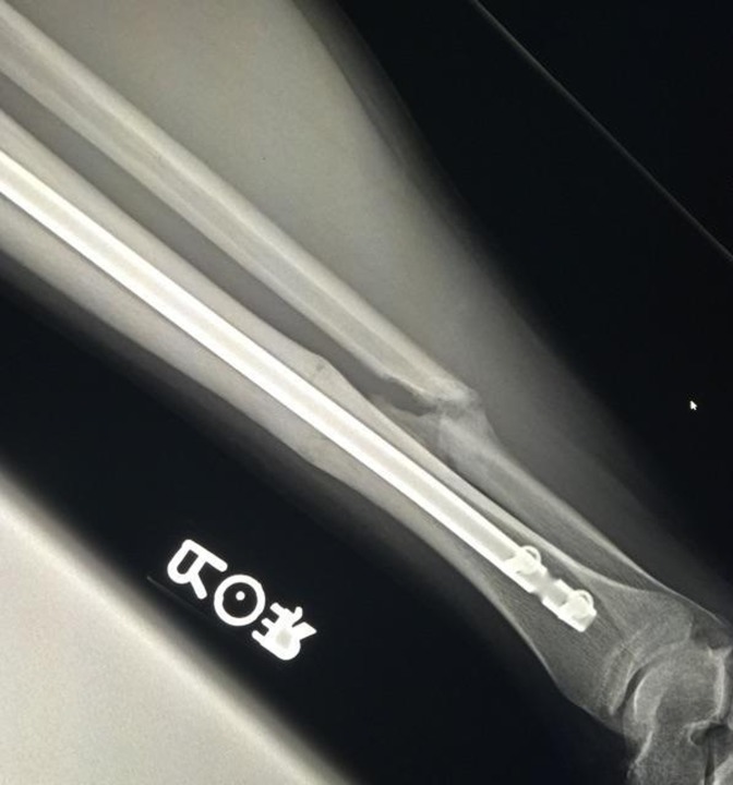
Radiographs confirmed a comminuted and displaced fracture of her right tibia and fibula (Figure 1). She was immediately admitted and underwent closed reduction and intramedullary nailing of the tibia, which is considered to be more advantageous for closed tibial fracture healing and function than casting (Figure 2).27-28 The fibula was reduced but was not fixated. There were no surgical complications.
Figure 1.
Lateral view radiograph of the tibia and fibula depicting a displaced fracture of both bones.
Figure 2.
Lateral view radiograph showing a non‐union of the fibula and near‐complete healing of the tibia.
Clinical Impression #1
At the time of her initial physical therapy evaluation the subject was informed that the data concerning her case would be submitted for publication. Subject confidentiality was protected according to the U.S. Health Insurance Portability and Accountability Act (HIPPA) and IRB approval for this case report was granted. The subject participated in a sport‐specific physical therapy program in the university athletic training facility days after being discharged from the hospital (Table 1). Rehabilitation for this injury follows a plan of care that is predicated on progressions through weight‐bearing activities, gradually increasing physiologic responses to exercise, and limiting any pain‐inducing activity. Impairments were assessed at the time of the initial evaluation and in conjunction with her physician's order.
Table 1.
Rehabilitation Protocol for Post‐Operative Leg Fractures
| PHASE | ACUTE | SUBACUTE | CHRONIC | FUNCTIONAL |
|---|---|---|---|---|
| Goals | Protect surgery Control inflammatory response Control pain, edema, spasm |
Full AROM 5/5 MMT Normalized gait SLS ≥ 30 seconds |
Pain‐free jogging Return to modified soccer specific activities Progression to plyometrics |
Return to sport |
| Weight‐bearing | WBAT | FWB | ||
| External Support | Axillary crutches Wrapping |
Aircast Ankle lace‐up |
Taping or bracing PRN | PRN |
| ROM | PROM → AAROM → AROM | Static stretching PNF stretching |
PNF stretching Ballistic stretching Self‐stretching |
Ballistic stretching Self‐stretching |
| Strengthening | Isometrics | Isotonics OKC Ankle Foot Hip Knee CKC Leg Press Squats Lunges |
Isotonics Olympic lifts OKC Ankle Foot Hip Knee CKC Leg Press Squats Lunges |
Olympic lifts Soccer program |
| PBA | SLS Tilt board |
Soccer‐specific ball drills Plyometrics |
Plyometrics | |
| Complimentary | Hip/ knee isotonic strengthening Upper body ergometry |
Core strengthening Stationary cycling Elliptical |
Treadmill Stair climber Jogging → Running |
Soccer conditioning |
| Modalities | Ice HVGS Effleurage |
Scar massage Contrast baths |
PRN | PRN |
AROM= Active range of motion; MMT= Manual muscle test;SLS= single limb stance; WBAT= weight bearing as tolerated; FWB= full weight bearing; PROM= passive range of motion; AAROM= active assisted range of motion; PNF= proprioceptive neuromuscular facilitation; OKC= open kinetic chain; CKC= closed kinetic chain; HVGS= high voltage galvanic stimulation.
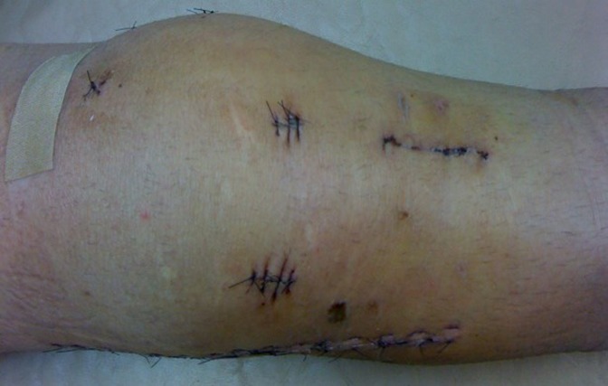
Her post‐operative presentation was normal for after this procedure. Due to the surgical sites for the procedure, most of her impairments were present at the knee (Figure 3). The subject participated in daily treatment sessions under the supervision of her physical therapist and athletic trainer. She was extremely eager to get back to soccer and was thus progressed quickly within the constraints of the protocol.
Figure 3.
Overhead view of post‐operative knee following IM fixation of the tibia.
Due to the traumatic nature of her injury, the treating therapist believed the subject may have been at an increased risk for developing kinesiophobia. However, this subject's extreme desire to return to soccer as soon as possible, as well as her intense work ethic, made her clinical impression unique. For these reasons, her kinesiophobia was assessed and tracked throughout her treatment.
Examination
The IKDC Subjective Knee Form measures pain, symptoms, function, and sport activity related to knee injuries.29-31The survey contains 18 items with a maximum of 87 points related to these domains. A score of 0 on an item demonstrates the least amount of function for the specified activity. Once a final score is determined, it can be plugged into an equation to give a percentile based on the person's age and gender category if the individual is between 18 and 65 years of age.32 Scoring is achieved through the summation of the first four subtest scores. These scores are then transformed into a scale ranging from 0 to 100 through a formula:
Higher scores are indicative of less disability.31 A score of 100 would indicate no disability.31 A change in score of greater than 9 points marks the threshold for the minimal detectable change of the IKDC.30 To distinguish between those who were or were not improved across a wide variety of knee conditions using the IKDC, revealed that a change score of 11.5 points had the highest sensitivity (.82), and a change score of 20.5 points had the highest specificity (.84).29 The IKDC has been shown to be reliable across a broad range of knee pathologies including ligament and meniscal injuries, articular cartilage lesions, and patellofemoral pain.31,33-38 The test battery of questions that comprise the IKDC give reliable results across all age ranges and genders.30
The TSK‐11 is a self‐report questionnaire used to measure fear associated with pain and fear associated with re‐injury.11 The TSK‐11 is scored on a 4‐point scale from 1 (strongly disagree) to 4 (strongly agree). Scores range from 11 to 44 points. Higher scores (>22) are indicative of fear‐related pain or re‐injury. Initially the (17‐item) Tampa Scale for Kinesiophobia (TSK) was used as a fear‐avoidance predictor for chronic low back pain, but it has been more recently studied as a strong predictor of knee, ankle, and shoulder reinjury. 8-9,12,16-18,24-25,39 The TSK‐11 has been proven to have similar reliability and validity to the original TSK.39 Kvist et al9 demonstrated that those patients who did not return to their previous level of activity were more afraid of re‐injury due to movement and had a worse knee‐related quality of life than those who had returned to their previous level of activity, as measured by the TSK. Woby et al39 found that a decrease in score by four points on the TSK increases the likelihood of identifying patients who have undergone an important reduction in fear or movement (sensitivity=66%; specificity=67%). A change of 3 points on the TSK‐11 is needed to be 95% confident that a change has occurred.7 Furthermore, a reduction of 4 or more points maximizes the likelihood of correctly identifying patients with an important decrease in their fear of movement or re‐injury.
Clinical Impression #2
The subject's extreme work ethic and strong desire to return to soccer became more evident as time progressed. Only days after her physical therapy began, it became clear that she was independently exercising beyond what was prescribed. Against medical advice, the subject was simultaneously involved in a self‐directed and intensive conditioning program. The conditioning program took place at the University Wellness Center and involved several hours of weight‐bearing activities on machines such as the elliptical, Jacobs LadderTM, stair climbers, and treadmills. While the subject did not freely admit to her extra conditioning activity, her teammates did report to the medical staff that it indeed was taking place.
The subject began reporting lateral leg pain during the 11th post‐operative week. Radiographs were taken at that time and revealed incomplete healing of both fractures. The subject was advised to reduce her workload and would be re‐evaluated if her symptoms persisted. However, the authors believe she again acted against medical advice and continued on her self‐directed conditioning program. During the 17th post‐operative week, the subject reported worsening of her symptoms and was revaluated. Radiographs showed a one centimeter translational deformity of the fibula and an intervening butterfly fragment revealing nonunion of the fibular fracture.
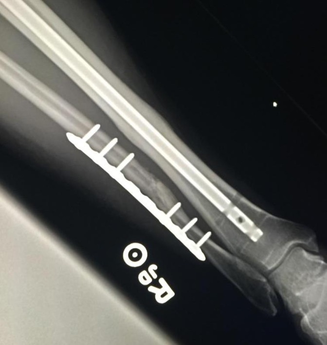
The nonunion required a second surgical intervention to fixate the fibula (Figure 4). This procedure was performed in the 21st week after her initial procedure. This time however, her recovery became complicated due to a surgical site infection with an eventual hospital admission for medical treatment. The subject was instructed to remain non‐weight bearing for the subsequent four weeks after the second surgical procedure. The authors believe this time the subject was compliant with her care and adhered to all precautions. She continued a rehabilitation program at the university training facility until eight months after the incident before leaving to coach and train independently without orthopedic restrictions. One year after the initial incident the subject was able to return to play internationally at the professional level without restrictions. For a detailed timeline of events throughout the course of her recovery, see Table 2.
Figure 4.
AP view radiograph after removal of interlocking screws in the tibial nail and bone grafting with internal fixation of the fibula.
Table 2.
Timeline of Events
| Week | Relevant Events |
|---|---|
| Week 0 | ‐ Initial incident ‐ Hospitalization ‐ Surgical Intervention: tibia reduction and fixation, fibula reduction |
| Week l | ‐ Weight bearing as tolerated |
| Week 11 | ‐ Reports pain with movement in the lateral aspect of the leg ‐ Radiographs show interval healing of both fractures |
| Week 17 | ‐ Patient reports movement and pain on the lateral leg ‐ Recommended for surgical intervention |
| Week 18 | ‐ Radiographs show complete union of tibia and a translation deformity of fibular with more than 1 cm translation and intervening butterfly fragment |
| Week 21 | ‐ Surgical reduction and fixation of fibula ‐ Tibia screws removed |
| Week 23 | ‐ Surgical site infection ‐ Admitted to hospital for IV antibiotics, discharged with home antibiotics |
| Week 24 | ‐ Infection resolved ‐ Radiographs show fibula and mortise in alignment, tibia healing ‐ Cleared to begin full weight bearing |
| Week 29 | ‐ Radiographs show early bridging of nonunion site and integration of bone graft ‐ Cleared to begin strength training as tolerated |
Outcome
In addition to typical impairment and physical function tests and measures, the main outcome measures discussed in this case study include the TSK‐11 and the IKDC (Table 3). The subject improved 52.87 points on the IKDC over the course of four months indicating a high reliability for improved self‐reported function. The decrease seen from November, 2012 to January, 2013 was a result of the second surgery performed to fixate the fibular head. Total TSK scores can range from between 11‐44. In this case, the patient's TSK‐11 scores ranged from 19‐20 throughout the treatment process. Although her scores did not decrease by four points her scores throughout were low, which is indicative of low kinesiophobia.
Table 3.
Post‐Operative TSK‐11 and IKDC Scores
| Post‐Operative TSK‐11 and IKDC Scores | ||||
|---|---|---|---|---|
| Week 3 | Week 7 | Week 17 | Week 21 | |
| TSK‐11 | 20/44 | 19/44 | 19/44 | n/a† |
| IKDC* | 8.05 (<5%) | 45.98 (5%) | 36.78 (<5%) | 60.92 (10%) |
Percentages based on the same age and gender group of the athlete.
Score was not taken due to her consistency with previous scores.
TSK= Tampa Scale for Kinesiophobia;
IKDC= International Knee Documentation Committee
Discussion
High velocity traumatic injuries are common in high level contact sports.4 Tibia‐fibula fractures are seen in soccer players.2,4 Researchers have found that surgical fixation of the tibia followed by physical therapy have been efficient in promoting successful union of the fibula.28,40-41 Specifically, immediate weight bearing of the lower extremity without discomfort or loss of position following lower leg fracture has been proven to have definite advantages in non‐surgical patients.42 Despite fractures of both the tibia and fibula, surgical fixation of the tibia without surgical intervention of the fibula is an acceptable protocol.40-41 Tyllianakis et al28 indicated that interlocking intramedullary nailing of the tibia is a reliable method of treatment associated with high rates of union and low incidence of complications. Non‐union fractures are often the result of non‐optimal healing environments (mechanical or biological).40 Excess movement at the fracture site, due to excess weight bearing in non‐physical therapy activities chosen by the subject of this case report may have inhibited the fibula's ability to heal properly.
Prognosis for return to sport in a tibia‐fibula fracture due to a soccer injury is approximately 40 weeks, however many factors can influence the outcome.2 Kinesiophobia, or pain related fear of movement, has recently received attention as a psychological factor that contributes to the timeline for progression of rehabilitation. An athlete's level of kinesiophobia, either high or low, may greatly impact their recovery timeline and their prognosis for return to competitive sport.7,9 Athletes with low levels of kinesiophobia should be monitored for signs of overtraining and lack of adherence to physical therapy restrictions. However, these athletes may also be more likely to ultimately return to their sport.
Athletes with high levels of kinesiophobia will likely be more hesitant to return to sport.7,9 These athletes may need special attention in order to encourage them throughout therapy and reassure them that it is safe to return to activities. At either extreme, an athlete may also benefit from sports psychological counseling during injury rehabilitation as an adjunct to physical therapy. The TSK‐11 can be a helpful tool in identifying and monitoring an athlete's level of kinesiophobia throughout rehabilitation.
The TSK and TSK‐11 are currently being used across a wide array of diverse patient populations. 5-8,12-13,15-26 Chmielewski et al7 used the TSK‐11 to measure fear of movement in patients undergoing ACL reconstruction rehabilitation and found that it was highest shortly after surgery and gradually decreased as more time passed. They also discovered that as the athletes started to return to sport the lower TSK scores were recorded in the higher functioning patients. Prugh et al10 studied TSK‐11 in throwing athletes with elbow injuries and found that a specific subscale of the TSK‐11 dealing with ‘fear of re‐injury’ has the potential to accurately depict fear of movement, however it was concluded that more research is required to better understand the scale's accuracy as well as the psychological component of an athlete's return to play.10
While the current study did not focus on performing psychometric tests related to the TSK‐11, the results shed light on how it can be clinically used in the rehabilitation of high‐level athletes. The subject's low level of fear after such a gruesome injury could have contributed to her desire to vigorously train outside of rehabilitation and ultimately could have led to the fibular nonunion fracture. This case report suggests a new use for the TSK‐11 in the athletic population. The TSK‐11 can help clinicians monitor how each individual psychologically perceives their injury in order to ensure the proper guidance and education necessary for optimal physical outcomes as needed.
Conclusion
While the subject's psychological state may have been contributory to be a setback initially, it ultimately contributed to her return to sport. Her low level of kinesiophobia may have negatively impacted her compliance with weight bearing restrictions. This non‐compliance may have been what led to her nonunion fracture and the need for a second surgery. Previous studies have found that psychological affect is more predictive of return to sport after injury than physical outcome measures.5 However, it is possible this subject's low level of kinesiophobia allowed her to return to a high level of play following a traumatic injury and subsequent complicated rehabilitation.
References
- 1.Hame SL Lafemina JM Mcallister DR, et al. Fractures in the collegiate athlete. Am J Sports Med. 2004;32(2):446‐51. [DOI] [PubMed] [Google Scholar]
- 2.Boden BP Lohnes JH Nunley JA, et al. Tibia and fibula fractures in soccer players. Knee Surg Sports Traumatol Arthrosc. 1999;7(4):262‐266. [DOI] [PubMed] [Google Scholar]
- 3.Joveniaux P Ohl X Harisboure A, et al. Distal tibia fractures: management and complications of 101 cases. Int Orthop. 2010;34(4):583‐8. [DOI] [PMC free article] [PubMed] [Google Scholar]
- 4.Robertson GA Wood AM Bakker‐Dyos J, et al. The epidemiology, morbidity, and outcome of soccer‐related fractures in a standard population. Am J Sports Med. 2012;40(8):1851‐7. [DOI] [PubMed] [Google Scholar]
- 5.Ardern CL Taylor NF Feller JA, et al. Psychological responses matter in returning to preinjury level of sport after anterior cruciate ligament reconstruction surgery. Am J Sport Med. 2013;41(7):1549‐58. [DOI] [PubMed] [Google Scholar]
- 6.Ardern CL Webster KE Taylor NF, et al. Return to sport following anterior cruciate ligament reconstruction surgery: a systematic review and meta‐analysis of the state of play. Br J Sports Med. 2011;45(7):596‐606. [DOI] [PubMed] [Google Scholar]
- 7.Chmielewski TL Jones D Day T, et al. The association of pain and fear of movement/reinjury with function during anterior cruciate ligament reconstruction rehabilitation. J Orthop Sports Phys Ther. 2008;38(12):746‐53. [DOI] [PubMed] [Google Scholar]
- 8.George SZ Lentz TA Zeppieri G, et al. Analysis of shortened versions of the tampa scale for kinesiophobia and pain catastrophizing scale for patients after anterior cruciate ligament reconstruction. Clin J Pain. 2012;28(1):73‐80. [DOI] [PMC free article] [PubMed] [Google Scholar]
- 9.Kvist J Ek A Sporrstedt K, et al. Fear of re‐injury: a hindrance for returning to sports after anterior cruciate ligament reconstruction. Knee Surg Sports Traumatol Arthrosc. 2005; 13(5):393‐397. [DOI] [PubMed] [Google Scholar]
- 10.Prugh J Zeppieri G George SZ Impact of psychosocial factors, pain, and functional limitations on throwing athletes who return to sport following elbow injuries: a case series. Physiother Theory Pract. 2012;28(8):633‐40. [DOI] [PubMed] [Google Scholar]
- 11.Kori SH Miller RP Todd DD Kinesiophobia: A new view of chronic pain behavior. Pain Manage. 1990; 35‐43. [Google Scholar]
- 12.Abbott AD, Tyni‐Lenné R Hedlund R The influence of psychological factors on pre‐operative levels of pain intensity, disability and health‐related quality of life in lumbar spinal fusion surgery patients. Physiotherapy. 2010;96(3):213‐221. [DOI] [PubMed] [Google Scholar]
- 13.Mintken PE Cleland JA Whitman JM, et al. Psychometric properties of the Fear‐Avoidance Beliefs Questionnaire and Tampa Scale of Kinesiophobia in patients with shoulder pain. Arch Phys Med Rehabil. 2010;91(7):1128‐1136. [DOI] [PubMed] [Google Scholar]
- 14.Lethem J Slade PD Troup JD, et al. Outline of a Fear‐Avoidance Model of exaggerated pain perception‐‐I. Behav Res Ther. 1983;21(4):401‐408. [DOI] [PubMed] [Google Scholar]
- 15.Burwinkle T Robinson JP Turk DC Fear of movement: factor structure of the tampa scale of kinesiophobia in patients with fibromyalgia syndrome. J Pain. 2005;6(6):384‐91. [DOI] [PubMed] [Google Scholar]
- 16.George SZ Wittmer VT Fillingim RB, et al. Comparison of graded exercise and graded exposure clinical outcomes for patients with chronic low back pain. J Orthop Sports Phys Ther. 2010:40(11); 694‐704. [DOI] [PMC free article] [PubMed] [Google Scholar]
- 17.George SZ Calley D Valencia C, et al. Clinical Investigation of Pain‐related Fear and Pain Catastrophizing for Patients With Low Back Pain. Clin J Pain. 2011; 27(2):108‐115. [DOI] [PubMed] [Google Scholar]
- 18.George SZ Valencia C Beneciuk JM A psychometric investigation of fear‐avoidance model measures in patients with chronic low back pain. J Orthop Sports Phys Ther. 2010;40(4):197‐205. [DOI] [PubMed] [Google Scholar]
- 19.Hudes K The Tampa Scale of Kinesiophobia and neck pain, disability and range of motion: a narrative review of the literature. J Can Chiropr Assoc. 2011;55(3):222‐32. [PMC free article] [PubMed] [Google Scholar]
- 20.Lentz TA Barabas JA Day T, et al. The relationship of pain intensity #physical |impairment, and pain‐related fear to function in patients with shoulder pathology. J Orthop Sports Phys Ther. 2009;39(4): 270‐277. [DOI] [PubMed] [Google Scholar]
- 21.Mielenz TJ Edwards MC Callahan LF First item response theory analysis on Tampa Scale for Kinesiophobia (fear of movement) in arthritis. J Clin Epidemiol. 2010;63(3):315‐20. [DOI] [PubMed] [Google Scholar]
- 22.Roelofs J Sluiter JK Frings‐Dresen MH, et al. Fear of movement and (re)injury in chronic musculoskeletal pain: Evidence for an invariant two‐factor model of the Tampa Scale for Kinesiophobia across pain diagnoses and Dutch, Swedish, and Canadian samples. Pain. 2007;131(1‐2):181‐90. [DOI] [PubMed] [Google Scholar]
- 23.Velthuis MJ, Van den Bussche E May AM, et al. Fear of movement in cancer survivors: validation of the modified Tampa scale of kinesiophobia‐fatigue. Psychooncology. 2012;21(7):762‐70. [DOI] [PubMed] [Google Scholar]
- 24.Vincent HK Omli MR Day T, et al. Fear of Movement, Quality of Life, and Self‐Reported Disability in Obese Patients with Chronic Lumbar Pain. Pain Med. 2010;12:154‐164. [DOI] [PubMed] [Google Scholar]
- 25.Vincent HK Lamb KM Day TI, et al. Morbid obesity is associated with fear of movement and lower quality of life in patients with knee pain‐related diagnoses. PMR. 2010;2(8):713‐722. [DOI] [PubMed] [Google Scholar]
- 26.Visscher CM Ohrbach R, Van Wijk AJ, et al. The Tampa Scale for Kinesiophobia for Temporomandibular Disorders (TSK‐TMD). Pain. 2010;150(3):492‐500. [DOI] [PubMed] [Google Scholar]
- 27.Schmidt AH Finkemeier CG Tornetta P Treatment of closed tibial fractures. Instr Course Lect. 2003; 85(2): 352‐386. [PubMed] [Google Scholar]
- 28.Tyllianakis M Megas P Giannikas D, et al. Interlocking intramedullary nailing in distal tibial fractures. Orthopedics. 2000;23(8):805‐8. [DOI] [PubMed] [Google Scholar]
- 29.Irrgang J Anderson A Boland A, et al. Responsiveness of the International Knee Documentation Committee Subjective Knee Form. Am J Sports Med. 2006;34(10):1567‐1573. [DOI] [PubMed] [Google Scholar]
- 30.Irrgang JJ Anderson AF Boland AL Harner CD Kurosaka M Neyret P Richmond JC Shelborne KD Development and validation of the international knee documentation committee subjective knee form. Am J Sports Med. 2001;29(5):600‐613. [DOI] [PubMed] [Google Scholar]
- 31.Irrgang JJ Ho H Harner CD Fu FH Use of the International Knee Documentation Committee guidelines to assess outcome following, anterior cruciate ligament reconstruction. Knee Surg Sports Traumatol Arthrosc. 1998;6:107‐114. [DOI] [PubMed] [Google Scholar]
- 32.Anderson AF Irrgang JJ Kocher MS, et al. The international knee documentation committee subjective knee evaluation form normative data. Am J Sports Med. 2006;34(1):128‐135. [DOI] [PubMed] [Google Scholar]
- 33.Anders JO Venbrocks RA Weinberg M Proprioceptive skills and functional outcome after anterior cruciate ligament reconstruction with a bone‐tendon‐bone graft. Int Orthop. 2008; 32(5):627‐633. [DOI] [PMC free article] [PubMed] [Google Scholar]
- 34.Chang HC Teh KL Leong KL, et al. Clinical evaluation of arthroscopic‐assisted allograft meniscal transplantation. Ann Acad Med Singapore. 2008;37(4):266‐72. [PubMed] [Google Scholar]
- 35.Cook C Hegedus E Hawkins R, et al. Diagnostic accuracy and association to disability of clinical test findings associated with patellofemoral pain syndrome. Physiother Can. 2010;62(1):17‐24. [DOI] [PMC free article] [PubMed] [Google Scholar]
- 36.Feller JA Webster KE Taylor NF, et al. Effect of physiotherapy attendance on outcome after anterior cruciate ligament reconstruction: a pilot study. Br J Sports Med. 2004: 38(1);74‐77. [DOI] [PMC free article] [PubMed] [Google Scholar]
- 37.Henderson IJ Tuy B Connell D, et al. Prospective clinical study of autologous chondrocyte implantation and correlation with MRI at three and 12 months. J Bone Joint Surg Br. 2003;85(7):1060‐1066. [DOI] [PubMed] [Google Scholar]
- 38.Lim AK Chang HC Hui JH Recurrent patellar dislocation: reappraising our approach to surgery. Ann Acad Med Singapore. 2008;37(4):320‐323. [PubMed] [Google Scholar]
- 39.Woby SR Roach NK Urmston M, et al. Psychometric properties of the TSK‐11: a shortened version of the Tampa Scale for Kinesiophobia. Pain. 2005;117(1‐2):137‐44. [DOI] [PubMed] [Google Scholar]
- 40.Megas P Panagiotis M Classification of non‐union. Injury. 2005;36 Suppl 4:S30‐7. [DOI] [PubMed] [Google Scholar]
- 41.Weber T Harrington R Henley M, et al. The Role of Fibular Fixation in Combined Fractures of the Tibia and Fibula: A Biomechanical Investigation. J Orthop Trauma. 1997;11(3): 206‐211. [DOI] [PubMed] [Google Scholar]
- 42.Dehne E Metz CW Defer PA, et al. Nonoperative treatment of the fractured tibia by immediate weight bearing. J Trauma. 1961;1(1):514‐535. [PubMed] [Google Scholar]


