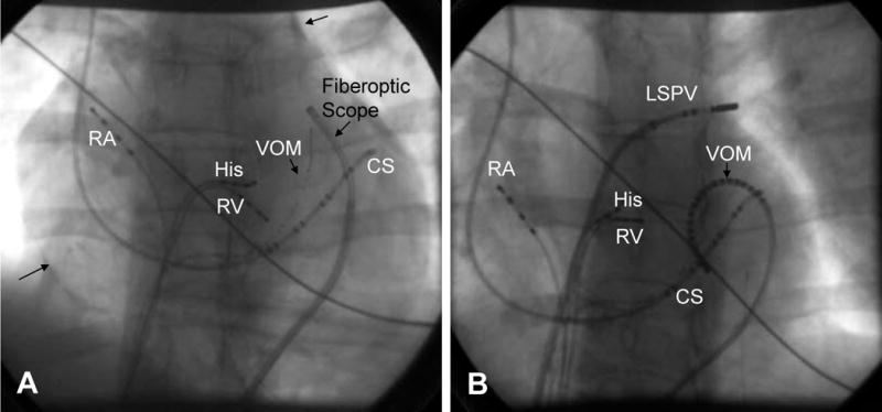
Figure 9.

Fiberoptic Pericardial Imaging, Epicardial Mapping and Ablation of the Vein of Marshall. (modified from ref 82). Panel A: Fiberoptic scope in the pericardium, RA=right atrial catheter, His= His bundle catheter, CS= coronary sinus catheter, RV=right ventricular catheter, VOM= vein of Marshall Catheter (1.5 French, multipolar catheter in the vein introduced via the CS). Arrows show contrast in the pericardial space. Panel B: Epicardial mapping of the Vein of Marshall, in this case a multi-electrode VOM catheter has been placed in the pericardial space. RA=right atrial catheter, His= His bundle catheter, CS= coronary sinus catheter, RV=right ventricular catheter, VOM= multi-electrode vein of Marshall Catheter
