Abstract
Considering the importance of professional plaque control for caries prevention, this study comprised an in vitro evaluation of wear by two prophylaxis methods (sodium bicarbonate jet – Profident and pumice and brush) on sound bovine enamel and with artificial carious lesions. Sixty enamel fragments were employed (4x4mm), which were divided into 4 groups: GI – 15 sound blocks treated with pumice and brush; GII – 15 sound blocks treated with Profident; GIII – 15 demineralized blocks treated with pumice and brush, and GIV – 15 demineralized blocks treated with Profident. In the fragments of Groups III and IV, artificial carious lesions were simulated by immersion in 0.05M acetic acid solution 50% saturated with bovine enamel powder at 37oC for 16h. The specimens were submitted to the prophylactic treatments for 10 seconds. Wear analysis was performed by profilometer and revealed the following results: 0.91μm – GI; 0.42μm – GII; 1.6μm – GIII, and 0.94μm – GIV. The two-way ANOVA and Tukey's test (p<0.05) revealed significant difference between all groups. Scanning electron microscopy images were employed to illustrate the wear pattern, with observation of larger alteration on the demineralized enamel surface (GIII; GIV), round-shaped wear on GI and GIII and blasted aspect on GII and GIV. The study indicated that the demineralized enamel presented more wear than the sound enamel, and the brush led to larger wear when compared to Profident.
Keywords: Dental prophylaxis, Dental enamel, wear
Abstract
Tendo em vista a importância do controle profissional da placa na prevenção da cárie, este estudo avaliou in vitro o desgaste de dois métodos de profilaxia (jato de bicarbonato de sódio-Profident e escova de Robinson com pasta de pedra pomes) sobre o esmalte bovino hígido e com lesões artificiais de cárie. Foram utilizados 60 fragmentos de esmalte (4 X 4mm), divididos em 4 grupos: GI-15 blocos hígidos tratados com escova de Robinson e pasta de pedra pomes; GII- 15 blocos hígidos tratados com Profident; GIII- 15 blocos desmineralizados tratados com escova de Robinson e pedra pomes e GIV- 15 blocos desmineralizados tratados com Profident. Nos fragmentos dos grupos III e IV foram simuladas lesões artificiais de cárie através da imersão em solução de ácido acético 0,05M, 50% saturada com pó de esmalte bovino, a 37oC por 16 h. Os espécimes foram submetidos aos tratamentos profiláticos durante 10 segundos. A análise do desgaste foi feita por meio de perfilometria, encontrando-se os seguintes resultados: 0,91μm-GI; 0,42μm-GII; 1,6μm-GIII e 0,94μm-GIV. Através do teste ANOVA a dois critérios e do teste de Tukey (p<0,05) detectou-se diferença significativa entre todos os grupos. Imagens de microscopia eletrônica de varredura foram utilizadas para ilustrar o padrão de desgaste, sendo observada uma maior alteração de superfície no esmalte desmineralizado (GIII; GIV), um desgaste com aspecto circular nos GI e GIII e aspecto jateado nos GII e GIV. O estudo indicou que o esmalte desmineralizado desgastou mais do que o esmalte hígido e a escova de Robinson foi responsável por um maior desgaste quando comparada ao Profident.
INTRODUCTION
Considering the important role played by dental plaque in the onset and progression of dental caries, the relevance of its control for prevention of this disease is acknowledged10,11,15.
The mechanical dental plaque control may be performed by both patient and professional7,21. The main methods include brushing and flossing, which are the home techniques that may be frequently employed by the patient18,19. However, the efficacy of control performed by the patient is related to interaction of several factors, such as motivation, level of oral hygiene instruction, manual dexterity and adequacy of oral hygiene instruments7. Moreover, some problems in Pediatric Dentistry are related to the patient, such as maturity and incomplete motor development, which impairs the learning and practice of techniques18. Thus, the mechanical control performed by the professional, by prophylaxis, plays an important role in this context, since it will compensate for the poor plaque control performed by the patient.
When regularly performed at intervals shorter than two months, professional prophylaxis is able to significantly reduce caries progression, being a very useful method for children unable to remove dental plaque by their own17.
However, considering the need of regular professional mechanical plaque control, one might question the amount of tooth structure removed at every session of dental prophylaxis. Thus, many investigators have attempted to quantify the wear caused by different methods of prophylaxis on sound enamel5,6,8,10,22,26,27; however, the action of prophylaxis methods on the demineralized enamel, i.e. with clinically signs of white spots, which is a reasonably weaker substrate less resistant to any type of mechanical action, is still unknown.
Thus, the present study comprised an in vitro evaluation of the wear yielded by professional prophylaxis with pumice and brush or sodium bicarbonate jet, in two different situations: sound bovine enamel and artificially demineralized bovine enamel.
MATERIAL AND METHODS
Sample
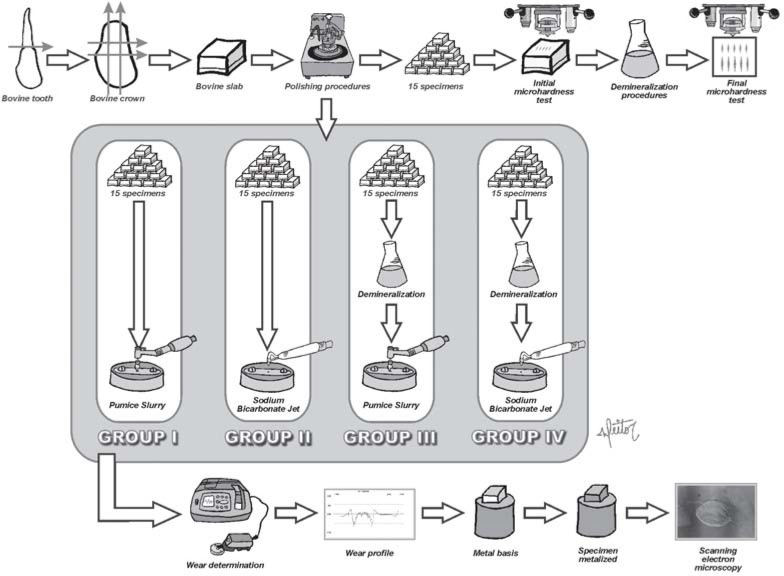
The wear tests were conducted on 60 enamel fragments achieved from crowns of bovine incisors. The fragments were randomly divided into 4 groups: Group I- 15 sound enamel blocks treated with pumice and brush, Group II- 15 sound enamel blocks treated with sodium bicarbonate jet, Group HI- 15 demineralized enamel blocks treated with pumice and brush, and Group IV- 15 demineralized enamel blocks treated with sodium bicarbonate jet. Microhardness tests were performed on further 15 specimens. The study design is illustrated in Figure 1.
FIGURE 1. Flowchart illustrating the study design.
Specimen preparation
The fragments were obtained from the flattest central portion of bovine incisors, which were sectioned with a precision sectioning machine and two double-faced diamond discs adapted to it, for achievement of specimens with 4x4mm dimensions.
Thereafter, the enamel blocks were flatten out and polished for achievement of absolutely flat surfaces parallel to the base, fundamental for the wear and microhardness tests. The dentinal aspects of the blocks were initially flatten with silicon carbide paper grit 320. Then, the enamel portion of the fragments was flatten with silicone carbide paper grits 600 and 1200, followed by final polishing with polishing cloth soaked with a 1-μm diamond suspension.
Demineralization procedures
For formation of the artificial carious lesions, the fragments were immersed in 32mL of 0.05M acetic acid solution 50% saturated with bovine enamel powder at pH 5.0 for 16 hours in an oven at 37oC23.
Microhardness tests
For investigation of the mineral loss of specimens after the process for formation of artificial carious lesions, microhardness tests were initially performed. The initial and final microhardness of specimens was measured with a microhardness tester and a Knoop diamond indenter, under static load of 50g (0.490N) applied for 5 seconds. Ten indents were made on each fragment: 5 before demineralization and further 5 after the demineralization process. There was a reduction in the surface hardness values of specimens before (mean 394 KHN) and after (mean 241 KHN) the demineralization process, with statistically significant difference (paired t test; p<0.05).
Prophylaxis simulation
For prophylaxis, the specimens were inserted in a metallic base over which a stainless steel strip with 1-mm diameter orifice coinciding with the central portion of the specimen was screwed, so that only a restricted area of the fragment would be exposed to the action of prophylactic agents.
Groups I and III received prophylaxis with a brush at low-speed handpiece and slurry (50g of water and 60g of powder). The brushes presented an acute end to allow access of bristles to the orifice in the stainless steel strip that protected the specimens. Prophylaxis was performed for 10 seconds, at 1500rpm with pressure resulting from the handpiece weight, and the procedure was stopped after the first 5 seconds for application of more pumice on the orifice.
The specimens in Groups II and IV received prophylaxis with sodium bicarbonate jet, at an approximate distance of 5mm from the stainless steel strip orifice for 10 seconds ceaselessly.
Wear determination
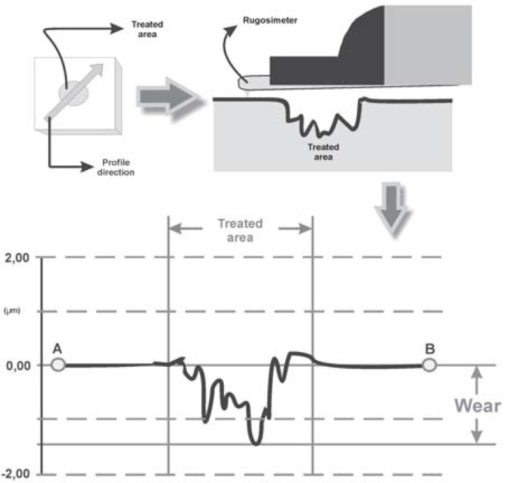
Quantification of wear was assessed in a roughness meter connected to a PC with specific software to provide the profiles of the surfaces tested. Five readings were performed on each specimen, and the average of wear was calculated. These profilometric traces were taken from the reference surface (protected area) on one side to the opposite reference surface, crossing the surface exposed to experimental procedures. Thus, the reading of wear was performed by the surface profile, measuring the distance in micrometers between the graph midline, corresponding to the specimen plane line (intact area), and the deepest valley corresponding to the ground area (Figure 2).
FIGURE 2. Schematic drawing demonstrating quantification of wear by the roughness meter.
Scanning Electron Microscopy
After the wear tests, a specimen of each group was randomly selected for observation under scanning electron microscopy. The specimens were taken to the metallizer to be gold-coated for posterior observation under SEM.
Statistical Analysis
Comparison of the variability between the wear groups was performed by application of two-way ANOVA and Tukey's test at a significance level of 5%.
RESULTS
Wear
The mean wear of the fragments of demineralized bovine enamel was also higher compared to the intact specimens, regardless of the prophylaxis method employed. In turn, the brush presented larger wear when compared to the bicarbonate jet, regardless of the tissue submitted to application.
The Table 1demonstrates the results of the mean wear values between groups, and statistical analysis revealed a statistically significant difference between the study variables: prophylaxis method and enamel conditions (p<0,05).
TABLE 1. Mean wear value (μm) and standard deviation of the studied groups.
| Enamel | Prophilaxis method | WEAR*(Mean values and standard deviation) | Number of specimens |
|---|---|---|---|
| Sound | pumice and brush – GI | 0,915 + 0,265 a | 15 |
| Profident – GII | 0,418 + 0,173 b | 15 | |
| Demineralized | pumice and brush – GIII | 1,620 + 0,309 c | 15 |
| Profident – GIV | 0,944 + 0,164 d | 15 |
Means followed by different letters in each column are significantly different (p<0.05).
Scanning Electron Microscopy
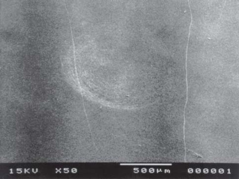
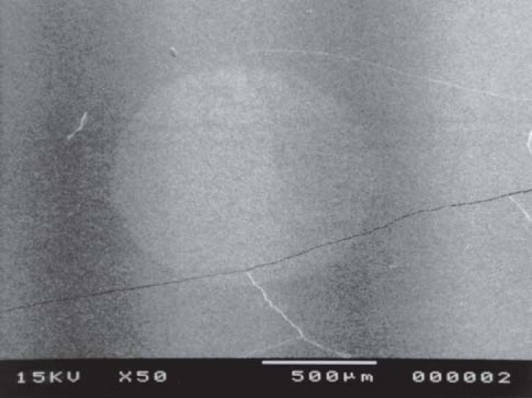
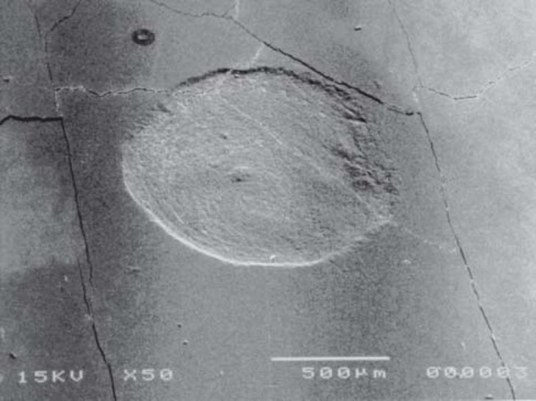
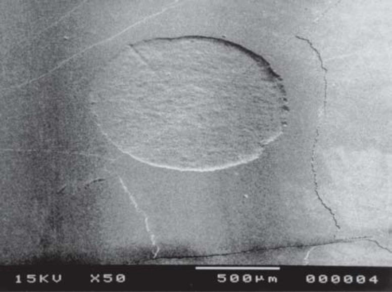
Group I revealed mild scratches yielded by the rotary movements of the brush bristles (Figure 3) whereas Group II revealed a well-delimited wear without round scratches (Figure 4). Groups III and IV presented the same pattern of wear when compared to Groups I and II respectively, yet with larger wear (Figure 5and 6).
FIGURE 3. Scanning electron microscopy of sound enamel after prophylaxis with pumice slurry-Group I (Original magnification, X 50).
FIGURE 4. Scanning electron microscopy of sound enamel after prophylaxis with sodium bicarbonate jet-Group II (Original magnification, X 50).
FIGURE 5. Scanning electron microscopy of demineralized enamel after prophylaxis with pumice slurry-Group III (Original magnification, X 50).
FIGURE 6. Scanning electron microscopy of demineralized enamel after prophylaxis with sodium bicarbonate jet – Group IV (Original magnification, X 50).
DISCUSSION
As in all pathologies, the search for prevention aims at combating the etiologic agents, and even though caries is a multifactorial disease, several studies have been indicating intervention directed to pathogenic microorganisms as a practical and effective means to avoid the development of the disease3,4,15,18, since based on the study of Holmen et al.15the mechanical suppression of microbial activities by regular physical disturbances is able to prevent enamel demineralization.
Even though the professional plaque removal is advocated as a safe means for caries prevention3,15, the fact that prophylactic procedures may promote wear of the tooth surface is unquestionable6,8,22,26,30; however, few studies have evaluated the influence of prophylaxis on demineralized tooth surfaces30.
Several methodologies have been employed to evaluate the wear of the dental tissue. Scanning electron microscopy allows qualitative analysis6,13,16,22,29, yet is unable to measure the structural loss. Radioactive labeling14, graduated focus microscopy5and weight loss12are quantitative methods that present the disadvantage of reducing the wear resistance of the dental tissue9, be based on personal interpretation of the loss values or do not allow measurement of microscopic wear, respectively. Many authors have quantified the wear resulting from prophylactic procedures by profilometry10,13,29because this process is regarded as more simple and direct for this type of measurement.
In this study, wear measurement was performed by profilometry with utilization of a roughness meter, which screens the entire surface to be tested with a small probe, tracing its profile. Due to its natural roughness, the profile of sound bovine enamel presents a highly variable and irregular design; if prophylaxis was performed on this surface, the profile after treatment would hardly allow observation of the structural loss resulting from the prophylactic procedure. Thus, the surface was regularized by polishing, so that the profile observed would have the aspect of a straight line, and any variation observed after prophylaxis would be surely related to the treatment applied. The wear resulting from the treatment performed was even more visible because of the manner through which the prophylaxis procedures were performed, since the wear did not occur on the entire fragment surface, but rather over the area exposed to prophylaxis. Therefore, when the probe of the roughness meter screened the surface of the specimen after treatment, it had contact with the surfaces worn or not by prophylaxis (control surface), yielding a profile similar to a straight line with a small irregular interval at the central region, and this methodology has been employed by other authors29.
The treatment performed with both methods (pumice slurry and brush and sodium bicarbonate jet) was applied for 10 seconds. Some studies performed prophylaxis for 30 seconds13,16,26,27,30, which according to Boyde6and Zuniga and Caldwell30simulates a cumulative effect of two or three sessions of prophylactic treatment. Thus, a hypothetic time of 10 seconds was standardized in the present study to simulate the mean time of prophylaxis on a given dental site.
The results of this study presented higher8,27or lower5,30wear rates compared to the literature, even though direct comparison is not viable because most studies were conducted on human enamel5,8,27, which is less porous than bovine enamel11,20. However, as the main objective of this study was to test the prophylaxis methods employed, the results were valid, since the substrate was the same for both prophylactic techniques and the only variable in this case was the treatment performed.
However, even though prophylaxis was performed only on bovine enamel, this study was tested in two conditions: sound and demineralized enamel. Thus, the results achieved in this study corroborate the findings in the literature30, since higher wear rates were observed for enamel surfaces with carious lesions than for the sound enamel. At first sight, these outcomes might seem obvious; however, it is important to perform a comparative analysis of wear yielded on the two types of substrate to observe, in practice, to which extent the decayed enamel is more worn than the sound enamel, which revealed an approximate ratio of 2X1, as also observed by Zuniga and Caldwell30.
The effect of prophylaxis varies considerably according to the integrity of the enamel surface, which allows a simple procedure applied on the sound tooth surface to be very harmful on the demineralized tooth30. The chemical and physical condition of the enamel surface changes during development of carious lesions and knowledge on its microstructure allows the statement that, when performing any type of prophylactic treatment on this surface, there is a greater chance to cause irreversible structural damages due to its lower resistance30.
Therefore, the concern is focused on the rupture of the surface layer of the white spot lesion, which would expose an even less resistant region: the lesion body. It is known that the surface layer thickness may vary, however some studies have reported values close to 10μm1,2.
Thus, considering the mean wear index observed for the demineralized enamel (1.62μm with pumice slurry and brush and 0.94μm with Profident), it may be assumed that there was no rupture of this thin surface in this study.
Concerning the routine utilization of professional prophylaxis, Primosch24contraindicates its routine application in the dental clinic, since it would yield excessive wear on the enamel surface. Shillingburg and Grace25found a mean value of 1.14mm of thickness for the enamel on the buccal aspect of maxillary central incisor (the human tooth most frequently employed in laboratory wear tests5,10,16,26,27). Thus, considering the wear observed in the present study on sound tooth structure and the mean enamel thickness of enamel on human incisors, it may be concluded that, if a patient is submitted to monthly prophylactic procedures with excessive periods of application for clinical practice, as in the present study, an individual would require 106 years to have the entire enamel surface worn by prophylaxis. Considering that human teeth are less porous than bovine enamel11,20, this value is multiplied. Therefore, even though the prophylactic technique presets disadvantages as wear, the benefit provided as to caries prevention, which may lead to a significant dental loss in short periods of time, seems to be rewarding.
The loss of tooth structure related to the two prophylaxis methods tested revealed nearly twice as much wear for procedures performed with pumice slurry and brush (0.91μm on sound enamel and 1.62μm on demineralized enamel) when compared to Profident (0.42μm on sound enamel and 0.94μm on demineralized enamel).
The scanning electron microscopy images also confirmed the profilometer findings of this study, since they demonstrate larger wear in demineralized tissue, regardless of the prophylaxis method employed. Moreover, the images present different patterns of wear of both prophylaxis methods investigated on the tooth structure, Willmann, Norling and Johnson29.
This difference in wear values may have occurred because of the excessive abrasiveness of pumice10,16. Davis and Winter10evaluated the wear of enamel and dentin submitted to brushing tests with several dentifrices and prophylactic pastes and observed that pumice yielded up to 20 times more wear than conventional dentifrices. Hosoya and Johnston16did not indicate utilization of pumice, since it might yield deep erosions on the enamel surface, leading to irreversible iatrogenia.
Many authors consider the sodium bicarbonate jet as an effective method for plaque removal, combined to its practical and easy utilization18,28. As the aforementioned studies, the present investigation also observed advantages of prophylactic procedures performed with sodium bicarbonate jet. In addition to the significantly lower wear, another aspect that called the attention was the standard deviation of the method.
The variability of wear of Profident (SD of GII: 0.173 and SD of GIV: 0.164) was always lower than the variation of pumice slurry and brush (SD of GI: 0.265 and SD of GIII: 0.309). Both methods were standardized during the study; however clinically there may be variations. For the treatment with Profident, the distance of specimens was maintained at 5mm, which is the distance clinically employed12,13,22,29. However, in the present study, the pressure corresponding to prophylaxis performed with pumice and brush was exclusively related to the weight of the handpiece; nevertheless, in clinical practice, this variation is extremely higher, since there is no standardization of the force applied by professionals.
Thus, in the conditions of this study, the wear caused by prophylaxis with pumice and brush was nearly twice larger than that of prophylaxis performed with sodium bicarbonate jet; however, clinically this difference may be even greater, since the first method is more sensitive to personal variations, and the second prophylactic techniques does not allow significant variations.
Therefore, even though the conditions of the present study were standardized for a comparative evaluation of wear yielded by these two prophylaxis methods on sound and demineralized enamel, further studies on this subject are wanted, especially to observe the performance of deciduous and permanent dental tissue under the same conditions.
CONCLUSIONS
The present results allowed the following conclusions:
Among the prophylaxis methods tested, pumice slurry and brush was more abrasive compared to sodium bicarbonate jet both on sound enamel and demineralized enamel;
The wear yielded by both methods employed for prophylaxis was statistically significantly higher for the demineralized enamel compared to the sound enamel.
REFERENCES
- 1.Arends J, Schuthof J, Jongebloed WG. Microhardness indentations on artificial white spot lesions. Caries Res. 1979;13(5):290–297. doi: 10.1159/000260412. [DOI] [PubMed] [Google Scholar]
- 2.Arends J, Schuthof J, Jongebloed WG. Lesion depth and microhardness indentations on artificial white spot lesions. Caries Res. 1980;14(4):190–195. doi: 10.1159/000260453. [DOI] [PubMed] [Google Scholar]
- 3.Axelsson P, Lindhe J. The effect of a preventive programme on dental plaque, gingivitis and caries in schoolchildren. Results after one and two years. J Clin Periodontol. 1974;1(2):126–138. doi: 10.1111/j.1600-051x.1974.tb01248.x. [DOI] [PubMed] [Google Scholar]
- 4.Axelsson P, Lindhe J, Waseby J. The effect of various plaque control measures on gingivitis and caries in schoolchildren. Community Dent Oral Epidemiol. 1976;4(6):232–239. doi: 10.1111/j.1600-0528.1976.tb00990.x. [DOI] [PubMed] [Google Scholar]
- 5.Bailey LR, Phillips RW. Effect of certain abrasive materials on tooth enamel. J Dent Res. 1950;29(6):740–748. doi: 10.1177/00220345500290060501. [DOI] [PubMed] [Google Scholar]
- 6.Boyde A. Airpolishing effects on enamel, dentine, cement and bone. Br Dent J. 1984;156(8):287–291. doi: 10.1038/sj.bdj.4805346. [DOI] [PubMed] [Google Scholar]
- 7.Buischi YP, Axelsson P. Promoção de saúde bucal. São Paulo: Artes Médicas; 1997. Controle mecânico da placa dental realizado pelo paciente; pp. 113–127. [Google Scholar]
- 8.Christensen RP, Bangerter VW. Immediate and long-term in vivo effects of polishing on enamel and dentin. J Prosthet Dent. 1987;57(2):150–160. doi: 10.1016/0022-3913(87)90138-7. [DOI] [PubMed] [Google Scholar]
- 9.Davis WB. Reduction in dentin wear resistence by irradiation and effects of storage in aqueous media. J Dent Res. 1975;54:1078–1081. doi: 10.1177/00220345750540051201. [DOI] [PubMed] [Google Scholar]
- 10.Davis WB, Winter PJ. Measurement in vitro of enamel abrasion by dentifrice. J Dent Res. 1976;55(6):970–975. doi: 10.1177/00220345760550064801. [DOI] [PubMed] [Google Scholar]
- 11.Featherstone JD, Mellberg JR. Relative rates of progress of artificial carious lesions in bovine, ovine and human enamel. Caries Res. 1981;15(1):109–114. doi: 10.1159/000260508. [DOI] [PubMed] [Google Scholar]
- 12.Galloway SE, Pashley DH. Rate of removal of root structure by the use of the Prophy-Jet device. J Periodontol. 1987;58(7):464–469. doi: 10.1902/jop.1987.58.7.464. [DOI] [PubMed] [Google Scholar]
- 13.Gerbo LR, Lacefield WR, Barnes CM, Russell CM. Enamel roughness after air-powder polishing. Am J Dent. 1993;6(2):96–98. [PubMed] [Google Scholar]
- 14.Grabenstetter RJ, Broge RW, Jackson FL, Radike AW. The measurement of the abrasion of human teeth by dentifrice abrasives: a test utilizing radiactive teeth. J Dent Res. 1958;37(6):1060–1068. doi: 10.1177/00220345580370060601. [DOI] [PubMed] [Google Scholar]
- 15.Holmen L, Mejare I, Malmgren B, Thylstrup A. The effect of regular professional plaque removal on dental caries in vivo. A polarized light and scanning electron microscope study. Caries Res. 1988;22(4):250–256. doi: 10.1159/000261116. [DOI] [PubMed] [Google Scholar]
- 16.Hosoya Y, Johnston JW. Evaluation of various cleaning and polishing methods on primary enamel. J Pedod. 1989;13(3):253–269. [PubMed] [Google Scholar]
- 17.Koch G, Arneberg P, Thylstrup A. Tratado de cariologia. Rio de Janeiro: Cultura Médica; 1988. Higiene oral e cárie; pp. 281–292. [Google Scholar]
- 18.Lima JEO. Um plano de prevenção para consultório odontopediátrico. RGO. 1992;40(6):395–399. [Google Scholar]
- 19.Lopes JCA, Duarte CA. Efeito do dentifrício no controle mecânico da placa bacteriana. RGO. 1994;42(5):266–268. [Google Scholar]
- 20.Manning RH, Edgar WM. Intra-oral models for studying de- and remineralization in man: methodology and measurement. J Dent Res. 1992;71:895–900. doi: 10.1177/002203459207100S20. [DOI] [PubMed] [Google Scholar]
- 21.Mestrinho HD, Carvalho JC, Figueiredo CS. Desempenho clínico das escovas infantis produzidas no Brasil. RGO. 1994;42(5):254–258. [Google Scholar]
- 22.Newman PS, Silverwood RA, Dolby AE. The effects of an airbrasive instrument on dental hard tissues, skin and oral mucosa. Br Dent J. 1985;159(1):9–12. doi: 10.1038/sj.bdj.4805629. [DOI] [PubMed] [Google Scholar]
- 23.Paes Leme AF, Tabchoury CPMC, Cury JA, Fu J, Zero DT. The effect of fluoridated dentifrice and/or acidulated phosphate fluoride application on early artificial carious lesions - an in vitro study. Am J Dent. 2003;16(2):91–95. [PubMed] [Google Scholar]
- 24.Primosch RE. Rubber cup prophylaxis: a reevaluation of its use in pediatric dental patients. Dent Hyg. 1980;54(11):525–527. [PubMed] [Google Scholar]
- 25.Shillingburg HT, Grace CS. Thickness of enamel and dentin. J Calif Dent Assoc. 1973;41:33–52. [PubMed] [Google Scholar]
- 26.Stookey GK. In vitro estimates of enamel and dentin abrasion associated with a prophylaxis. J Dent Res. 1978;57(1):36–36. doi: 10.1177/00220345780570011501. [DOI] [PubMed] [Google Scholar]
- 27.Stookey GK, Schemehorn BR. A method for assessing the relative abrasion of prophylaxis materials. J Dent Res. 1979;58(2):588–592. doi: 10.1177/00220345790580020801. [DOI] [PubMed] [Google Scholar]
- 28.Strand GV, Raadal M. The efficiency of cleaning fissures with an air-polishing instrument. Acta Odontol Scand. 1988;46(2):113–117. doi: 10.3109/00016358809004756. [DOI] [PubMed] [Google Scholar]
- 29.Willmann DE, Norling BK, Johnson WN. A new prophylaxis instrument: effect on enamel alterations. J Am Dent Assoc. 1980;101(6):923–925. doi: 10.14219/jada.archive.1980.0458. [DOI] [PubMed] [Google Scholar]
- 30.Zuniga MA, Caldwell RC. Pastes on normal and "white-spot" enamel the effect of fluoride-containing prophylaxis. ASDC J Dent Child. 1969;36(5):55–59. [PubMed] [Google Scholar]


