

Abstract
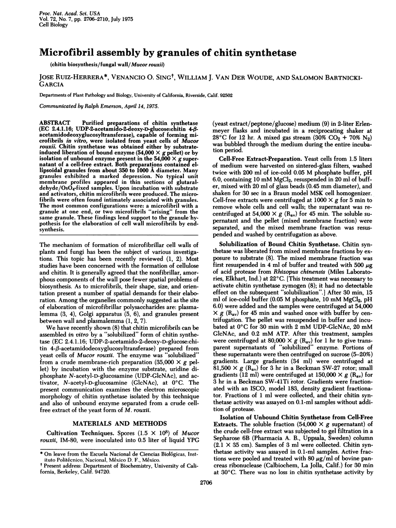
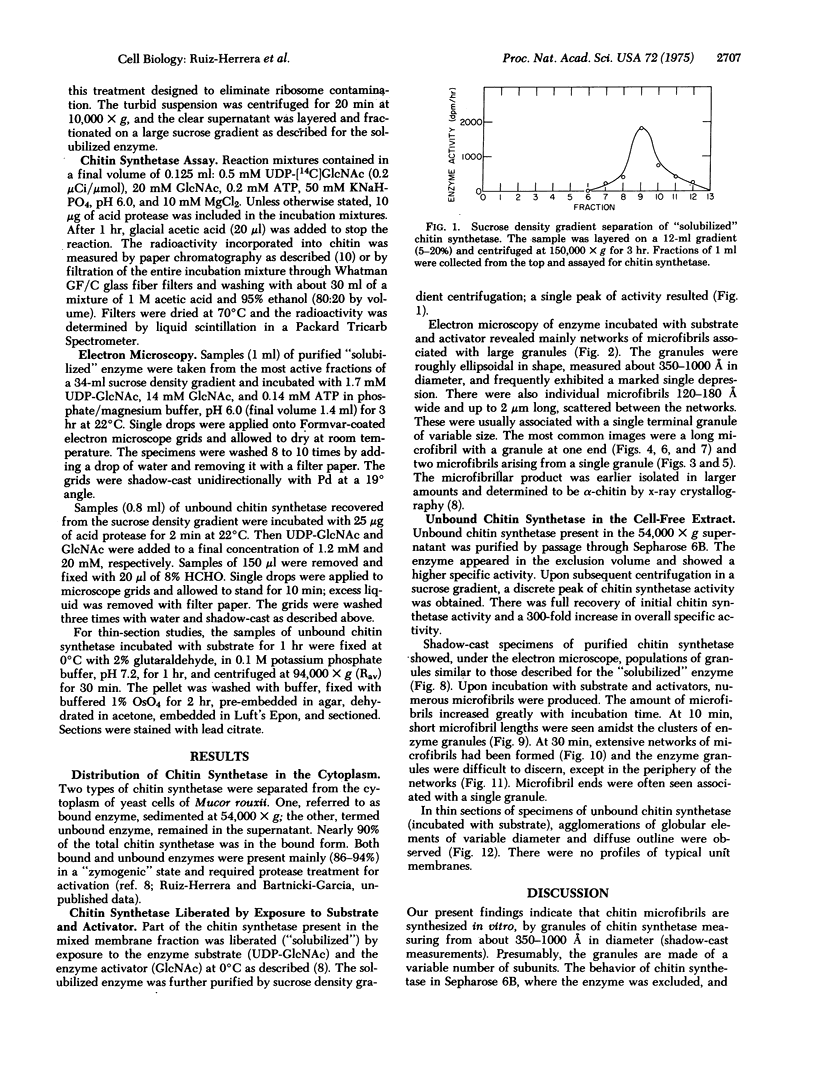
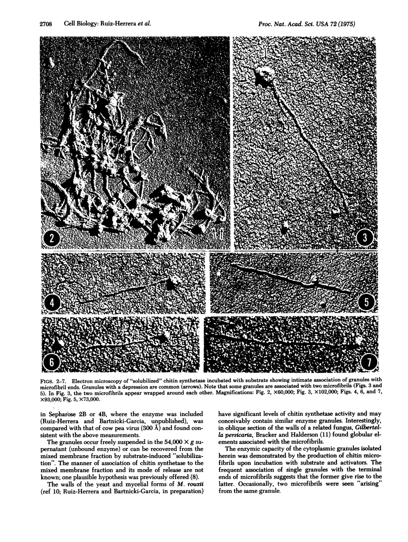
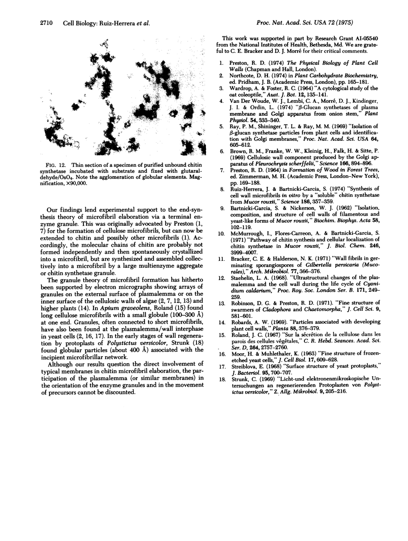
Purified preparations of chitin synthetase (EC 2.4.1.16; UDP-2-acetamido-2-deoxy-D-glucose:chitin 4-beta-acetamidodeoxyglucosyltransferase), capable of forming microfibrils in vitro, were isolated from yeast cells of Mucor rouxii. Chitin synthetase was obtained either by substrate-induced liberation of bound enzyme (54,000 x g pellet) or by isolation of unbound enzyme present in the 54,000 x g supernatant of a cell-free extract. Both preparations contained ellipsoidal granules from about 350 to 1000 A diameter. Many granules exhibited a marked depression. No typical unit membrane profiles appeared in thin sections of glutaraldehyde/OsO4-fixed samples. Upon incubation with substrate and activators, chitin microfibrils were produced. The microfibrils were often found intimately associated with granules. The most common configurations were: a microfibril with a granule at one end, or two microfibrils "arising" from the same granule. These findings lend support to the granule hypothesis for the elaboration of cell wall microfibrils by end-synthesis.
Full text
PDF
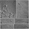
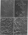
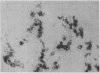
Images in this article
Selected References
These references are in PubMed. This may not be the complete list of references from this article.
- BARTNICKI-GARCIA S., NICKERSON W. J. Isolation, composition, and structure of cell walls of filamentous and yeast-like forms of Mucor rouxii. Biochim Biophys Acta. 1962 Mar 26;58:102–119. doi: 10.1016/0006-3002(62)90822-3. [DOI] [PubMed] [Google Scholar]
- Brown R. M., Jr, Franke W. W., Kleinig H., Falk H., Sitte P. Cellulosic wall component produced by the golgi apparatus of Pleurochrysis scherffelii. Science. 1969 Nov 14;166(3907):894–896. doi: 10.1126/science.166.3907.894. [DOI] [PubMed] [Google Scholar]
- McMurrough I., Flores-Carreon A., Bartnicki-Garcia S. Pathway of chitin synthesis and cellular localization of chitin synthetase in Mocor rouxii. J Biol Chem. 1971 Jun 25;246(12):3999–4007. [PubMed] [Google Scholar]
- Ray P. M., Shininger T. L., Ray M. M. ISOLATION OF beta-GLUCAN SYNTHETASE PARTICLES FROM PLANT CELLS AND IDENTIFICATION WITH GOLGI MEMBRANES. Proc Natl Acad Sci U S A. 1969 Oct;64(2):605–612. doi: 10.1073/pnas.64.2.605. [DOI] [PMC free article] [PubMed] [Google Scholar]
- Robinson D. G., Preston R. D. Fine structure of swarmers of Cladophora and Chaetomorpha. I. The plasmalemma and Golgi apparatus in naked swarmers. J Cell Sci. 1971 Nov;9(3):581–601. doi: 10.1242/jcs.9.3.581. [DOI] [PubMed] [Google Scholar]
- Ruiz-Herrera J., Bartnicki-Garcia S. Synthesis of cell wall microfibrils in vitro by a "soluble" chitin synthetase from Mucor rouxii. Science. 1974 Oct 25;186(4161):357–359. doi: 10.1126/science.186.4161.357. [DOI] [PubMed] [Google Scholar]
- Staehelin L. A. Ultrastructural changes of the plasmalemma and the cell wall during the life cycle of Cyanidium caldarium. Proc R Soc Lond B Biol Sci. 1968 Nov 5;171(1023):249–259. doi: 10.1098/rspb.1968.0067. [DOI] [PubMed] [Google Scholar]
- Streiblová E. Surface structure of yeast protoplasts. J Bacteriol. 1968 Feb;95(2):700–707. doi: 10.1128/jb.95.2.700-707.1968. [DOI] [PMC free article] [PubMed] [Google Scholar]
- Strunk C. Licht- und elektronenmikroskopische Untersuchungen an regenerierenden Protoplasten von Polystictus versicolor. Z Allg Mikrobiol. 1969;9(3):205–216. doi: 10.1002/jobm.3630090306. [DOI] [PubMed] [Google Scholar]
- Van Der Woude W. J., Lembi C. A., Morré D. J. beta-Glucan Synthetases of Plasma Membrane and Golgi Apparatus from Onion Stem. Plant Physiol. 1974 Sep;54(3):333–340. doi: 10.1104/pp.54.3.333. [DOI] [PMC free article] [PubMed] [Google Scholar]