Abstract
This review analytically examines the published data for erionite-related malignant pleural mesothelioma (E-MPM) and any data to support a genetically predisposed mechanism to erionite fiber carcinogenesis. Adult patients of age ≥18 years with erionite-related pleural diseases and genetically predisposed mechanisms to erionite carcinogenesis were included, while exclusion criteria included asbestos- or tremolite-related pleural diseases. The search was limited to human studies though not limited to a specific timeframe. A total of 33 studies (31042 patients) including 22 retrospective studies, 6 prospective studies, and 5 case reports were reviewed. E-MPM developed in some subjects with high exposures to erionite, though not all. Chest CT was more reliable in detecting various pleural changes in E-MPM than chest X-ray, and pleural effusion was the most common finding in E-MPM cases, by both tests. Bronchoalveolar lavage remains a reliable and relatively less invasive technique. Chemotherapy with cisplatin and mitomycin can be administered either alone or following surgery. Erionite has been the culprit of numerous malignant mesothelioma cases in Europe and even in North America. Erionite has a higher degree of carcinogenicity with possible genetic transmission of erionite susceptibility in an autosomal dominant fashion. Therapeutic management for E-MPM remains very limited, and cure of the disease is extremely rare.
Keywords: Asbestos, malignant mesothelioma, pleural effusion
INTRODUCTION
Mesothelioma is a unique cancer that originates from the mesothelial cells that line the pleural, pericardial, and peritoneal surfaces.1 There are approximately 2500 cases and deaths from malignant mesothelioma (MM) annually in the United States, with most related to prior asbestos exposure.2
Asbestos is a chemically inert, insoluble, and odorless mineral that occurs naturally in metamorphic deposits located around the world.3 Asbestos is fire resistant, and it does not conduct heat or electricity. However, asbestos does not exist in a single form; rather, it is a group of six fibrous minerals of the hydrous magnesium silicate variety. The six types include tremolite asbestos, actinolite asbestos, anthophyllite asbestos, chrysotile asbestos, amosite asbestos, and crocidolite asbestos.3 The combination of properties exhibited by asbestos has made it a valuable element in variety of household products and construction materials.
Several studies reported an increased incidence of malignant pleural mesothelioma (MPM) in residents of southeast Turkey.2,4,5 Most of the affected population had been exposed by inhalation since childhood to a material containing a fibrous mineral, erionite, that caused up to 50% of all deaths in three small villages.6 Furthermore, many of the affected subjects had migrated to a number of European countries, including Germany and Sweden, with public health reports of MM induced by erionite in Turkish immigrants in those countries as well.7,8
Asbestos-related diseases are less likely without prior occupational exposure. Indeed, most studies report that about 80% of mesotheliomas develop in asbestos-exposed individuals.9 However, the relationship between exposure and development of mesothelioma varies from about 10% to 100% depending on the study and on the criteria used to establish definite prior exposure.9 Additionally, in comparison to other carcinogens there is no linear dose-response relationship between exposure to asbestos and the incidence of mesothelioma.1
The goal of this study was to critically examine and characterize the published data regarding erionite-related malignant pleural mesothelioma (E-MPM). Additionally, we aimed to elucidate whether sufficient evidence exists to support a genetically predisposed mechanism to erionite fiber carcinogenesis.
STUDIES ABOUT ERIONITE
A comprehensive search was performed to collect relevant published and unpublished studies of fiber-related pleural diseases, mesothelioma, erionite, and tremolite asbestos. The search strategies were adapted to accommodate the unique search features of each database, including databases for PubMed/Medline and Index Medicus, where database-specific MESH- and EMTREE-controlled vocabulary terms were used. The search was limited to human studies published in English; however, there were no limits for date or publication status. Relevant bibliographies were also scanned for additional studies.
In an attempt to minimize publication bias, the following databases were also searched to collect relevant unpublished studies: the Australian and New Zealand Clinical Trials Registry, the World Health Organization (WHO) International Clinical Trials Registry Platform, Cochrane Library, ClinicalTrials.gov, MINDCULL.com, Current Controlled Trials, and Google.
Inclusion criteria included 1) adult patients (≥18 years of age) with erionite-related diseases and 2) a genetically predisposed mechanism to erionite carcinogenesis in high risk families with MM. Exclusion criteria included 1) asbestos-related diseases and 2) tremolite-related diseases.
All studies were independently evaluated and scored according to a scale adapted from Jadad, et al. (Table 1).10 Evidence was rated according to the Oxford Scheme.11 Inclusion and exclusion criteria, study parameters, Jadad score, and evidence level were assessed by three evaluators (ED, CG, and MR). Any discrepancy was resolved independently by a fourth evaluator (EE).
Table 1.
Criteria for Scoring Included Manuscripts as Published by Jadad, et al.10

*Score based on initial three-item tool (1-3) with addendum criteria (4-7). "Yes" (1 or -1 point) and "No" (0 points) answers are totaled for a potential maximum score of 5 points.
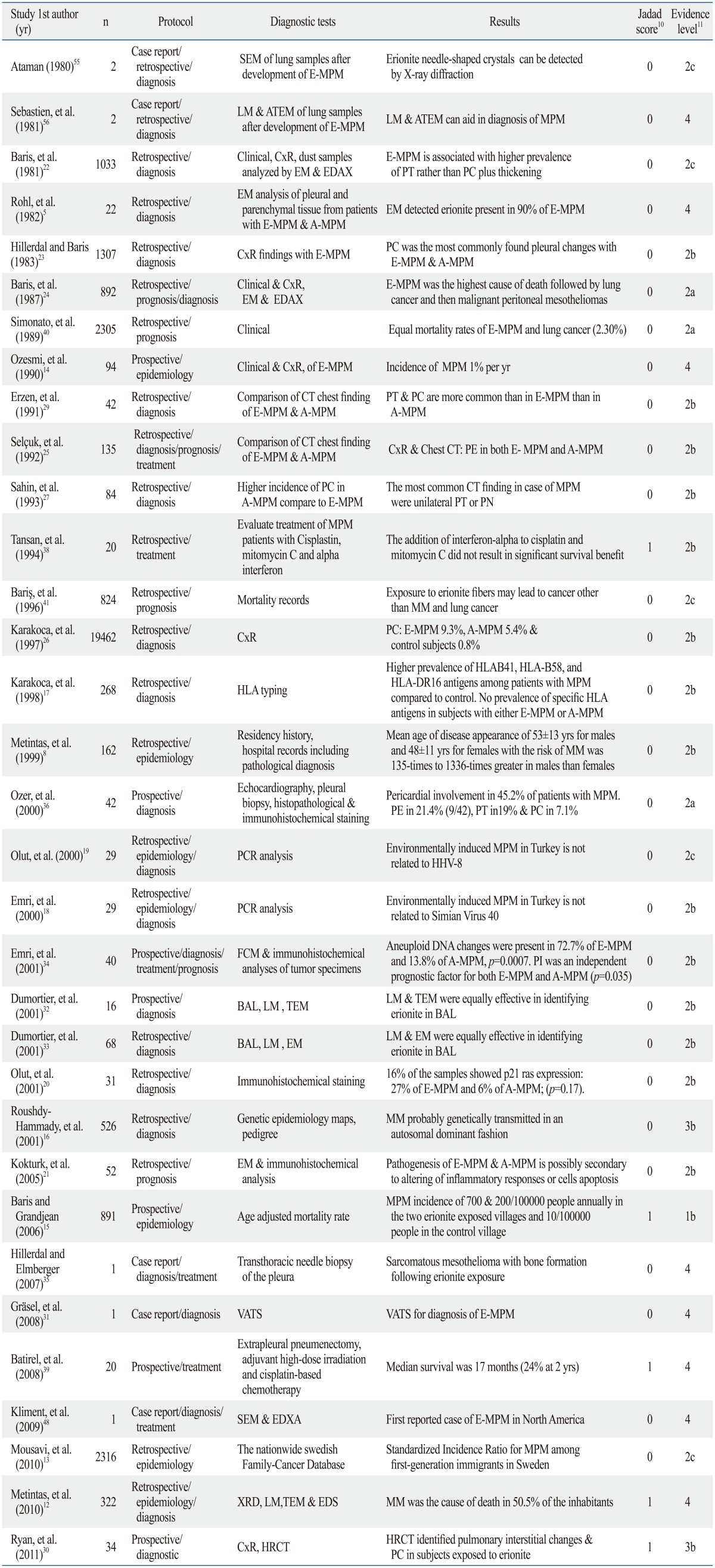
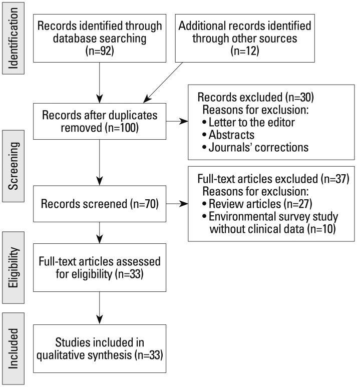
Thirty-three studies out of 100 publications met the inclusion criteria for this review, thereby including a total of 31042 patients (Fig. 1). These publications included six prospective studies (1137 patients), 22 retrospective observational studies (29898 patients), and five case reports (7 patients) (Fig. 2). Descriptions of the included studies are listed in Table 2.
Fig. 1.
Flow diagram of included and excluded studies.
Fig. 2.
Characteristics of erionite studies that met the inclusion criteria.
Table 2.
Review of the Studies Assessing the Clinical and Prognostic Features of Erionite-Induced Malignant Mesothelioma

SEM, scanning electron microscope; LM, light microscopy; TEM, transmission electron microscope; EM, electron microscopy; EDAX, energy-dispersion analysis of X-rays; PT, pleural thickening; PC, pleural calcification; XRD, X-ray diffraction; EDS, energy dispersive X-ray spectrometry; PCR, polymerase chain reaction; BAL, bronchoalveolar lavage; FCM, flow cytometric; VATS, video-assisted thoracoscopic biopsy; HRCT, high resolution CT; ATEM, analytical transmission electron microscopy; MPM, malignant pleural mesothelioma; E-MPM, erionite-related MPM; A-MPM, asbestos-related MPM; CxR, chest X-ray; PN, pleural nodule; MM, malignant mesothelioma; HLA, human leukocyte antigens; HHV-8, human herpes virus 8.
EPIDEMIOLOGY
Overall, six retrospective studies (3384 patients) and four prospective (1305 patients) studies (4689 patients in total) assessed the epidemiology of E-MPM.
Incidence
One retrospective study (162 patients) reported the mean age of disease appearance as 53±13 years (range: 37-71 years) for males and 48±11 years (range: 28-67 years) for females with a reported MM risk of 135-times to 1336-times greater in males than in females.8 A more recent retrospective study (322 patients) reported MM cohorts as having a mean age of 53.3±18.5 years (range: 22-95 years), with MM reported as the cause of death in 50.5% (52 of 322) of the inhabitants.12 Another retrospective study (2316 patients) reported the Standardized Incidence Ratio (SIR) for MPM among first-generation immigrants in Sweden. The database included 2128 cases of MPM among native Swedish residents and 188 cases among immigrants. The median ages at immigration and at diagnosis were 26 and 57 for male Turkish immigrants and 33 and 55 for females, respectively.13 Male SIR was 2.82 [95% confidence interval (CI): 1.22-5.56] while female SIR was 23.76 (95% CI: 10.86-45.11), and the SIR of both genders was 5.29 (95% CI: 3.08-8.47).
A prospective study of 94 patients with a mean age 36 years (range: 17-64 years) estimated the incidence of MPM as 1% per year in an immigrant cohort in Sweden who were previously exposed to erionite.14 Additionally, a long term prospective study of residents of two villages exposed to erionite and one nearby control village on the Anatolian plateau in Turkey was initiated in 1979 and continued through December 31, 2003.15 The study included 891 men and women aged 20 years or older. Of these subjects, 74% (661/891) resided in the villages with known exposure to erionite. Interestingly, only two cases of mesothelioma occurred in the control village. When standardized to the entire world population, the MPM incidences were approximately 700 and 200 cases per 100000 people annually in the two erionite exposed villages and only 10 cases per 100000 people in the control village.
Biological & genetic factors
Two studies investigated the probability of genetic factors behind the development of E-MPM.16,17 A retrospective study by Roushdy-Hammady, et al.16 investigated a six-generation extended pedigree of 526 individuals who lived in one village and indicated that MM was genetically transmitted, likely in an autosomal dominant way. Conversely, a prospective study of 268 subjects evaluated a possible relation between MPM and the presence and distribution of human leukocyte antigens (HLA) in patients environmentally exposed to erionite and asbestos in rural Anatolia, Turkey.17 There was a higher prevalence of HLAB41, HLA-B58, and HLA-DR16 antigens among patients with MPM than among healthy inhabitants from the same village and renal donors (control). However, there was no difference in the prevalence of any specific HLA antigen among the study subjects with MPM following exposure to either erionite or asbestos.
A possible link between exposure to Simian virus 40 (SV40) and the development of MPM was evaluated in a retrospective study. SV40 (DNA tumor virus) contaminated polio vaccine supplies that were distributed worldwide between the late 1950s and early 1960s.18 Polymerase Chain Reaction (PCR) analysis failed to identify SV40 in any of the 29 pleural biopsy specimens from patients diagnosed with MPM. The authors conclude that the pathogenesis of environmentally induced MPM in Turkey was most likely related to exposure to inorganic fibers, asbestos, or erionite rather than previous exposure to SV40.
Another retrospective study investigated the presences of human herpes virus 8 (HHV-8) in pleural biopsy samples from patients with MPM (29 patients: 14 E-MPM, 15 A-MPM) through PCR analysis.19 HHV-8 has been shown to be associated with Kaposi sarcoma lesions and other body cavity-originating B-cell lymphomas. In addition, HHV-8 upregulates levels of interleukin-6, which is also secreted by mesothelioma cells. However, the PCR analysis failed to identify HHV-8 in any of the 29 pleural biopsy specimens, leading the investigators to conclude that the environmentally-induced mesotheliomas were not related to HHV-8 exposure.19
Three Ras proteins, H-RAS, K-RAS, and N-RAS, are 21 kDa oncoproteins that play a pivotal role in growth factor mutagenesis. It is estimated that close to 10-20% of all human tumors contain mutated versions of Ras proteins.20 A retrospective study investigated the presence of Ras proteins in pleural biopsy samples from patients with MPM (31 patients: 8 E-MPM, 21 A-MPM, & 2 fibrous MPM).20 A total of 16% (5 of 31) of the samples showed Ras expression: 27% (4 of 15) of E-MPM samples and 6% (1 of 16) of A-MPM samples were associated with p21 expression; however the difference was not statistically significant (p=0.17).
A recent retrospective study by Kokturk, et al.21 (52 patients) was conducted to determine the expression of various apoptosis-regulating proteins and their prognostic significance in E-MPM and A-MPM. The pathogenesis of E-MPM and A-MPM is possibly secondary to direct involvement of inflammatory cells or tumor growth allowed by escaping from the normal cell apoptosis. Such apoptosis has two main pathways: the mitochondrial pathway through antiapoptotic proteins (such as Bcl-2) or proapoptotic proteins (such as Bax) and the death receptor pathway through the Fas (Apo-1/CD95) to Fas Ligand (Fas L) interaction.21 These apoptotic pathways (Bcl-2/Bax and Fas/Fas L) were assessed by immunohistochemistry for expression patterns in tissue sections from patients who had E-MPM and A-MPM, with adenocarcinoma sections serving as control group samples (Table 3). Bcl-2 and Fas staining were found to be almost negative in MPM and adenocarcinoma sections.21
Table 3.
Immunohistochemistry Expression of Bax and Fas Ligand in Tissue Sections from Patients with E-MPM and A-MPM21

Bcl-2, B-cell lymphoma 2; Bax, Bcl-2-associated X protein; E-MPM, erionite-related malignant pleural mesothelioma; A-MPM, asbestos-related malignant pleural mesothelioma.
Fas (TNF receptor superfamily, member 6).
DIAGNOSIS
Overall, 11 retrospective studies (22793 patients) and five prospective (175 patients) studies and two case reports of one patient each (total 22970 patients) assessed the various diagnostic modalities of E-MPM.
Radiology
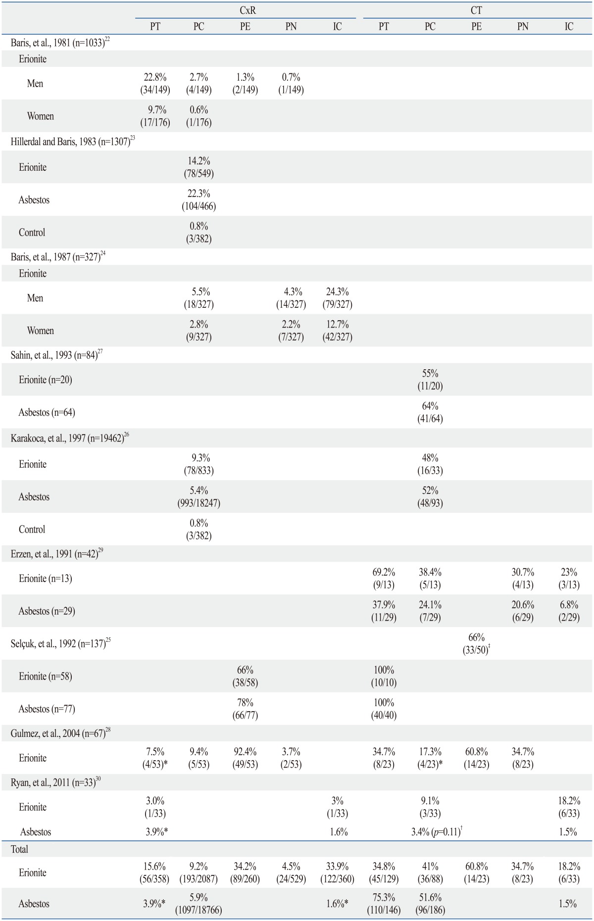
A total of seven retrospective studies (22492 patients)22,23,24,25,26,27,28 and two prospective studies (76 patients),29,30 in addition to a case report of one patient (total 22569 patients),31 are summarized in Table 4. The studies investigated various radiological findings such as pleural effusion (PE), pleural calcification (PC), and pleural nodules (PN) in E-MPM through plain chest X-ray (CxR) and thoracic computed tomography (CT).
Table 4.
Comparisons of Radiological Findings between Subjects with E-MPM and Those with A-MPM

CxR, chest X-ray; CT, thoracic computed tomography; PT, pleural thickening; PC, pleural calcification; PE, pleural effusion; PN, pleural nodule; IC, interstitial changes; E-MPM, erionite-related malignant pleural mesothelioma; A-MPM, asbestos-related malignant pleural mesothelioma.
*No asbestos data; results are not included in final analysis.
†Historical asbestos data; results are not included in final analysis.
‡Combined E-MPM and A-MPM data; results are not included in final analysis.
Bronchoscopy, pleural biopsy, and thoracoscopy
Four retrospective studies (301 patients), three prospective studies (98 patients), and one case report of one patient (total 400 patients) assessed the diagnostic utilities of bronchoscopy, pleural biopsy, and thoracoscopy in E-MPM.
A retrospective study by Dumortier, et al. (68 subjects)32 and a prospective study (16 subjects)33 reported the utility of bronchoalveolar lavage (BAL) followed by light and electron microscopy in identifying erionite in Turkish subjects who were born and lived in exposed villages. Another prospective study by Emri, et al. (40 patients)34 utilized thoracotomy in 75% of cases (30 of 40) or video-assisted thoracoscopy (VATS) (25%, 10 of 40) followed by flow cytometric DNA analysis. All patients had either T3 or T4 tumors without distant metastases.
Two other retrospective studies (202 patients)25,28 utilized pleural biopsy as the main diagnostic tool for the E-MPM-induced pleural plaques, followed by thoracotomy and then thoracoscopy. Another retrospective study and a case report (32 patients) reported similar results without reporting superiority of a specific procedure.20,35
A prospective study of 42 patients with MPM utilized transthoracic echocardiography (TTE) for documentation of pericardial involvement and percutaneous biopsy.36 TTE was followed by percutaneous pericardial biopsy, thoracotomy, or video-associated thoracoscopy and then histopathological and immunohistochemical staining. Through such an approach, these researchers were able to determine pericardial involvement in 45.2% of patients (19 of 42) with MPM, and pericardial effusion was detected in 21.4% (9 of 42), followed by pericardial thickening in 19% (8 of 42) and pericardial calcification in 7.1% (2 of 42). However, there was no difference in terms of pericardial involvement or tumor stage between patients who were exposed to asbestos and to erionite (p>0.05).
TREATMENT
One retrospective study (67 patients) and three prospective (79 patients) studies (146 patients total) assessed the therapeutic utilities of chemotherapy and pleural resection in treatment of MPM.
In one such prospective study (19 subjects), Tansan, et al. assessed response and toxicity according to WHO criteria37 after treating 3 E-MPM and 16 A-MPM patients with cisplatin, mitomycin C, and alpha-2b-interferon versus a well-matched historical control treated with cisplatin and mitomycin C alone.38 The addition of alpha-2b-interferon to cisplatin and mitomycin C did not result in an objective response difference when compared to the cytotoxic agents alone (10.5%; 95% CI: 1.3-19.7%). However, the difference in patient median survival when compared with a well-matched historical group was significantly borderline (Wilcoxon test; χ2=3.64; p=0.0564), which suggests possible benefits from the inclusion of alpha-2b-interferon. Due to the small number of subjects, the study did not comment on the treatment response difference or survival benefits in E-MPM versus A-MPM. Another prospective study by Emri, et al.34 involved 40 patients with a median age of 50 years (range: 30-68 years) and investigated three combinations of chemotherapy drugs: 40% (16 of 40) received cisplatin and mitomycin C, 10% (4 of 40) received ifosfamide and interferon-α, and 8% (5 of 40) were given carboplatin and paclitaxel combinations. None of the patients were able to undergo curative resection as all had either T3 or T4 tumors without distant metastases. Only 30% of patients (12 of 40) had palliative radiotherapy while 37.5% (15 of 40) refused chemotherapy following surgery. The objective response rate in patients who received chemotherapy was 20%, with a median overall survival (OS) of 10±2 months (95% CI: 6-14) and a 1-year survival rate of 45.2%.
Additionally, in a retrospective study of 67 patients with MPM, 52.2% (35 of 67) received a chemotherapy regimen of ifosfamide, mesna, and interferon-α for 6 months.28 Follow-up data were available for the 33% of patients (22 of 67) who received ≥2 cycles of chemotherapy; results included a 2-year survival rate of 22% and a 2-year progression-free interval of 15.7%. Although the study did not report the number of E-MPM cases in the remaining 22 patients, the authors commented that there were no survival differences between the E-MPM and A-MPM patients. The estimated durations of OS and progression-free survival were 12±3.8 months and 9±3.1 months, respectively. In addition, pleurodesis and pleurectomy/decortications were performed as palliative treatment in 19 and 12 MM patients, respectively, who did not receive chemotherapy; however, no survival data were reported for these patients.
Finally, Batirel, et al.39 conducted a prospective feasibility study of extrapleural pneumonectomy followed by adjuvant high-dose irradiation and cisplatin-based chemotherapy to reduce local and distant recurrence rates, thus prolonging survival in E-MPM and A-MPM subjects. Out of 20 patients enrolled in the study, 80% (16 of 20; 3 E-MPM & 13 A-MPM) underwent extrapleural pneumonectomy with en-bloc removal of ipsilateral lung. In addition, 60% (12 of 20) received hemithoracic radiotherapy and cisplatin following surgery. Sixty percent of the study patients (12 of 20) completed all three treatments with a median follow-up of 16 months (range: 1-43 months). The overall median survival was 17 months (24% at 2 years), and 40% of patients (8 of 20) had extrapleural lymph node involvement. However, patients without lymph node metastasis had better overall median survival (no metastasis: 24 months; metastasis: 13 months; p=0.052).
PROGNOSIS & SURVIVAL
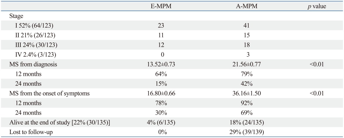
Five retrospective studies (4208 patients) and one prospective study (891 patients) (5099 patients total) assessed prognosis and survival of E-MPM. Four retrospective studies reported an E-MPM-related mortality of 2.30% to 20.6% among the inhabitants of three villages in the Cappadocia region of Central Anatolia followed by lung cancer in 2.30% to 12.1% and then malignant peritoneal mesotheliomas in 2.8% (4 of 141).24,25,40,41 One of the retrospective studies by Selçuk, et al.25 staged patients according to a modified system proposed by Butchart, et al.42 (Table 5) with a median survival of 64% from diagnosis for E-MPM patients at 12 months.25 The latter findings were supported by two recent studies. The first was a retrospective study by Kokturk, et al. (52 patients),21 who studied the impact of apoptotic pathways Bcl-2/Bax and Fas/Fas L on survival in tissue sections from patients with E-MPM and A-MPM, with adenocarcinoma sections serving as a control group. Overall, the median survival times for the E-MPM and A-MPM groups were similar even when considering different histological subtypes (epithelial vs. non-epithelial), stage, and different treatment modalities [palliative surgical debulking (PSD) vs. non-PSD]. However, E-MPM patients with negative Bax had longer survival than Bax-positive patients (Bax-negative: 18 months; Bax-positive: 14 months; p=0.06). While Fas Ligand-positive patients had statistically better survival than Fas Ligand-negative patients in the two MPM groups (Fas L-positive: 15 months; Fas L-negative: 12 months; p=0.05).21 The second study was a long-term prospective study of residents (891 subjects) of two villages exposed to erionite and one nearby control village on the Turkish Anatolian plateau conducted between 1979 and December 31, 2003.15 Over the 23-year follow-up, 42% of the subjects (372 of 891) died; 32% (119 of 372) of these deaths were from mesothelioma, which was the cause of 44.5% of all deaths in the erionite-exposed villages. Interestingly, only two cases of mesothelioma occurred in the control village.
Table 5.
Staging and Survival for E-MPM and A-MPM Patients25

MS, median survival (in months); E-MPM, erionite-related malignant pleural mesothelioma; A-MPM, asbestos-related malignant pleural mesothelioma.
KEY POINTS ABOUT ERIONITE
Although the link between asbestos exposure and mesothelioma was established over 50 years ago, it is still unclear whether all types of asbestos can cause mesothelioma.9,43,44 The median latency from time of asbestos exposure to the disease is about 32 years; however, in many instances it ranges from 20 to 50 years.1,45,46 Unfortunately, the median survival is about 1 year from diagnosis, as current therapies have had only marginal effects in altering the natural course of the disease.46
In contrast to industrialized Western countries, in which MM is typically related to previous occupational exposure to asbestos, MM in central and eastern Turkey, Greece, and Cyprus is instead associated with environmental exposure to tremolite asbestos or erionite, a natural fibrous zeolite.25,47 Interestingly, recent environmental studies also report the presence of erionite fibers in North America, with one case report of E-MPM in Mexico.30,48 Furthermore, other enviromentally-related MM cases were reported following exposure to asbestos and other fibrous minerals such as winchite, richterite, fluoroedenite, and antigorite.49,50,51
Erionite belongs to the mineralogical group of zeolites, a complex group of hydrated aluminosilicates of alkali and alkaline earths that includes about 40 natural minerals.33 Zeolites naturally occur in volcanic rocks cavities and other hydrothermal environments.33 Most naturally-occurring zeolites are non-fibrous, in contrast to erionite, clinoptilolite, and mordenite, which are fibrous.52
Various epidemiological studies support the high incidence of E-MPM among the inhabitants and emigrants of three villages in the Cappadocia region of Central Anatolia. In contrast to A-MPM, E-MPM occurred at a younger age, with a median age at diagnosis in the mid-50 s (57 for males and 55 for females), and the male-to-female ratio of MM in Turkish immigrants was 1.1.13 Such a ratio was quite different from that observed in industrialized countries, where occupational exposure to asbestos is common in men.53 Furthermore, some data raised the possibility of extrapleural and peritoneal cancers as additional causes of mortality linked to erionite-fiber exposure.41 Interestingly, asbestos fiber concentrations in erionite-affected villagers were not different from those in Turks without environmental exposure to tremolite.22,33
The development of E-MPM in some but not all subjects who resided in the three Cappadocian villages with high exposure to erionite raise the probability of genetic factors behind such selectivity.16,17,20 Possible support for this finding is the absence of MM cases in nearby villages, whose inhabitants had similar amounts of erionite in stone samples from their houses.16 Furthermore, it is possible that genetic predisposition, viral infection, and even exposure to other environmental factors, such as radiation, may act singularly or in combination to induce E-MPM.54
When considering the diagnosis of E-MPM, radiological assessments play an essential role in diagnosis, staging, and management decisions for E-MPM. Table 4 summarizes the yields of various studies that examined the utility of CxR and chest CT in diagnosis of E-MPM in addition to comparing the CxR and chest CT findings between E-MPM and A-MPM. Six studies reported CxR findings in 2178 patients who were exposed to erionite, comparing them with 18766 patients who were exposed to asbestos.22,23,24,25,26,30 PE (34.2%) and interstitial changes (IC; 33.9%) were the most common findings. These were followed by pleural thickening (PT; 15.7%) and PC (9.2%), with PN reported in only 4.5% of E-MPM patients. Limited CxR data were available in one study due to low incidences of PT and IC, which hindered comparison between the CxR findings in patients with E-MPM and A-MPM.30
Five recent studies that included 109 E-MPM and 186 A-MPM patients examined the role of chest CT in diagnosis of E-MPM.25,26,27,28,29,30 Three of these studies were able to compare CxR and chest CT findings in patients with E-MPM and A-MPM.25,28,30 PE was the most common chest CT finding at 60.8%, followed by PC (41%), PT (34.8%), and PN (34.7%), while IC was reported in 18.2% of the E-MPM patients. As expected, chest CT was more reliable in detecting various pleural changes in E-MPM than CxR (Table 4), and PE was the most common finding in E-MPM, by both CxR and chest CT, followed by PT and IC.
Various sampling techniques were utilized in identifying erionite in the lung and pleural tissues. Bronchoscopy with BAL remains an effective and relatively less invasive technique in establishing E-MPM in patients from areas with known high exposure.32,33 By measuring the concentration of erionite particles in BAL from subjects, it is possible to establish past exposure to erionite even 20 years after the exposure had stopped.33 In other instances, pleural sampling can be achieved through pleural biopsy, thoracoscopy, or thoracotomy, with a 29% to 95.7% yield for E-MPM by light microscopy.20,25,28,33,35 Limited data are available for assessing the role of electron microscopy,32 scanning electron microscopy,48,55 analytical transmission electron microscopy,56 and immunohistochemical analysis in diagnosis of E-MPM.20
Regarding therapeutic options, several studies investigated the utility of chemotherapeutic agents, radiotherapy, and surgical treatment in achieving local control of E-MPM and perhaps limiting distant metastases.34,38 When considering chemotherapy, cisplatin, and mitomycin remain the main chemotherapeutic agents, administered either alone or following surgery. However, objective response rate to chemotherapy remains low, in the range of 20%, with median OS close to 10 months.34 Although the addition of interferon-α in one study was clinically tolerable, it did not result in an objective response difference when compared to cisplatin and mitomycin alone.38 Pleurodesis, VATS, thoracotomy, and pleurectomy/decortications could be attempted in selected patients at specialized medical facilities.28,34 However, most patients present at a late stage (T3-T4) and are therefore unable to undergo a curative resection.34 In such patients, extrapleural pneumonectomy, followed by adjuvant high-dose irradiation and cisplatin-based chemotherapy may improve local and distant control rates and possibly prolong their survival.39
Survival in patients with E-MPM remains low when compared to patients with A-MPM (Table 5). Such a difference could be related in part to genetic differences in originally acquiring E-MPM16,20 or to delay in clinical presentation and evaluation. However, the expression of various apoptosis-regulating proteins may prove to be of utmost prognostic significance in some patients with E-MPM.21
In summary, erionite, a non-asbestos fibrous zeolite, has been the cause of numerous MM cases in Turkey and even in North America. Furthermore, many of the affected subjects had migrated to various European countries such as Germany and Sweden, which has impacted public healthcare in those countries.
Available data indicate that neither SV40 nor HHV-8 were identified as co-factors in the pathogenesis of environmentally-induced MM. However, the observed difference could be due to a higher degree of erionite carcinogenicity or genetic transmission of erionite susceptibility in an autosomal dominant fashion among the residents of the erionite-affected villages. Further studies are necessary to identify the gene that may predispose individuals to erionite-induced MM. Therapeutic options for E-MPM remain very limited, and cure of the disease is extremely rare.
Limitations of the studies about erionite
This review provides a summary of multiple relevant studies and an independent triplicate assessment of trial validity. However, the heterogeneity of patients and varied diagnostic and therapeutic regimens precluded a systematic meta-analysis of the results. We advise readers to review the original publications for any further details.
Blinding both patients and investigators to therapeutic intervention is of utmost importance when evaluating subjective outcomes. Among several of the studies reviewed, blinding was neither routinely employed nor reported. Accordingly, interpretation of variables such as chemotherapeutic response difference or survival benefits in E-MPM versus A-MPM may be biased.
Finally, all co-interventions, including thoracotomy tube insertion for E-MPM-induced pleural effusion, use of therapies (other than those being directly studied), and timing of end-of-life decisions were neither standardized nor consistently reported. Monitoring and reporting such measures will help reduce bias in endpoint interpretation, e.g., estimating durations of OS and progression-free survival following various therapies.
Footnotes
The authors have no financial conflicts of interest.
References
- 1.Carbone M, Kratzke RA, Testa JR. The pathogenesis of mesothelioma. Semin Oncol. 2002;29:2–17. doi: 10.1053/sonc.2002.30227. [DOI] [PubMed] [Google Scholar]
- 2.Senyiğit A, Babayiğit C, Gökirmak M, Topçu F, Asan E, Coşkunsel M, et al. Incidence of malignant pleural mesothelioma due to environmental asbestos fiber exposure in the southeast of Turkey. Respiration. 2000;67:610–614. doi: 10.1159/000056289. [DOI] [PubMed] [Google Scholar]
- 3.Boffetta P. Health effects of asbestos exposure in humans: a quantitative assessment. Med Lav. 1998;89:471–480. [PubMed] [Google Scholar]
- 4.Artvinli M, Bariş YI. Malignant mesotheliomas in a small village in the Anatolian region of Turkey: an epidemiologic study. J Natl Cancer Inst. 1979;63:17–22. [PubMed] [Google Scholar]
- 5.Rohl AN, Langer AM, Moncure G, Selikoff IJ, Fischbein A. Endemic pleural disease associated with exposure to mixed fibrous dust in Turkey. Science. 1982;216:518–520. doi: 10.1126/science.7071597. [DOI] [PubMed] [Google Scholar]
- 6.Carbone M, Emri S, Dogan AU, Steele I, Tuncer M, Pass HI, et al. A mesothelioma epidemic in Cappadocia: scientific developments and unexpected social outcomes. Nat Rev Cancer. 2007;7:147–154. doi: 10.1038/nrc2068. [DOI] [PubMed] [Google Scholar]
- 7.Boman G, Schubert V, Svane B, Westerholm P, Bolinder E, Rohl AN, et al. Malignant mesothelioma in Turkish immigrants residing in Sweden. Scand J Work Environ Health. 1982;8:108–112. doi: 10.5271/sjweh.2489. [DOI] [PubMed] [Google Scholar]
- 8.Metintas M, Hillerdal G, Metintas S. Malignant mesothelioma due to environmental exposure to erionite: follow-up of a Turkish emigrant cohort. Eur Respir J. 1999;13:523–526. doi: 10.1183/09031936.99.13352399. [DOI] [PubMed] [Google Scholar]
- 9.Powers A, Carbone M. The role of environmental carcinogens, viruses and genetic predisposition in the pathogenesis of mesothelioma. Cancer Biol Ther. 2002;1:348–353. [PubMed] [Google Scholar]
- 10.Jadad AR, Moore RA, Carroll D, Jenkinson C, Reynolds DJ, Gavaghan DJ, et al. Assessing the quality of reports of randomized clinical trials: is blinding necessary. Control Clin Trials. 1996;17:1–12. doi: 10.1016/0197-2456(95)00134-4. [DOI] [PubMed] [Google Scholar]
- 11.Oxford Centre for Evidence-based Medicine. Levels of Evidence (March 2009) Available at: http://www.cebm.net/index.aspx?o=1025.
- 12.Metintas M, Hillerdal G, Metintas S, Dumortier P. Endemic malignant mesothelioma: exposure to erionite is more important than genetic factors. Arch Environ Occup Health. 2010;65:86–93. doi: 10.1080/19338240903390305. [DOI] [PubMed] [Google Scholar]
- 13.Mousavi SM, Sundquist J, Hemminki K. Is risk of pleural mesothelioma an environmental risk outside Turkey? A study on immigrants to Sweden. Lung Cancer. 2010;68:125–126. doi: 10.1016/j.lungcan.2010.01.014. [DOI] [PubMed] [Google Scholar]
- 14.Ozesmi M, Hillerdal G, Svane B, Widström O. Prospective clinical and radiologic study of zeolite-exposed Turkish immigrants in Sweden. Respiration. 1990;57:325–328. doi: 10.1159/000195865. [DOI] [PubMed] [Google Scholar]
- 15.Baris YI, Grandjean P. Prospective study of mesothelioma mortality in Turkish villages with exposure to fibrous zeolite. J Natl Cancer Inst. 2006;98:414–417. doi: 10.1093/jnci/djj106. [DOI] [PubMed] [Google Scholar]
- 16.Roushdy-Hammady I, Siegel J, Emri S, Testa JR, Carbone M. Genetic-susceptibility factor and malignant mesothelioma in the Cappadocian region of Turkey. Lancet. 2001;357:444–445. doi: 10.1016/S0140-6736(00)04013-7. [DOI] [PubMed] [Google Scholar]
- 17.Karakoca Y, Emri S, Bagci T, Demir A, Erdem Y, Baris E, et al. Environmentally-induced malignant pleural mesothelioma and HLA distribution in Turkey. Int J Tuberc Lung Dis. 1998;2:1017–1022. [PubMed] [Google Scholar]
- 18.Emri S, Kocagoz T, Olut A, Güngen Y, Mutti L, Baris YI. Simian virus 40 is not a cofactor in the pathogenesis of environmentally induced malignant pleural mesothelioma in Turkey. Anticancer Res. 2000;20:891–894. [PubMed] [Google Scholar]
- 19.Olut AI, Ertugrul DT, Kocagoz T, Er M, Emri S. HHV-8 is not a cofactor in the pathogenesis of environmentally induced malignant pleural mesothelioma. Monaldi Arch Chest Dis. 2000;55:110–113. [PubMed] [Google Scholar]
- 20.Olut A, Firat P, Ertugrul D, Gungen Y, Emri S. Ras oncoprotein expression in erionite- and asbestos-induced Turkish malignant pleural mesothelioma patients--a pilot study. Respir Med. 2001;95:697–698. doi: 10.1053/rmed.2001.1122. [DOI] [PubMed] [Google Scholar]
- 21.Kokturk N, Firat P, Akay H, Kadilar C, Ozturk C, Zorlu F, et al. Prognostic significance of Bax and Fas ligand in erionite and asbestos induced Turkish malignant pleural mesothelioma. Lung Cancer. 2005;50:189–198. doi: 10.1016/j.lungcan.2005.05.025. [DOI] [PubMed] [Google Scholar]
- 22.Baris YI, Saracci R, Simonato L, Skidmore JW, Artvinli M. Malignant mesothelioma and radiological chest abnormalities in two villages in Central Turkey. An epidemiological and environmental investigation. Lancet. 1981;1:984–987. doi: 10.1016/s0140-6736(81)91742-6. [DOI] [PubMed] [Google Scholar]
- 23.Hillerdal G, Baris YI. Radiological study of pleural changes in relation to mesothelioma in Turkey. Thorax. 1983;38:443–448. doi: 10.1136/thx.38.6.443. [DOI] [PMC free article] [PubMed] [Google Scholar]
- 24.Baris I, Simonato L, Artvinli M, Pooley F, Saracci R, Skidmore J, et al. Epidemiological and environmental evidence of the health effects of exposure to erionite fibres: a four-year study in the Cappadocian region of Turkey. Int J Cancer. 1987;39:10–17. doi: 10.1002/ijc.2910390104. [DOI] [PubMed] [Google Scholar]
- 25.Selçuk ZT, Cöplü L, Emri S, Kalyoncu AF, Sahin AA, Bariş YI. Malignant pleural mesothelioma due to environmental mineral fiber exposure in Turkey. Analysis of 135 cases. Chest. 1992;102:790–796. doi: 10.1378/chest.102.3.790. [DOI] [PubMed] [Google Scholar]
- 26.Karakoca Y, Emri S, Cangir AK, Bariş YI. Environmental pleural plaques due to asbestos and fibrous zeolite exposure in Turkey. Indoor Built Environ. 1997;6:100–105. [Google Scholar]
- 27.Sahin AA, Cöplü L, Selçuk ZT, Eryilmaz M, Emri S, Akhan O, et al. Malignant pleural mesothelioma caused by environmental exposure to asbestos or erionite in rural Turkey: CT findings in 84 patients. AJR Am J Roentgenol. 1993;161:533–537. doi: 10.2214/ajr.161.3.8394641. [DOI] [PubMed] [Google Scholar]
- 28.Gulmez I, Kart L, Buyukoglan H, Er O, Balkanli S, Ozesmi M. Evaluation of malignant mesothelioma in central Anatolia: a study of 67 cases. Can Respir J. 2004;11:287–290. doi: 10.1155/2004/204793. [DOI] [PubMed] [Google Scholar]
- 29.Erzen C, Eryilmaz M, Kalyoncu F, Bilir N, Sahin A, Baris YI. CT findings in malignant pleural mesothelioma related to nonoccupational exposure to asbestos and fibrous zeolite (erionite) J Comput Assist Tomogr. 1991;15:256–260. doi: 10.1097/00004728-199103000-00012. [DOI] [PubMed] [Google Scholar]
- 30.Ryan PH, Dihle M, Griffin S, Partridge C, Hilbert TJ, Taylor R, et al. Erionite in road gravel associated with interstitial and pleural changes--an occupational hazard in western United States. J Occup Environ Med. 2011;53:892–898. doi: 10.1097/JOM.0b013e318223d44c. [DOI] [PubMed] [Google Scholar]
- 31.Gräsel B, Kaya A, Stahl U, Rauber K, Kuntz C. [Erionite-induced pleural plaques. Exposition to urban pollution in a female Turkish migrant in Germany] Chirurg. 2008;79:584–588. doi: 10.1007/s00104-008-1515-9. [DOI] [PubMed] [Google Scholar]
- 32.Dumortier P, Göcmen A, Laurent K, Manço A, De Vuyst P. The role of environmental and occupational exposures in Turkish immigrants with fibre-related disease. Eur Respir J. 2001;17:922–927. doi: 10.1183/09031936.01.17509220. [DOI] [PubMed] [Google Scholar]
- 33.Dumortier P, Coplü L, Broucke I, Emri S, Selcuk T, de Maertelaer V, et al. Erionite bodies and fibres in bronchoalveolar lavage fluid (BALF) of residents from Tuzköy, Cappadocia, Turkey. Occup Environ Med. 2001;58:261–266. doi: 10.1136/oem.58.4.261. [DOI] [PMC free article] [PubMed] [Google Scholar]
- 34.Emri S, Akbulut H, Zorlu F, Dinçol D, Akay H, Güngen Y, et al. Prognostic significance of flow cytometric DNA analysis in patients with malignant pleural mesothelioma. Lung Cancer. 2001;33:109–114. doi: 10.1016/s0169-5002(00)00249-x. [DOI] [PubMed] [Google Scholar]
- 35.Hillerdal G, Elmberger G. Malignant mediastinal tumor with bone formation--mesothelioma or sarcoma? J Thorac Oncol. 2007;2:983–984. doi: 10.1097/JTO.0b013e318153fd76. [DOI] [PubMed] [Google Scholar]
- 36.Ozer N, Shehu V, Aytemir K, Ovünç K, Emre S, Kes S. Echocardiographic findings of pericardial involvement in patients with malignant pleural mesothelioma with a history of environmental exposure to asbestos and erionite. Respirology. 2000;5:333–336. [PubMed] [Google Scholar]
- 37.Miller AB, Hoogstraten B, Staquet M, Winkler A. Reporting results of cancer treatment. Cancer. 1981;47:207–214. doi: 10.1002/1097-0142(19810101)47:1<207::aid-cncr2820470134>3.0.co;2-6. [DOI] [PubMed] [Google Scholar]
- 38.Tansan S, Emri S, Selçuk T, Koç Y, Hesketh P, Heeren T, et al. Treatment of malignant pleural mesothelioma with cisplatin, mitomycin C and alpha interferon. Oncology. 1994;51:348–351. doi: 10.1159/000227363. [DOI] [PubMed] [Google Scholar]
- 39.Batirel HF, Metintas M, Caglar HB, Yildizeli B, Lacin T, Bostanci K, et al. Trimodality treatment of malignant pleural mesothelioma. J Thorac Oncol. 2008;3:499–504. doi: 10.1097/JTO.0b013e31816fca1b. [DOI] [PubMed] [Google Scholar]
- 40.Simonato L, Baris R, Saracci R, Skidmore J, Winkelmann R. Relation of environmental exposure to erionite fibres to risk of respiratory cancer. IARC Sci Publ. 1989:398–405. [PubMed] [Google Scholar]
- 41.Bariş B, Demir AU, Shehu V, Karakoca Y, Kisacik G, Bariş YI. Environmental fibrous zeolite (erionite) exposure and malignant tumors other than mesothelioma. J Environ Pathol Toxicol Oncol. 1996;15:183–189. [PubMed] [Google Scholar]
- 42.Butchart EG, Ashcroft T, Barnsley WC, Holden MP. Pleuropneumonectomy in the management of diffuse malignant mesothelioma of the pleura. Experience with 29 patients. Thorax. 1976;31:15–24. doi: 10.1136/thx.31.1.15. [DOI] [PMC free article] [PubMed] [Google Scholar]
- 43.Tweedale G, McCulloch J. Chrysophiles versus chrysophobes: the white asbestos controversy, 1950s-2004. Isis. 2004;95:239–259. doi: 10.1086/426196. [DOI] [PubMed] [Google Scholar]
- 44.McDonald JC, McDonald A. Mesothelioma and asbestos exposure. In: Pass HI, Vogelzang NJ, Carbone M, editors. Malignant Mesothelioma. New York: Springer; 2005. pp. 267–292. [Google Scholar]
- 45.Peto J, Hodgson JT, Matthews FE, Jones JR. Continuing increase in mesothelioma mortality in Britain. Lancet. 1995;345:535–539. doi: 10.1016/s0140-6736(95)90462-x. [DOI] [PubMed] [Google Scholar]
- 46.Pass HI, Vogelzang N, Hahn SM, Carbone M. Malignant pleural mesothelioma. In: DeVita VT, Hellman S, Rosenberg SA, editors. Cancer, Principles & Practice of Oncology. Philadelphia: Lippincott Williams and Wilkins; 2005. pp. 1687–1712. [Google Scholar]
- 47.Barış YI. Asbestos and Erionite Related Chest Diseases. Ankara, Turkey: Semih Offset Matbaacilik; 1987. [Google Scholar]
- 48.Kliment CR, Clemens K, Oury TD. North American erionite-associated mesothelioma with pleural plaques and pulmonary fibrosis: a case report. Int J Clin Exp Pathol. 2009;2:407–410. [PMC free article] [PubMed] [Google Scholar]
- 49.Sullivan PA. Vermiculite, respiratory disease, and asbestos exposure in Libby, Montana: update of a cohort mortality study. Environ Health Perspect. 2007;115:579–585. doi: 10.1289/ehp.9481. [DOI] [PMC free article] [PubMed] [Google Scholar]
- 50.Groppo C, Tomatis M, Turci F, Gazzano E, Ghigo D, Compagnoni R, et al. Potential toxicity of nonregulated asbestiform minerals: balangeroite from the western Alps. Part 1: Identification and characterization. J Toxicol Environ Health A. 2005;68:1–19. doi: 10.1080/15287390590523867. [DOI] [PubMed] [Google Scholar]
- 51.Putzu MG, Bruno C, Zona A, Massiccio M, Pasetto R, Piolatto PG, et al. Fluoro-edenitic fibres in the sputum of subjects from Biancavilla (Sicily): a pilot study. Environ Health. 2006;5:20. doi: 10.1186/1476-069X-5-20. [DOI] [PMC free article] [PubMed] [Google Scholar]
- 52.Thomas JA. Toxicological assessment of zeolites. Int J Toxicol. 1992;11:259–273. [Google Scholar]
- 53.Yang H, Testa JR, Carbone M. Mesothelioma epidemiology, carcinogenesis, and pathogenesis. Curr Treat Options Oncol. 2008;9:147–157. doi: 10.1007/s11864-008-0067-z. [DOI] [PMC free article] [PubMed] [Google Scholar]
- 54.Carbone M, Ly BH, Dodson RF, Pagano I, Morris PT, Dogan UA, et al. Malignant mesothelioma: facts, myths, and hypotheses. J Cell Physiol. 2012;227:44–58. doi: 10.1002/jcp.22724. [DOI] [PMC free article] [PubMed] [Google Scholar]
- 55.Ataman G. [The role of erionite (zeolite) in pulmonary mesothelioma] C R Seances Acad Sci D. 1980;291:167–169. [PubMed] [Google Scholar]
- 56.Sebastien P, Gaudichet A, Bignon J, Baris YI. Zeolite bodies in human lungs from Turkey. Lab Invest. 1981;44:420–425. [PubMed] [Google Scholar]


