

Abstract
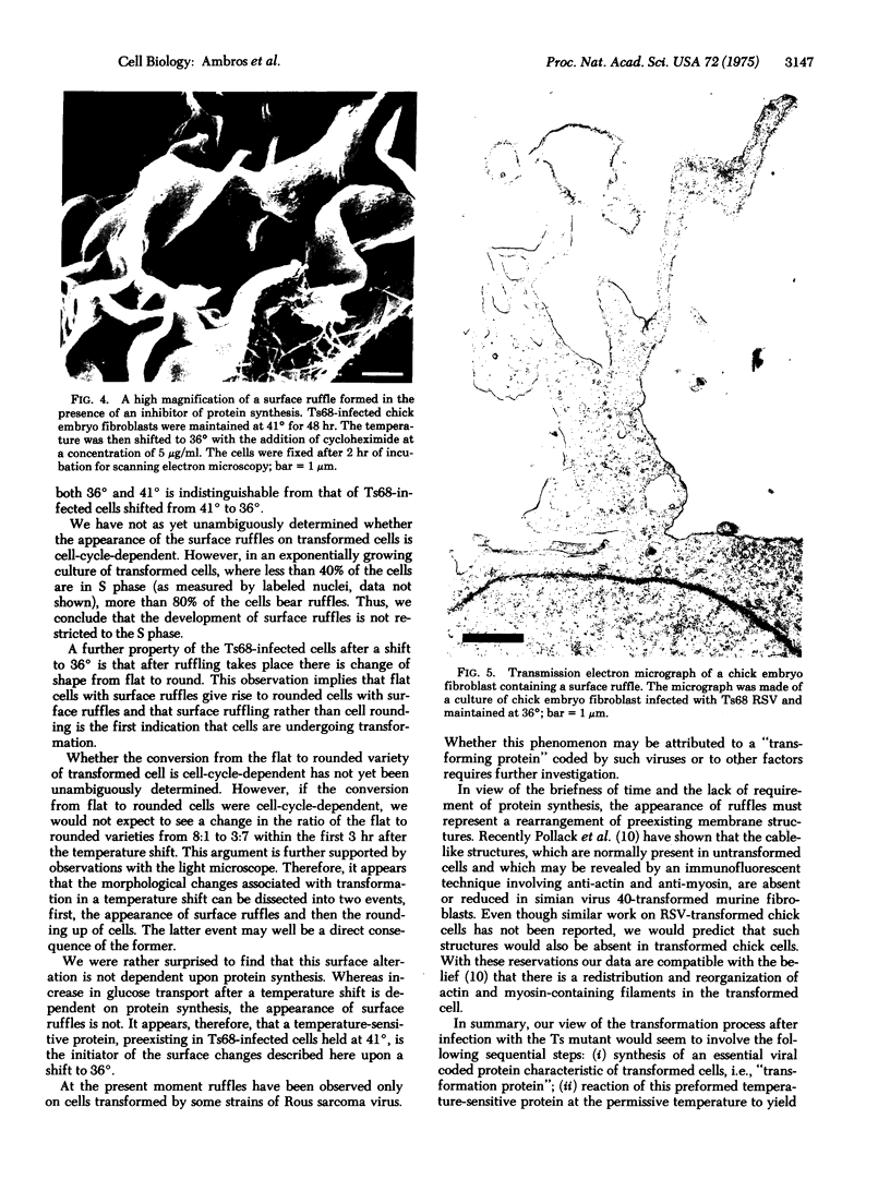
Confluent chick embryo fibroblasts infected with the Ts68 mutant of Rous sarcoma virus were examined by scanning electron microscopy at the permissive (36 degrees) and nonpermissive (41 degrees) temperatures for transformation. Infected cells shifted from 41 degrees to 36 degrees undergo a change in shape from elongated to rounded. This process is preceded by the appearance of surface ruffles on the cell. These surface ruffles are not observed on cells maintained at 41 degrees, appear as early as 0.5 hr after a shift to 36 degrees, and are common on cells maintained at 36 degrees. By 3.5 hr after the shift from 41 degrees to 36 degrees, cultures appear fully transformed by the criteria of cell roundedness and the presence of surface ruffles. This surface alteration of cells is the earliest event of those so far reported during the transformation process and is not dependent upon protein synthesis and extracellular plasminogen during the period of temperature shift.
Full text
PDF


Images in this article


Selected References
These references are in PubMed. This may not be the complete list of references from this article.
- Chen L. B., Buchanan J. M. Plasminogen-independent fibrinolysis by proteases produced by transformed chick embryo fibroblasts. Proc Natl Acad Sci U S A. 1975 Mar;72(3):1132–1136. doi: 10.1073/pnas.72.3.1132. [DOI] [PMC free article] [PubMed] [Google Scholar]
- Hale A. H., Winkelhake J. L., Weber M. J. Cell surface changes and Rous sarcoma virus gene expression in synchronized cells. J Cell Biol. 1975 Feb;64(2):398–407. doi: 10.1083/jcb.64.2.398. [DOI] [PMC free article] [PubMed] [Google Scholar]
- Kawai S., Hanafusa H. The effects of reciprocal changes in temperature on the transformed state of cells infected with a rous sarcoma virus mutant. Virology. 1971 Nov;46(2):470–479. doi: 10.1016/0042-6822(71)90047-x. [DOI] [PubMed] [Google Scholar]
- Pollack R., Osborn M., Weber K. Patterns of organization of actin and myosin in normal and transformed cultured cells. Proc Natl Acad Sci U S A. 1975 Mar;72(3):994–998. doi: 10.1073/pnas.72.3.994. [DOI] [PMC free article] [PubMed] [Google Scholar]
- Porter K. R., Todaro G. J., Fonte V. A scanning electron microscope study of surface features of viral and spontaneous transformants of mouse Balb-3T3 cells. J Cell Biol. 1973 Dec;59(3):633–642. doi: 10.1083/jcb.59.3.633. [DOI] [PMC free article] [PubMed] [Google Scholar]
- Robbins P. W., Wickus G. G., Branton P. E., Gaffney B. J., Hirschberg C. B., Fuchs P., Blumberg P. The chick fibroblast cell surface after transformation by Rous sarcoma virus. Cold Spring Harb Symp Quant Biol. 1975;39(Pt 2):1173–1180. doi: 10.1101/sqb.1974.039.01.135. [DOI] [PubMed] [Google Scholar]
- Stone K. R., Smith R. E., Joklik W. K. Changes in membrane polypeptides that occur when chick embryo fibroblasts and NRK cells are transformed with avian sarcoma viruses. Virology. 1974 Mar;58(1):86–100. doi: 10.1016/0042-6822(74)90143-3. [DOI] [PubMed] [Google Scholar]
- Wickus G. G., Robbins P. W. Plasma membrane proteins of normal and Rous sarcoma virus-transformed chick-embryo fibroblasts. Nat New Biol. 1973 Sep 19;245(142):65–67. doi: 10.1038/newbio245065a0. [DOI] [PubMed] [Google Scholar]