Abstract
Purpose.
We evaluated the roles of luminance and fixation in the pathophysiology of dissociated vertical divergence (DVD).
Methods.
Vertical eye position was measured in 6 subjects with DVD (ages 11–47 years, 5 females) and 6 controls (ages 16–40 years, 5 females) using video-oculography (VOG) under conditions of change in fixation and luminance.
Results.
Subjects with DVD showed the following VOG responses. When fixation was precluded with a translucent filter and bright light was shone into one eye to produce a marked binocular luminance disparity, we found some subjects had a small induced vertical divergence causing the illuminated eye to be lower than the nonilluminated eye (mean −1.6° ± 1.5°, P = 0.06 compared to no vertical divergence using the signed rank test). When fixation was precluded with a translucent filter, while alternate occlusion produced a mild binocular luminance disparity, we found a smaller vertical divergence of the eyes that was not statistically significant (1.2° ± 2.1°, P = 0.3). When alternate occlusion produced reversal of monocular fixation in the dark (with essentially no change in peripheral luminance disparity), there was a significant vertical divergence movement causing the covered eye to be relatively higher than the uncovered eye (7.2° ± 3.1°, P = 0.03). The amplitude of this vertical divergence was similar to that measured under conditions of alternate occlusion in a lighted room (where there also was a significant average relative upward movement of the covered eye of 8.1° ± 2.9°, P = 0.03). Control subjects showed no vertical divergence under any testing conditions.
Conclusions.
Dissociated vertical divergence is mediated primarily by changes in fixation and only to a minor degree by binocular luminance disparity.
Keywords: dissociated vertical divergence, strabismus, DVD, videooculography
Dissociated vertical divergence is mediated primarily by changes in fixation, and only to a minor degree by binocular luminance disparity.
Introduction
Over a century after its initial description,1,2 dissociated vertical divergence (DVD) remains one of the most controversial ocular motor disturbances. It is characterized clinically by the slow ascent of either eye followed, after a variable period of time, by a slow descent back to its neutral position. It generally is associated with infantile esotropia, but also can accompany other forms of binocular misalignment that develop in early infancy.3
Bielschowsky4 defined the essential role of luminance disparity in DVD and summarized it as follows: “If one puts in front of the fixating eye a darkening glass wedge (Zeiss) moving it in such a manner that the fixed lamp is gradually darkened the covered eye which is first elevated upward will be seen behind the cover to move downward below the horizontal plane, sometimes in almost exact proportion to the fixating eye, which is continuously keeping fixation during the examination.”5 This Bielschowsky phenomenon is unique to DVD and to the related dissociated horizontal deviation (DHD).6
The importance of fixation was demonstrated by Posner,7 who induced DVD by placement of a small occluder in front of one eye to preclude central fixation while inducing only minimal luminance imbalance. When a second cover then was placed equidistant in front of the fixating eye, the occluded hyperdeviated eye descended slowly back to its neutral position, indicating that fixation is just as important as illumination in producing DVD. Bielschowsky5 and Posner7 emphasized the momentary fluctuations in the hyperdeviation that depend in part upon the patient's level of attention during visual fixation. In 1955, Ohm8 summarized the dual contributions of luminance and fixation in producing this delicate binocular balancing movement, as reviewed by Ohm,8 and Mattheus and Kommerell.9
Nevertheless, it remains unknown whether a binocular luminance disparity can trigger DVD in the absence of a preexisting binocular fixational stimulus. Because this issue is crucial to understanding the pathophysiology of DVD, we performed video-oculographic (VOG) eye movement recordings in subjects with DVD while independently controlling for luminance and fixational disparity in the two eyes.
Methods
We enrolled 6 subjects with DVD and 6 control subjects. The diagnosis of DVD was based on the following clinical criteria: a clinical history of infantile strabismus, and the finding of a hyperdeviation of each eye when covered on alternate cover testing in primary position (i.e., no hypodeviation). Exclusion criteria included inability to perform the eye movement recording protocol (age younger than 8 years), a refractive error greater than 3 diopters (unless corrected with contact lenses), or a history of vertical muscle surgery. All control subjects were orthotropic at distance and near fixation, had no history of strabismus, and no evidence of DVD on alternate cover testing.
This study was approved by the Institutional Review Board and written informed consent was obtained for the testing of all subjects, with parental consent and child assent in the cases of children. All testing was conducted in a manner compliant with the Health Insurance Portability and Accountability Act and adhered to the tenets of the Declaration of Helsinki.
Subjects with DVD ranged in age from 11 to 47 years (mean 28 years) with 5 subjects being female. All subjects had a history of infantile esotropia with all 6 subjects having had previous horizontal muscle surgery. Corrected visual acuity ranged from 20/20 to 20/50 in the better-seeing eye and from 20/20 to 20/80 in the worse-seeing eye. Coexistent latent nystagmus was diagnosed in 3 patients clinically and in 5 on VOG. Control subjects ranged in age from 16 to 40 years (mean 28 years) with 5 subjects being female. Visual acuity was 20/15 to 20/20 in the better eye and ranged from 20/15 to 20/25 in the fellow eye.
Simultaneous horizontal, torsional, and vertical eye movements were recorded using the SensoMotoric infrared VOG system (3D-VOG; SensoMotoric Instruments, Teltow, Germany), which is a measurement system for acquisition of eye movements based on noninvasive video image processing technology using head-mounted infrared video cameras.10 This system uses infrared cameras to track eye movements by detecting the pupil center within each video image.10
The testing protocol for all subjects involved four specific conditions, performed in the following order, in succession with less than a minute between conditions.
Alternate Occlusion Without Fixation (Monocular Darkening)
First, to test the effect of a change in luminance when no fixation was possible, a translucent filter was placed before both eyes for 30 seconds (precluding fixation), and then the left eye was occluded for 15 seconds (reducing the luminance to that eye), followed by occlusion of the right eye for 15 seconds. The translucent filter reduced the room light luminance from 475 to 375 lux.
Alternate Increased Luminance Without Fixation (Monocular Flashlight)
Second, to test the effect of increasing the luminance to one eye, when no fixation was possible, a flashlight was introduced 33 cm in front of the filter, first in front of the left eye for 15 seconds, and then in front of the right eye for 15 seconds. The monocular flashlight increased luminance through the translucent filter to 2500 lux.
Alternate Occlusion With Fixation in Darkness (Crossbar)
Third, to test the effect of a change in fixation when there was no or minimal change in luminance, the subject was allowed to fixate on a distant red cross in a dark room for 30 seconds and then the left eye was occluded for 15 seconds, followed by occlusion of the right eye for 15 seconds.
Alternate Occlusion With Fixation (Room Light)
Finally, to test the effect of a change in luminance and fixation the subject fixated binocularly on a distance target in a lighted room for 15 seconds and then the left eye was occluded for 15 seconds, followed by occlusion of the right eye for 15 seconds.
Data Analysis
To calculate the position of each eye during each experimental condition, the VOG positional recordings at 60 Hz were analyzed. Because DVD can cause minor variability in the baseline vertical alignment, the induced vertical divergence of the eyes was determined by comparing the relative positions of the eyes during left eye illumination, darkening, or occlusion with right eye illumination, darkening, or occlusion for each of the 4 testing conditions. To obtain representative values, we analyzed VOG data from the last 12 seconds before and the first 12 seconds after the shift of the stimulus from one eye to the other.
To reduce noise from the data, we removed all the values that corresponded to a loss of fixation (assigned zero by the VOG software) and all values that exceeded a 30° movement which most likely represented a blink artifact or fixation loss. We calculated the 90th percentile of the remaining values of the vertical recordings, during each 12-second segment studied, to represent the maximum vertical deviation.
We used these 90th percentile values to represent the vertical position of each eye and to calculate the amount of vertical divergence under each testing condition. Regardless of which eye was measured as higher, we subtracted the vertical position of the left eye from the position of the right eye during the 12 seconds before and 12 seconds after the illumination, darkening, or fixation stimulus was shifted from the left to the right eye. We then derived a total vertical divergence value for each condition by calculating the difference in these values, which yielded a single value for each subject for each of the 4 conditions (5 values for the increased luminance condition due to missing data in one subject). The average vertical divergence movement of the eyes for each subject under each testing condition of illumination, darkening, or fixation, was then calculated by dividing the single value by 2.
We determined whether these values were statistically different from 0 by using the signed rank test. If the difference calculated was significantly different from 0, we concluded that the experimental testing condition had induced a vertical divergence of the eyes. In contrast, if the value was not significantly different from 0, we concluded that the testing condition did not induce a vertical divergence of the eyes. We calculated 95% confidence intervals (CI) to represent a reasonable range of values that our data represent.
Results
Alternate Occlusion Without Fixation (Monocular Darkening)
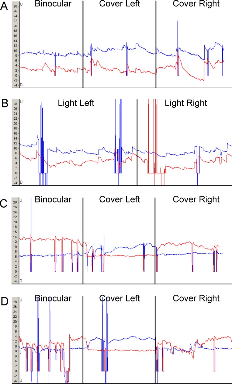
When fixation was precluded with a translucent filter while alternate occlusion produced an isolated binocular luminance disparity, the average vertical divergence of the eyes was not significant in subjects with DVD (1.2° ± 2.1°; P = 0.3; 95% CI, −1.1°–3.4°; Table 1; Fig. 1A). In control subjects, under these conditions, there was no vertical divergence. (0.09° ± 0.3°; P = 0.5; 95% CI, −0.2°–0.4°; Table 2).
Table 1.
Average Vertical Movement of the Eyes for Each DVD Patient Under Each Condition (Values in Degrees)
|
Subject |
Age, y |
Alternate Occlusion Without Fixation (Monocular Darkening) |
Alternate Increased Luminance Without Fixation (Monocular Flashlight) |
Alternate Occlusion With Fixation in Darkness (Crossbar) |
Alternate Occlusion With Fixation (Room Light) |
| 1 | 18 | 4.80 | −0.85 | 6.35 | 6.80 |
| 2 | 25 | 1.65 | −0.33 | 4.35 | 3.75 |
| 3 | 28 | −0.75 | −3.31 | 4.05 | 8.95 |
| 4 | 11 | 1.95 | −0.35 | 12.40 | 12.15 |
| 5 | 47 | −1.05 | NP | 8.15 | 7.10 |
| 6 | 36 | 0.50 | −3.15 | 7.95 | 10.15 |
| Mean ± SD | 28 ± 13 | 1.18 ± 2.15 | −1.60 ± 1.51 | 7.21 ± 3.08 | 8.15 ± 2.93 |
| P value | 0.3 | 0.06 | 0.03 | 0.03 |
P value reflects significance of magnitude of change. NP, not performed.
Table 2.
Average Vertical Movement of the Eyes for Each Control Subject Under Each Condition (Values in Degrees)
|
Subject |
Age, y |
Alternate Occlusion Without Fixation (Monocular Darkening) |
Alternate Increased Luminance Without Fixation (Monocular Flashlight) |
Alternate Occlusion With Fixation in Darkness (Crossbar) |
Alternate Occlusion With Fixation (Room Light) |
| 1 | 16 | 0.55 | 0.65 | 0.35 | 0.00 |
| 2 | 26 | −0.20 | 0.15 | 0.00 | 0.05 |
| 3 | 34 | 0.05 | −0.10 | −0.20 | 0.10 |
| 4 | 40 | 0.08 | 0.05 | 0.65 | 0.55 |
| 5 | 30 | −0.05 | 0.20 | −0.25 | 0.15 |
| 6 | 24 | 0.10 | −0.15 | 0.30 | 0.35 |
| Mean ± SD | 28 ± 8 | 0.09 ± 0.25 | 0.13 ± 0.29 | 0.14 ± 0.35 | 0.20 ± 0.21 |
| P value | 0.5 | 0.3 | 0.3 | 0.06 |
value reflects significance of magnitude of change.
Alternate Increased Luminance Without Fixation (Monocular Flashlight)
When fixation was precluded with the opaque filter, and luminance was increased with a flashlight, there was a vertical divergence causing the illuminated eye to be lower, which was borderline significant in subjects with DVD (−1.6° ± 1.5°; P = 0.06; 95% CI, −3.5°–0.3°; Table 1; Fig. 1B). In control subjects, under these conditions, there was essentially no vertical divergence (0.1° ± 0.3°; P = 0.3; 95% CI, −0.2°–0.4°; Table 2).
Figure 1.
Representative eye movement recordings from a subject with DVD. Red line signifies right eye, blue line signifies left eye. U, upward position; D, downward position. Four layered horizontal panels (A–D) depict changes in DVD (as demonstrated by changing distances between blue and red tracings) in the order that the tests were performed. (A) Alternate occlusion without fixation (monocular darkening). The VOG tracing shows a baseline left hyperdeviation (blue line higher) with mild superimposed DVD caused by isolated luminance disparity. (B) Alternate increased luminance without fixation (monocular flashlight). The VOG tracing shows a lesser left hyperdeviation (relative left hypodeviation) with increased luminance to the left eye than with increased luminance to the right eye. (C) Alternate occlusion with fixation in darkness (crossbar). The VOG tracing shows baseline right hyperdeviation with a hyperdeviation of either eye when that eye is occluded. (D) Alternate occlusion with fixation. The VOG tracing shows baseline right hyperdeviation with a hyperdeviation of either eye when that eye is occluded.
Alternate Occlusion With Fixation in Darkness (Crossbar)
When alternate occlusion produced reversal of monocular fixation in the dark (with essentially no change in peripheral luminance disparity), the average vertical divergence of the eyes was significant causing the occluded eye to be relatively higher than the nonoccluded eye (7.2° ± 3.1°; P = 0.03; 95% CI, 4.0°–10.4°; Table 1; Fig. 1C). In control subjects, there was no measurable vertical divergence (0.1° ± 0.4°; P = 0.3; 95% CI, −0.2°–0.5°; Table 2).
Alternate Occlusion With Fixation (Room Light)
Under conditions of alternate occlusion in a lighted room, where there was change in luminance and fixation, there was a significant average vertical divergence of the eyes causing the occluded eye to be relatively higher than the nonoccluded eye (8.1° ± 2.9°; P = 0.03; 95% CI, 5.1°–11.2°; Table 1; Fig. 1D). In control subjects, a negligible vertical divergence of the eyes under these conditions was found (0.2° ± 0.2°; P = 0.06; 95% CI, −0.02°–0.4°; Table 2).
Discussion
Based in part on the Bielschowsky phenomenon, Brodsky11,12 hypothesized that DVD arises from a subcortical dorsal light reflex that occurs in fish and other lateral-eyed animals when unequal luminance to the 2 eyes evokes a body tilt toward the side of the illuminated eye.13–15 In the case of a restrained fish, the same stimulus evokes a vertical divergence of the eyes, with ventral rotation of the illuminated eye and a dorsal rotation of the unilluminated eye.13 In lower animals, this righting reflex serves to restore vertical orientation, since the sky is a space-stable luminance hemisphere that always is aligned with the gravitational vertical.12–15 Brodsky11,12 proposed that this reflex exists in vestigial form in humans, but becomes suppressed in the service of single binocular vision and stereopsis, only to resurge when cortical binocular vision fails to develop in infancy. This model explained the requisite dorsal rotation of the visually-disadvantaged eye that characterizes DVD, the momentary descent of the uncovered eye below midline when the occluder is switched to the other eye, and the noncompensatory head tilt toward the side of the fixating eye that sometimes accompanies DVD as arising from unbalanced visuo-vestibular input from the two eyes.
To test this hypothesis, Wang and Bedell16 examined normal subjects as they fixated a light 4 meters away in a dark room. Either eye then was covered with a vertically oriented Maddox rod and an illuminated or a dark occluder as the other eye fixating the penlight. Following removal of the occluder, subjects were asked to estimate the directional separation of the horizontal line from the penlight in space. Using this paradigm, the authors found normal human subjects exhibit little or no vestige of the dorsal light reflex. However, their study was confounded by several methodologic problems. First, it used subjects with normal binocularity and stereopsis, which may have precluded the clinical expression of a human dorsal light reflex. Second, its methodology did not isolate binocular luminance disparity, since covering one eye produced a simultaneous fixational imbalance as well. Finally, although cortical suppression of the hyperdeviating eye is a requisite finding in DVD, this study relied upon the absence of cortical suppression to detect binocular displacement in normal subjects.
ten Tusscher and van Rijn argued that cortical visual pathways must be involved in the pathogenesis of DVD.17 They contended that binocular visual balancing of form and luminance is known to be processed at the level of the visual cortex in humans.18 They also noted that the Bielschowsky phenomenon involves fixation in addition to luminance disparity, and that this binocular luminance effect is explained best by the enhancement of cortical interocular suppression, which has been shown on functional MR imaging to accompany a paucity of horizontal intraocular connections within the primary visual cortex.19 They further argued that the neuroanatomical structures that modulate the dorsal light reflex in fish11 are not present in the human cerebellum.17 Gallegos-Duarte et al.20,21 presented additional evidence for an active role for cortical input in the pathogenesis of DVD.
Since the time of Posner,7 it has been clear that the visual cortex has an integral role in the pathogenesis of DVD given the fact that DVD occurs spontaneously, is driven by fixation, and is modulated by the patient's level of visual attention.22 Guyton23 observed that DVD manifests or increases in amplitude as subjects continue to read smaller letters on the visual acuity chart, confirming Posner's earlier conclusion that DVD is in part dependent upon fixation and attention. In the current study, we found that isolated binocular luminance disparity induces only a tiny amount of DVD, and that DVD can be elicited by alternating fixation of a small crossbar in darkness. Surprisingly, the mean amplitude of the DVD with change in fixation when viewing the crossbar in darkness approximated that seen with a change of fixation at baseline luminance levels. These findings confirmed the fundamental role of fixation (or fixational effort) as the primary stimulus for DVD, and suggested that the superimposition of binocular luminance disparity upon monocular fixation at the cortical level can produce the Bielschowsky phenomenon that characterizes DVD, as suggested by ten Tusscher and van Rijn.17
The absence of diplopia in humans with DVD necessitates a tight link between foveal fixation with one eye and cortical suppression of the peripheral retina in the nonfixating eye. The degree of cortical binocular disparity appears to correlate with the amplitude of DVD, as evidenced by the Bielschowsky phenomenon, in which placement of neutral density filters of increasing density before the fixating eye leads to an incrementally decreasing amplitude of the measured DVD.24 This physiological mechanism also explains the delayed onset of DVD, which reflects the more gradual development of cortical visual pathways and their dynamic suppression. The degree of cortical binocular disparity appears to correlate with the amplitude of DVD, as evidenced by the Bielschowsky phenomenon, in which placement of neutral density filters of increasing density before the fixating eye leads to an incrementally decreasing amplitude of the measured DVD in the nonfixating eye. Occlusion of one eye provides an exteroceptive stimulus, producing complete suppression of cortical input from the covered eye, whereas spontaneous cortical suppression of one eye by the other provides an interoceptive stimulus, producing a lesser degree of fluctuating cortical suppression, which may explain the observed variability in the amplitude of spontaneous DVD.
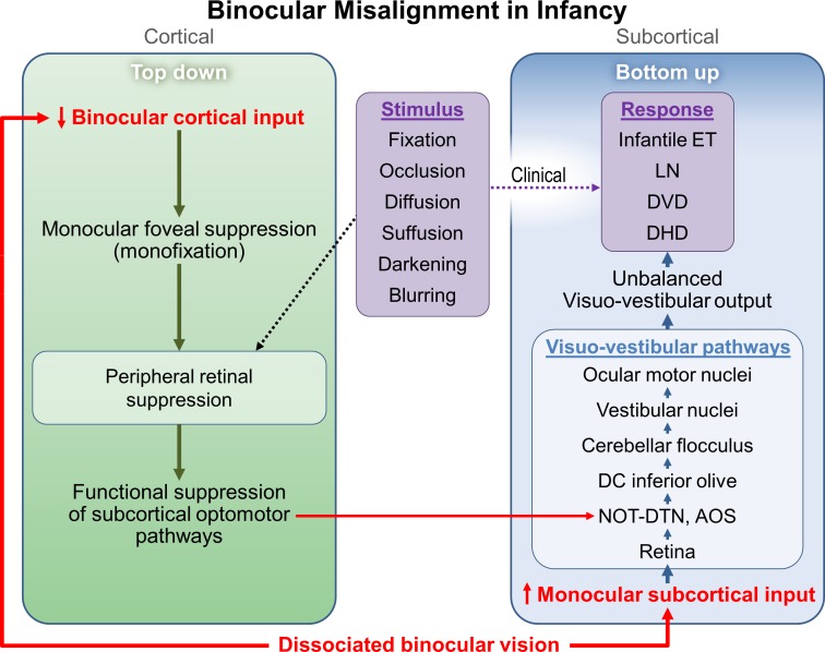
Thirty years ago, Schor proposed that selective maldevelopment of cortical binocular vision could provide a competitive advantage to reinforce the activation of direct subcortical projections from the nasal retina of either eye to the contralateral nucleus of the optic tract and the dorsal terminal nucleus of the accessory optic system (NOT-DTN) and thereby potentiate their function.25 These subcortical visuo-vestibular pathways ultimately channel binocular visual input through the inferior olivary nucleus and the cerebellar flocculus before they reach the vestibular nucleus (Fig. 2).26,27 The prominent torsional rotations that accompany latent nystagmus and DVD implicate downstream activation of these visuo-vestibular pathways as the neural generator of these movements.26,27 This dual cortical/subcortical mechanism is supported by the neuroanatomy of latent nystagmus, which has been postulated to derive from an old visuo-vestibular reflex in lateral-eyed animals26 yet can be driven by (even voluntary) cortical suppression in humans.28 Such changes in subcortical plasticity have recently been demonstrated within the superior olivary nucleus following unilateral auditory deprivation.29
Figure 2.
Proposed dissociated binocular vision involvement in the cortical and subcortical visual pathways. Flow diagram depicting how dissociated binocular vision can alter the balance of excitation and inhibition in the cortical and subcortical visual pathways to give rise to infantile esotropia and DVD. Purple frames depict portions of the response that are observed clinically. DC, dorsal cap of inferior olive; AOS, accessory optic system; LN, latent nystagmus.
This formulation provides a model of infantile strabismus in which human visuo-vestibular eye movements are actively driven by unequal binocular visual input through the visual cortex. According to this model, as cortical ocular motor control systems are engrafted on older subcortical control systems, primitive visual reflexes involving luminance and optokinesis are subsumed within newer cortical reflexes involving foveal fixation and cortical pursuit.30 In the process, binocular cortical systems feed into older subcortical centers (such as the NOT-DTN for latent nystagmus),25–27 to rechannel older visuo-vestibular reflexes through the visual cortex and generate latent nystagmus and DVD (Fig. 2). Primitive visual reflexes that rely on full-field binocular input to subserve visual balance then are retained in latent form and reactivated by the visual cortex only in the setting of dissociated binocular vision. This mechanism explains the small hypodeviation in the illuminated eye that our subjects displayed under conditions that precluded fixation, which supports a real but minor role of luminance in the pathogenesis of DVD.
There are several limitations to our study. First, calibration for the VOG system uses iris markings, which move when a luminance change induces a corresponding change in pupil size. Consequently, some recordings had brief segments of lost data, but we based our conclusions on all available data over a 12-second recording period. Second, the exquisite sensitivity of VOG to small shifts in eye position allowed us to recognize that there is no true “baseline” for DVD under binocular conditions, since DVD is characterized by some degree of inherent variability due to the fluctuating cortical suppression that drives it. Consequently, VOG often would measure a slightly different vertical misalignment under binocular conditions at different stages of the testing paradigm. It also is possible that the dissociating effects of each stimulus may have altered the fusional status of the subjects and influenced our results as the test progressed. For these reasons, we evaluated relative vertical positions of the eyes when the left eye and then right eye was illuminated or occluded under the experimental condition, using 90th percentiles of data after blink artifacts were removed and we based our conclusions on mean values. Third, the length of the VOG paradigm may have induced patient fatigue, which also could have altered our findings by diminishing visual attention and fixational effort toward the end of the test. To minimize these potentially confounding effects, our protocol allowed subjects to refixate binocularly in normal room light between each stimulus condition. Fourth, occlusion of either eye over the translucent filter may have reduced the binocular luminance disparity to a level that was insufficient to elicit a significant vertical divergence movement of the eyes. Finally, our protocol did not enable us to distinguish the act of fixation from the effort of fixation as the final common stimulus for DVD.
In conclusion, our results confirm that DVD is driven primarily by fixation, and only to a very minor degree by isolated binocular luminance disparity. Accordingly, they suggest that peripheral binocular luminance disparity modulates DVD at the cortical level primarily when superimposed upon fixation (as seen with the Bielschowsky phenomenon). The DVD develops when fixation (or fixational effort) evokes facultative cortical suppression of the peripheral retina, which suggests that this secondary effect provides the cortical output signal that ultimately triggers DVD. These findings may explain how primitive visual reflexes can be driven through the human visual cortex and provide a novel neurological template for the reemergence of subcortical visual reflexes in the setting of higher cortical dysfunction.
Acknowledgments
Supported in part by Research to Prevent Blindness, Inc. (Unrestricted Grant to Mayo Clinic [Rochester, MN, USA] Department of Ophthalmology and an Olga Keith Weiss Scholarship [JMH]), the Knights Templar Foundation (MCB), the National Institutes of Health (Bethesda, MD, USA) Grant EY018810 (JMH), and the Mayo Foundation. The authors alone are responsible for the content and writing of the paper.
Disclosure: R. Ghadban, None; L. Liebermann, None; L.D. Klaehn, None; J.M. Holmes, None; M.C. Brodsky, None
References
- 1. Schweiger C. Die Erfolge der Schieloperation [The success of strabismus surgery]. Arch Augenheilk. 1893–1894; 28–29: 207. [Google Scholar]
- 2. Stevens GT. Memoirs on double vertical strabismus and symmetrical vertical deviations of degrees less than strabismus. Ann D'Oculiat. 1895; 113: 225–232. [Google Scholar]
- 3. von Noorden GK. Current concepts of infantile esotropia (Bowman Lecture). Eye. 1988; 2: 343–353. [DOI] [PubMed] [Google Scholar]
- 4. Bielschowsky A. Die einseitigen und gegensinnigen ("dissoziierten”) Vertikalbewegungen der Augen [The one-sided and in opposite directions ("dissociated”) vertical movements of the eyes]. Graefes Arch Clin Exp Ophthalmol. 1931; 125: 493–509. [Google Scholar]
- 5. Bielschowsky A. Lectures on Motor Anomalies. Hanover, NH: Dartmouth College Publications; 1940: 128. [Google Scholar]
- 6. Wilson ME, McClatchey SK. Dissociated horizontal deviation. J Pediatr Ophthalmol Strabismus. 1991; 28: 90–95. [DOI] [PubMed] [Google Scholar]
- 7. Posner A. Noncomitant heterophorias; considered as aberrations of the postural tonus of the muscular apparatus. Am J Ophthalmol. 1944; 27: 1275–1279. [Google Scholar]
- 8. Ohm J. Nystagmus und Schielen bei Sehschwachen und Blinden [Nystagmus and Strabismus in Sighted and Blind]. Stuttgart, Germany: F. Enke; 1958: 102. [Google Scholar]
- 9. Mattheus S, Kommerell G. Reversed fixation test as a means to differentiate between dissociated and non-dissociated strabismus. Strabismus. 1996; 4: 3–9. [DOI] [PubMed] [Google Scholar]
- 10. Wassill KH, Kaufmann H. Binocular 3-D video-oculography. Strabismus. 2001; 9: 29–32. [DOI] [PubMed] [Google Scholar]
- 11. Brodsky MC. Dissociated vertical divergence: a righting reflex gone wrong. Arch Ophthalmol. 1999; 117: 1216–1222. [DOI] [PubMed] [Google Scholar]
- 12. Brodsky MC. Dissociated vertical divergence: perceptual correlates of the human dorsal light reflex. Arch Ophthalmol. 2002; 120: 1174–1178. [DOI] [PubMed] [Google Scholar]
- 13. Graf W, Meyer DL. Central mechanisms counteract visually induced tonus asymmetries: A study of ocular responses to unilateral illumination in goldfish. J Comp Physiol. 1983; 150: 473–481. [Google Scholar]
- 14. von Holst E. Die Gleichgewichtssinne der Fische [The equilibrium sense of fish]. Verh Dtsch Zool Ges. 1935; 37: 143–148. [Google Scholar]
- 15. von Holst E. Über den Lichtrückenreflex bei Fischen [About the dorsal light reflex in fish]. Pubbl Stn Zool Napoli II. 1935; 15: 143–148. [Google Scholar]
- 16. Wang M, Bedell HE. Asymmetrical vertical phorias in normal subjects: the influence of unbalanced illumination. Strabismus. 2005; 13: 123–128. [DOI] [PubMed] [Google Scholar]
- 17. ten Tusscher MP, van Rijn RJ. A hypothetical mechanism for DVD: unbalanced cortical input to subcortical pathways. Strabismus. 2010; 18: 98–103. [DOI] [PubMed] [Google Scholar]
- 18. Ronchi LR. Balancing visual weights. Ophthalmic Physiol Opt. 2002; 22: 416–419. [DOI] [PubMed] [Google Scholar]
- 19. Alvarez-Linera PJ, Rios-Lago M, Martin-Alvarez H. Functional magnetic resonance imaging of the visual cortex: relation between stimulus intensity and bold response. (Resonancia magnetica funcional de la corteza visual: estudio de las relaciones entre la intensidad del estimulo y la respuesta BOLD). Rev Neurol. 2007; 105: 147–151. [PubMed] [Google Scholar]
- 20. Gallegos-Duarte M. Dissociated vertical divergence. Strabismus. 2012; 20: 31–32, author reply 33–36. [DOI] [PubMed] [Google Scholar]
- 21. Gallegos-Duarte M, Mendiola-Santibanez J, Ortiz-Retana JJ, de Celis-Monteverde BR, Vidal-Pineda R, Sigala-Zamora A. Dissociated deviation. A strabismus of cortical origin. Cir Cir. 2007; 75: 241–247. [PubMed] [Google Scholar]
- 22. Kowal L. Dissociated deviations: fixation linked or caused by inattention. Binocul Vis Quart. 1997; 12: 12. [Google Scholar]
- 23. Guyton DL, Cheeseman EW Jr, Ellis FJ, Straumann D, Zee DS. Dissociated vertical deviation: an exaggerated normal eye movement used to damp cyclovertical latent nystagmus. Trans Am Ophthalmol Soc. 1998; 96: 389–424, discussion 424–429. [PMC free article] [PubMed] [Google Scholar]
- 24. Brodsky MC. Dissociated vertical divergence: cortical or subcortical in origin? Strabismus. 2011; 19: 67–68, author reply 69–70. [DOI] [PubMed] [Google Scholar]
- 25. Schor CM. Subcortical binocular suppression affects the development of latent and optokinetic nystagmus. Am J Optom Physiol Opt. 1983; 60: 481–502. [DOI] [PubMed] [Google Scholar]
- 26. Brodsky MC, Tusa RJ. Latent nystagmus: vestibular nystagmus with a twist. Arch Ophthalmol. 2004; 122: 202–209. [DOI] [PubMed] [Google Scholar]
- 27. Brodsky MC. An expanded view of infantile esotropia: bottoms up! Arch Ophthalmol. 2012; 130: 1199–1202. [DOI] [PubMed] [Google Scholar]
- 28. Kommerell G, Zee DS. Latent nystagmus. Release and suppression at will. Invest Ophthalmol Vis Sci. 1993; 34: 1785–1792. [PubMed] [Google Scholar]
- 29. Maslin MR, Munro KJ, Lim VK, Purdy SC, Hall DA. Investigation of cortical and subcortical plasticity following short-term unilateral auditory deprivation in normal hearing adults. Neuroreport. 2013; 24: 287–291. [DOI] [PubMed] [Google Scholar]
- 30. Keiner GBJ. New Viewpoints on the Origin of Squint. The Netherlands; Springer; 1951: 222. [Google Scholar]


