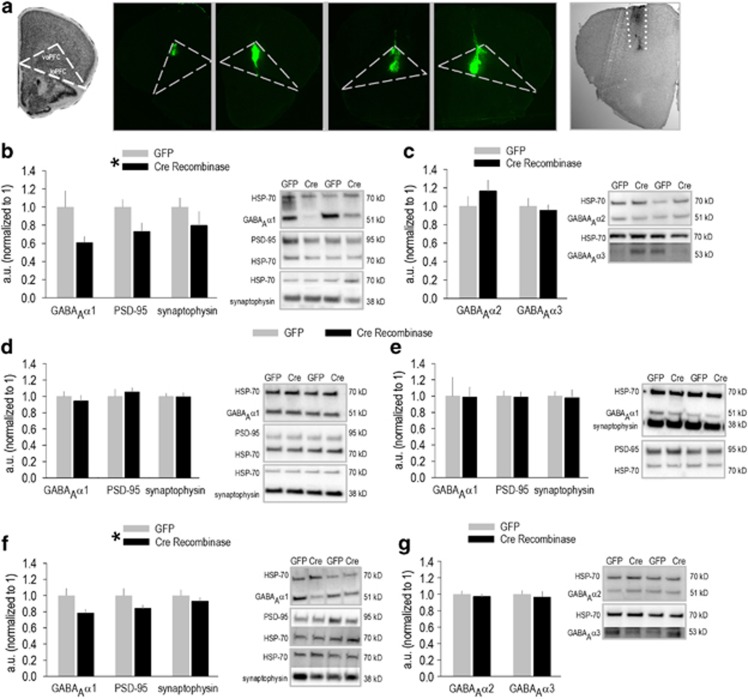
Figure 1.
GABAAα1 silencing regulates synaptic marker expression. (a) A coronal section from the Mouse Brain Library (Rosen et al, 2000) is shown adjacent to images of Green Fluorescent Protein (GFP) expression within the orbitofrontal prefrontal cortex (oPFC) of multiple mice. ‘voPFC' refers to the ventral oPFC, and ‘loPFC' refers to the lateral oPFC. Expression patterns contrast those following M2-targeted infusions. For example, Cre recombinase (Cre) immunoreactivity in M2 (between the dotted white lines) is shown at far right. (b) Regional GABAAα1 protein expression was decreased in homogenized oPFC tissue punches collected 11 weeks following viral vector infusion; PSD-95 and synaptophysin were also reduced. (c) By contrast, expression of the GABAAα2 and GABAAα3 subunits was unaffected. (d) No effects were identified off-target, in the hippocampus, or (e) dorsal striatum (n=5/group). (f) GABAAα1 and synaptic marker expression were also reduced in M2 following Cre delivery, and effects were detectable 2 weeks after infusion. (g) As in the oPFC, GABAAα2 and GABAAα3 were unaffected (n=10–11/group). Representative blots are adjacent throughout. Mean+SEM, *p<0.05, main effect of group.

