Abstract
Context:
Dental evidence is a valuable tool in identifying individuals, especially when disasters befall. Reference points in faciomaxillary region such as interpupillary distance, intercanthal distance, interalar distance and bizygomatic width can significantly contribute toward reconstruction of two-dimensional (2D) facial profiles. This study was researched upon to determine the relationship between the maxillary intercanine width and the different reference points of the face.
Aim:
The aim of the following study is to ascertain whether maxillary intercanine width can be used to detect interpupillary distance, intercanthal distance, interalar distance and bizygomatic distance and to evaluate the role of maxillary intercanine width in the 2D reconstruction of the face.
Materials and Methods:
The study was carried out by consent and involved 90 subjects-45 males and 45 females who satisfied the inclusion criteria. Subjects were divided into three age groups, i.e. 18-24, 25-28, 29-35. Four parameters were measured– intercanine width, interpupillary distance, intercanthal distance and interalar distance. All the measurements were carried out with a digital Vernier caliper. The bizygomatic width was measured from posterior-anterior view. Two empiricists were assigned for the task. Each test was carried out twice to validate the soundness of the findings and to reduce bias.
Statistical Analysis:
Analysis of variance and Pearson correlation was established. Regression analysis was performed to predict the study variables by intercanine width.
Results:
Intercanine width showed a significant relationship with different points. The width varied with age and gender.
Conclusion:
Inter canine width can be used as a valuable parameter in the reconstruction of face in two dimensional as it shows significant relationship with faciomaxillary reference point such as interpupillary distance, intercanthal distance, interalar distance and bizygomatic width.
Keywords: Facial reconstruction anthropometry, forensic science, odontomertic analysis
Introduction
Personal identification forms an integral part of forensic science, especially when it deals with crimes or with mutilated bodies that have undergone damage beyond recognition. There are numerous techniques of identification: Some of them are constructive and others comparative.[1,2,3,4,5,6] Identification of an Individual is classified as certain, probable, possible or excluded.[7] The aim of all these techniques is the positive identification done by ante mortem and post-mortem comparisons (e.g. Finger Prints, X-rays, Odontology or deoxyribonucleic acid) as well as cranio facial reconstruction (CFR) techniques. This may only be a lead toward a proposal for identification. CFR has emerged as an important tool in forensic identification of human remains. It may be two-dimensional (2D), three-dimensional and/or computer assisted.[8]
Facial reconstruction is the building of the face of an individual on to the skull and has been used primarily in the forensic field for identification of skeletal remains.[9,10,11] Krogman and Iscan stated that “The skull is the matrix of the living head; it is the bony core of the fleshy head and face in life”.[12] This has been the principal behind forensic facial reconstruction. Practitioners in this field take help from soft-tissue prediction guidelines that have been published.[13,14] However, many of these guidelines are subjective and have not been systematically evaluated using empirical methods. Teeth are the hardest and chemically the most stable tissues in the body. They are known to resist post-mortem, mechanical, chemical, physical and thermal types of destruction.[15]
Besides, they are also readily accessible and do not need special dissection. Therefore, teeth are invaluable elements used in identifying anthropological, genetic, odontologic, evolutionary and forensic investigations among living and non-living population.[16,17]
Teeth are used for sex determination by way of odontomertic analysis. Mandibular canines are found to exhibit the greatest sexual dimorphism among all teeth.[18,19] “Sexual dimorphism” refers to those differences in size, stature and appearance between males and females. These differences can be applied to dental identification, since no two mouths are alike.
The study of permanent Maxillary Canine teeth offers certain advantages. These teeth are less affected by periodontal diseases and are also less exposed to plaque. Moreover, they are less prone to calculus and abrasion from brushing. Over and above, maxillary canines are the last to be extracted with respect to age.[20]
The construction of complete dentures is based on valuable clinical parameters such as the combined width of maxillary anterior teeth, especially when pre-extraction records are not available. According to previous studies intercanine width is known to correlate well with several facial measurements. However, there seem to be conflicting views on the value of such estimation.
The objective of this study was to discover if intercanine width can be used to determine interpupillary distance, intercanthal distance, interalar distance, bizygomatic distance and then to evaluate the possible application of these findings for use in forensics and thereby 2D reconstruction of the face.
Materials and Methods
The study enrolled 90 patients visiting the Department of Oral Medicine and Radiology in Raja Rajeshwari Dental College and Hospital. Out of the 90 subjects, 45 were males and 45 were females. Both sexes were divided into three age groups, i.e. 18-24, 25-28, 29-35 respectively.
Inclusion criteria for the study were participants between 18 and 35 years, because facial growth is complete at the end of this period. Participants who had complete permanent dentition with ideal arch form and alignment and in whom there was an absence of morphological developmental anomalies (peg lateral, supernumerary teeth, mesiodens, retained deciduous teeth) were selected.
Patients with history of orthodontic treatment or extraction, congenital facial defects, presence of class III or class IV caries, presence of restoration, presence of crowding, diastema, rotation, tooth fracture, proclination and retroclination and gingival inflammation or hypertrophy were excluded from the study. Prior consent was taken from the institution and ethical clearance was obtained from the institutional ethical committee. Nature of the study was explained explicitly and consent of the subjects was obtained.
The three parameters, inner intercanthal distance, outer intercanthal distance, inter alar width were measured with digital Vernier caliper having a resolution of 0.01 m. Inter canine width was measured on the casts made by a high quality alginate impression using a digital Vernier caliper (NSK) having a resolution of 0.01 mm. Two investigators measured three parameters independently and each subject was investigated upon twice.
Determination of inner inter canthal distance
The subjects were seated comfortably on the dental chair in a relaxed state in an upright position with the head resting firmly against the head rest. The inner inter canthal distance was measured from the medial angle to the medial angle of the palpebral fissure. The distance between these two points was measured using a digital Vernier caliper of resonance 0.01 mm. The experiment was done by bringing the recording parts of the caliper just in contact with the medial angle of the palpebral fissure, without applying pressure [Figure 1].
Figure 1.
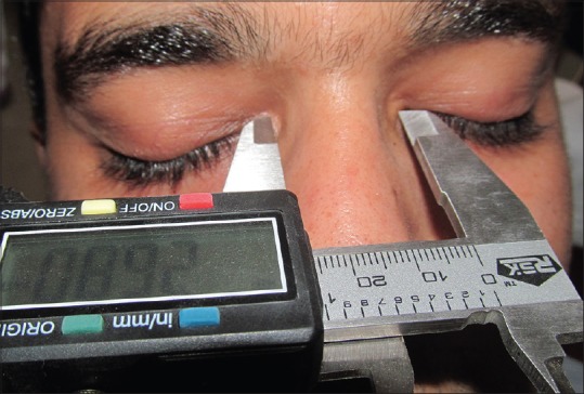
Measurement of inner inter canthal distance
Determination of outer inter canthal distance
The subjects were seated comfortably on the dental chair in a relaxed state in an upright position with the head resting firmly against the head rest. The outer canthal distance was measured from the lateral angle to the lateral angle of the palpebral fissure. The distance between these two points was measured using a digital Vernier caliper by placing two scales vertically just in contact with the lateral angle of the palpebral fissure, without applying pressure [Figure 2].
Figure 2.
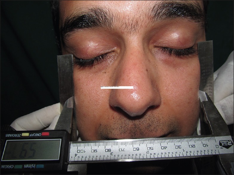
Measurement of outer inter canthal distance
Determination of interpupillary distance
The subjects were seated comfortably on the dental chair in a relaxed position with the head resting firmly against the head rest. Subjects were advised to look straight without laying much pressure on the eyes. The distance between the center of the pupils was measured using a digital Vernier caliper, by placing two scales vertically, just at the position of the center of the pupil [Figure 3].
Figure 3.
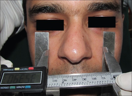
Measurement of interpupillary distance
Determination of inter-alar width
As in the above experiments, the subjects were seated comfortably on the dental chair in a relaxed state in an upright position with the head resting firmly against the head rest. The inter-alar width was determined by using the external width of the nose at the widest point. The distance between the two points was measured without applying pressure on the nose. The recording part of the caliper was in contact with the outer surface of the nose. While measuring, the patients were asked to stop breathing momentarily, in order to avoid any changes in the shape of the nose. The inter alar width was measured using the external width of the nose at the widest point and the distance between these two points was determined [Figure 4].
Figure 4.
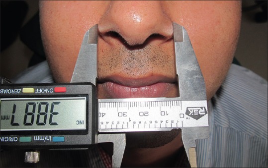
Measurement of inter-alar width
Determination of inter-canine width
Inter-canine width was measured from the casts made by a high quality alginate impression using a digital Vernier caliper having a resolution of 0.01 mm, between incisal edges of canines [Figure 5].
Figure 5.
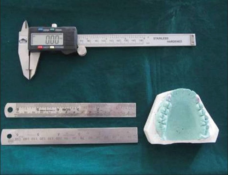
Instrumentation used
Determination of bizygomatic width
Bizygomatic width was measured from posteroanterior view by measuring the distance between most lateral positions in zygomatic arch (SIDEXIS next generation software) [Figures 6 and 7].
Figure 6.
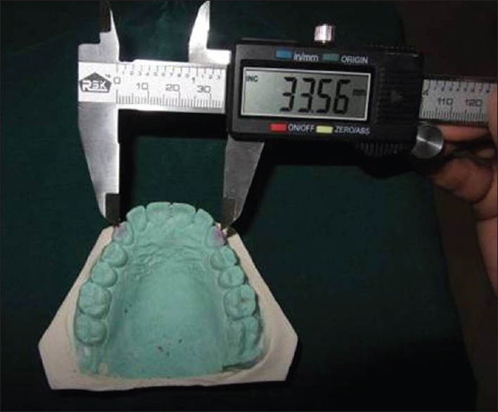
Measurement of inter-canine width
Figure 7.
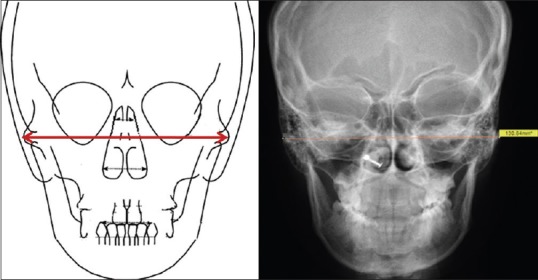
Measurement of bizygomatic width
Statistical analysis
Analysis of variance and Pearson correlation of study variables was established. Regression analysis was performed to predict the study variables by inter canine width. The Statistical software namely SAS 9.2, SPSS 15.0, Stata 10.1, MedCalc 9.0.1, Systat 12.0 and R environment version 2.11.1 were used for the analysis of the data.
Results
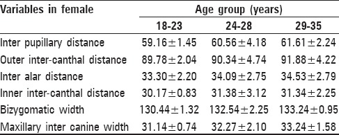
The mean of different variables i.e. interpupillary distance, inter-canthal distance, interalar distance, bizygomatic distance and intercanine width in different age groups is shown in Tables 1 and 2. Most of the reference points were comparatively more in males than in females except inner inter-canthal distance in the age group 24-28 and 29-35 years [Tables 1 and 2].
Table 1.
Mean values of inter pupillary distance, outer intercanthal distance, inter alar distance, inner inter-canthal distance, bizygomatic width and maxillary inter canine width in male
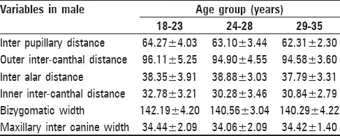
Table 2.
Mean values of inter pupillary distance, outer intercanthal distance, inter alar distance, inner inter-canthal distance, bizygomatic width and maxillary inter canine width in female
Pearson correlation in males showed
There was a highly significant association of intercanine width with inter pupillary distance, outer inter-canthal distance and interalar width. There was the non-significant association with inner inter-canthal distance and bizygomatic distance in the age group 18-23 [Table 3]
There was a highly significant association of intercanine width with inter pupillary distance, outer inter-canthal distance, moderately significant with bizygomatic width and non-significant with inner inter-canthal distance and inter-alar distance in the age group 24-28 [Table 3]
There was a highly significant association of intercanine width with inter pupillary distance, suggestive significant with outer inter-canthal distance and non-significant with inner inter-canthal distance, inter-alar distance and bizygomatic width in the age group 29-35 [Table 3].
Table 3.
Pearson correlation of interpupillary distance, interalar distance, inner-canthal distance, outer-canthal distance, bizygomatic width, with intercanine width in different age and gender

In females Pearson correlation showed
There was a moderately significant association of intercanine width with inner inter-canthal distance, suggestive significant association with bizygomatic width and non-significant with outer inter-canthal distance and interalar width in the age group 18-23 [Table 4]
There was a highly significant association of intercanine width with inter pupillary distance. The outer inter-canthal distance was moderately significant with inter-alar distance and non-significant with inner inter-canthal distance and bizygomatic distance in the age group 24-28 [Table 4]
There was a highly significant association of intercanine width with outer inter-canthal distance and bizygomatic width, moderately significant with inter pupillary distance, inner inter-canthal distance and non-significant with inter-alar distance in the age group of 29-35 [Table 4].
Table 4.
Pearson correlation of interpupillary distance, interalar distance, inner-canthal distance, outer-canthal distance, bizygomatic width, with intercanine width in different age and gender

Prediction analysis showed the regression equation to predict the various reference points with intercanine width
Reference point to be detected = Constant + Beta-coefficient × intercanine width
Where constant and Beta-coefficient are fixed values [as by Tables 5 and 6] and intercanine width varies from subject to subject.
Table 5.
Prediction analysis of interpupillary distance, interalar distance, inner-canthal distance, outer-canthal distance, bizygomatic width, with intercanine width in different age and gender
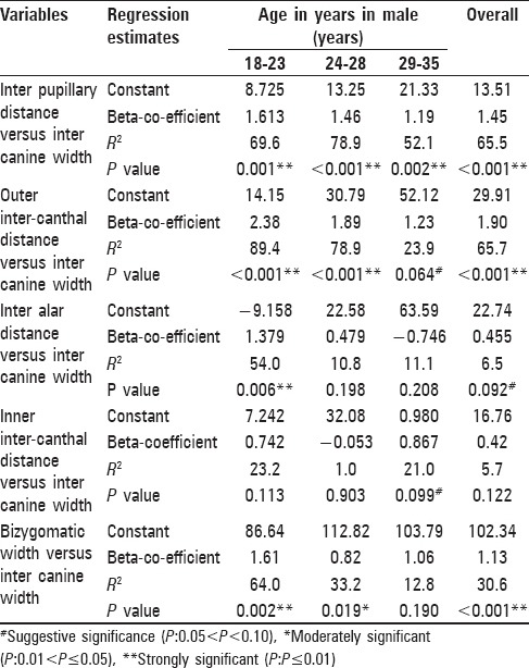
Table 6.
Prediction analysis of interpupillary distance, interalar distance, inner-canthal distance, outer-canthal distance, bizygomatic width, with intercanine width in different age and gender
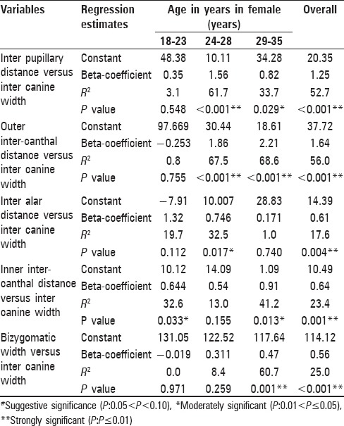
For example, for females in 18-23 age group-
Inter papillary distance = 48.38 + 0.35 × inter canine width [Tables 5 and 6].
Discussion
This study is based on the principle of Prosthodontics where different facial reference points are used to determine the width of anterior teeth and intercanine width for the purpose of teeth setting.[21,22,23,24] In some studies, there was a significant relationship between different reference points with intercanine width.[21,22,23,24] Hence this study is done contrary to this principle. We aimed to see whether intercanine width can be used to determine various reference points. Age groups selected in our study is similar to age groups used in other studies.
We had divided subjects into three age groups in both males and females because literatures showed variability in intercanine width with age.[25,26] Our study showed very little difference of intercanine width with age in males but comparatively, the difference substantially increased in females. However, this criterion cannot be reliable, as intercanine width can vary in different facial profiles and it does not depend on the age group but depend on the gender.
In males, Pearson correlation of maxillary intercanine width with Interpupillary, outer inter-canthal distance, inter-alar distance of age group 18-23, Interpupillary, outer inter-canthal distance for age group 24-28 and Interpupillary distance in the age group 29-35 showed a high degree of significance with P < 0.01.
Pearson correlation of maxillary intercanine width with bizygomatic width for age group 24-28 showed a moderate significance and suggestive significance with outer inter-canthal distance for the 29-35 age groups in males.
In females Pearson correlation of maxillary intercanine width with Interpupillary, outer inter-canthal distance for age group 24-28 and outer inter-canthal distance, bizygomatic width in the age group of 29-35 showed a high degree of significance with P < 0.01.
Pearson correlation of maxillary intercanine width with inner inter-canthal distance for age group 18-23, inter alar distance for age group 24-28, inner inter-canthal distance for age group 25-35, showed a moderate significance. It showed suggestive significance with bizygomatic width for age group of 18-23 in females.
In the present study, subjects were divided into two groups-males and females in order to determine the dimensions on both the sexes. It was found that there was a statistically, highly significant difference in maxillary inter canine width, outer inter-canthal distance and inter alar distance, whereas only a significant difference was observed in the Interpupillary distance. These findings reveal that they are influenced by the differences in the size of the jaws, the teeth and the overall facial form.
The limitation of this study was resiliency of the soft-tissues. Hence additional studies are required where bony landmarks can also be taken as reference points, in which case, it will be perhaps more reliable.
Another limitation is that, this study was carried out within the institutional set up and subjects in the 18-35 age group were evaluated. Hence, the result may be applicable to just a small population in the said age range.
The results of the study should be validated by including a large population size spread over the entire Indian subcontinent. This would help to generate multiple factors for various anthropological measurements for use among the Indian population.
The correlation between intercanine distance and other cephalometric or anthropometric parameters like facial type and vertical dimensions thus obtained, can be a future prospectus for the basis of scientific co-relation of these parameters.
Conclusion
Analysis of measurements showed that inter canine width can be used as a predictor for different facial reference points but not in all geographical areas, as this study was purely limited to south Indian population. This is the first study of its kind. Hence further research should necessarily be done on different ethnic groups to confirm the empirical observations.
Footnotes
Source of Support: Nil
Conflict of Interest: None declared
References
- 1.Indira A, Gupta M, David MP. Usefullness of palatal rugae patterns in establishing identity: Preliminary results from Bengaluru city, India. J Forensic Dent Sci. 2012;4:2–5. doi: 10.4103/0975-1475.99149. [DOI] [PMC free article] [PubMed] [Google Scholar]
- 2.Olze A, Bilang D, Schmidt S, Wernecke KD, Geserick G, Schmeling A. Validation of common classification systems for assessing the mineralization of third molars. Int J Legal Med. 2005;119:22–6. doi: 10.1007/s00414-004-0489-5. [DOI] [PubMed] [Google Scholar]
- 3.Mühler M, Schulz R, Schmidt S, Schmeling A, Reisinger W. The influence of slice thickness on assessment of clavicle ossification in forensic age diagnostics. Int J Legal Med. 2006;120:15–7. doi: 10.1007/s00414-005-0010-9. [DOI] [PubMed] [Google Scholar]
- 4.Schmeling A, Schulz R, Danner B, Rösing FW. The impact of economic progress and modernization in medicine on the ossification of hand and wrist. Int J Legal Med. 2006;120:121–6. doi: 10.1007/s00414-005-0007-4. [DOI] [PubMed] [Google Scholar]
- 5.Schmeling A, Baumann U, Schmidt S, Wernecke KD, Reisinger W. Reference data for the Thiemann-Nitz method of assessing skeletal age for the purpose of forensic age estimation. Int J Legal Med. 2006;120:1–4. doi: 10.1007/s00414-005-0002-9. [DOI] [PubMed] [Google Scholar]
- 6.Schulz R, Mühler M, Mutze S, Schmidt S, Reisinger W, Schmeling A. Studies on the time frame for ossification of the medial epiphysis of the clavicle as revealed by CT scans. Int J Legal Med. 2005;119:142–5. doi: 10.1007/s00414-005-0529-9. [DOI] [PubMed] [Google Scholar]
- 7.Quatrehomme G, Subsol G. Classical non-computer-assisted craniofacial reconstruction. In: Clement JG, Marks MK, editors. Computer Graphic Facial Reconstruction. Amsterdam: Elsevier, Academic; 2005. pp. 15–32. [Google Scholar]
- 8.Quatrehomme G, Balaguer T, Staccini P, Alunni-Perret V. Assessment of the accuracy of three-dimensional manual craniofacial reconstruction: A series of 25 controlled cases. Int J Legal Med. 2007;121:469–75. doi: 10.1007/s00414-007-0197-z. [DOI] [PubMed] [Google Scholar]
- 9.Gatliff BP. Facial sculpture on the skull for identification. Am J Forensic Med Pathol. 1984;5:327–32. doi: 10.1097/00000433-198412000-00009. [DOI] [PubMed] [Google Scholar]
- 10.Helmer R, Rohricht S, Petersen D, Mohr F. Assessment of the reliability of facial reconstruction. In: Iscan MY, Helmer RP, editors. Forensic Analysis of the Skull. Ch. 17. New York: Wiley Liss Publications; 1993. pp. 229–34. [Google Scholar]
- 11.Prag J, Neave RA. Making Faces. London: British Museum Press; 1997. [Google Scholar]
- 12.Wilkinson CM, Neave RA. Skull re-assembly and the implications for forensic facial reconstruction. Sci Justice. 2001;41:5–6. [Google Scholar]
- 13.Krogman WM. The Human Skeleton in Forensic Medicine. Illinois: Charles C. Thomas; 1962. [Google Scholar]
- 14.Krogman WM, Iscan MY. The Human Skeleton in Forensic Medicine. Illinois: Charles C. Thomas; 1986. [Google Scholar]
- 15.Teschler-Nicola M, Prossinger H. Sex determination using tooth dimensions. In: Alt KW, Roising FW, Teschler-Nicola M, editors. Dental Anthropology, Fundamentals. Limits and Prospects. Wien: Springer-Verlag; 1998. pp. 479–501. [Google Scholar]
- 16.Bindu A, Vasudeva K, Subash K, Usha C, Singla S. Gender based comparison of intercanine distance of mandibular permanent canine in different populations editorial. J Punjab Acad Forensic Med Toxicol. 2008;2:6–9. [Google Scholar]
- 17.Williams PL, Bannister LH, Berry MM, Collin SP, Dyson M, et al. The Teeth in Gray's Anatomy. 38th ed. New York: Churchill Livingstone; 2000. pp. 1699–721. [Google Scholar]
- 18.Kaushal S, Patnaik VV, Agnihotri G. Mandibular canines in sex determination. J Anat Soc India. 2003;52:119–24. [Google Scholar]
- 19.Garn SM, Lewis AB, Swindler DR, Kerewsky RS. Genetic control of sexual dimorphism in tooth size. J Dent Res. 1967;46:963–72. doi: 10.1177/00220345670460055801. [DOI] [PubMed] [Google Scholar]
- 20.Anderson DL, Thompson GW. Interrelationships and sex differences of dental and skeletal measurements. J Dent Res. 1973;52:431–8. doi: 10.1177/00220345730520030701. [DOI] [PubMed] [Google Scholar]
- 21.Patel JR, Sethuraman R, Naveen YG, Sha MH. A comparative evaluation of the relationship of inner-canthal distance and inter-alar width to the inter-canine width amongst the Gujarati population. J Adv Oral Res. 2011;2:31–8. [Google Scholar]
- 22.Abdullah MA, Stipho HD, Talic YF, Khan N. The significance of inner canthal distance in prosthodontics. Saudi Dent J. 1997;9:36–9. [Google Scholar]
- 23.Cesario VA, Jr, Latta GH., Jr Relationship between the mesiodistal width of the maxillary central incisor and interpupillary distance. J Prosthet Dent. 1984;52:641–3. doi: 10.1016/0022-3913(84)90133-1. [DOI] [PubMed] [Google Scholar]
- 24.Al Wazzan KA. The relationship between intercanthal dimension and the widths of maxillary anterior teeth. J Prosthet Dent. 2001;86:608–12. doi: 10.1067/mpr.2001.119682. [DOI] [PubMed] [Google Scholar]
- 25.Paulino V, Paredes V, Cibrian R, Gandia JL. Dental arch changes from adolescence to adulthood in a Spanish population: A cross-sectional study. Med Oral Patol Oral Cir Bucal. 2011;16:e607–13. doi: 10.4317/medoral.16.e607. [DOI] [PubMed] [Google Scholar]
- 26.Bishara SE, Treder JE, Damon P, Olsen M. Changes in the dental arches and dentition between 25 and 45 years of age. Angle Orthod. 1996;66:417–22. doi: 10.1043/0003-3219(1996)066<0417:CITDAA>2.3.CO;2. [DOI] [PubMed] [Google Scholar]


