

Abstract
Introduction:
Age estimation is of immense importance not only for personal identification but also for treatment planning in medicine and dentistry. Chronologic age conveys only a rough approximation of the maturational status of a person, hence dental and skeletal ages have been explored as maturity indicators since decades. The tooth maturation provides a valuable indicator of dental age and serves as a better index of the maturation of a child as compared to other maturity indicators.
Aims and Objectives:
To test the applicability of Demirjian's and Willem's dental age assessment methods as well as Greulich and Pyle skeletal age assessment method in children residing in Gandhinagar district.
Materials and Methods:
The study consisted of randomly selected 180 subjects (90 males and 90 females) ranging from 6 to 16 years age and residing in Gandhinagar district. Dental age estimation was performed from radiovisuograph (RVG) images of mandibular teeth of left quadrant by both Demirjian's and Willem's methods. Skeletal age estimation was done from right hand wrist radiograph by Greulich and Pyle method. The differences between the chronological age and the estimated dental and skeletal ages were statistically tested using paired ‘t’ test. The correlation between chronological age, dental and skeletal age estimation methods was confirmed statistically by Pearson's correlation. The reproducibility of the estimations was statistically tested using the Pearson's Chi-square test.
Results:
Amongst the age estimation methods used in this study, the Willem's dental age estimation method proved to be the most accurate and consistent.
Conclusion:
Although various age estimation methods do exist, the results are varied in different populations due to ethnic differences. However, till new tables are formulated, the Willem's method (Modified Demirjian method) can be accurately applied to estimate chronological age for the population residing in Gandhinagar district.
Keywords: Dental age estimation, Demirjian's method, Greulich and Pyle method, skeletal age estimation, Willem's method
Introduction and Review
Age is defined as the length of time an organism or individual has survived after birth.[1] Age estimation, a sub-discipline of the forensic sciences, is of immense importance in forensic medicine for identification of deceased victims as well as in connection with crimes and accidents.[2,3,4,5,6,7] The importance of age determination also pertains to many medical and paramedical fields such as treatment planning in orthodontics and pediatric dentistry,[8,9] pediatric medicine and endocrinology.[5,9]
As human growth is characterized by considerable variation in the rate of progress towards physiological maturity, chronological age has little or no place in the assessment of the maturational state of a child.[10] Physiologic age is the registry of the rate of progress towards maturity that can be estimated by somatic, sexual, skeletal and dental maturity.[11,12] Somatic maturity is recognized by the annual growth increments in height or weight. Sexual maturity is indicated by the changes of secondary sexual characteristics such as voice changes in boys and menarche in girls. These maturity indicators have limited value because they can be applied only after serial recordings. Skeletal maturity estimation consists of examination of the initial appearance and subsequent ossification of various bones.[11]
Assessing skeletal maturation status, whether pubertal growth spurt of the patient has been reached or not, can have a considerable influence on diagnosis, treatment goals, treatment planning and the eventual outcome of orthodontic treatment.[10,11,12,13,14,15,16,17,18] Skeletal maturation is generally determined by stages in the ossification of bones of hand-wrist because of the quantity of different types of bones available in the area[14,17] and easy accessibility with minimum expense and time.[8,16,19] Various skeletal maturity indicators such as epiphysis–diaphysis fusion, hand-wrist examination, cervical vertebrae assessment, sternoclavicular bones, changes in the pubic symphysis and fusion of cranial sutures, have their advantages and disadvantages.[14,17] The most frequently used method to evaluate skeletal age from hand-wrist radiographs is the atlas of Greulich and Pyle.[8,10,14]
The age-related changes in the dentition could be divided into three categories: Formative, degenerative and histological. The formative or developmental changes such as tooth eruption and tooth calcification are good predictors of age in the years until adulthood. Degenerative changes like attrition, periodontal diseases, secondary dentin deposition, root translucency, cementum apposition, root resorption, color changes and increase in root roughness are appreciable in teeth with increasing age. But, quantification of these changes nearly always requires extraction and sectioning of teeth, which is impractical and unethical in living individuals. Hence, the techniques which are being developed for age estimation in living individuals rely mostly on radiological imaging of teeth.[6,7] Tooth eruption has been reported to be more variable than the calcification sequence in the dentition.[13] Tooth development is a useful measure of maturity since it represents a series of recognizable events that occurs in the same sequence from an initial event to a constant end-point of reportedly low variability.[4,5,20] Dental development milestones, therefore, can be utilized in age estimation.[2,5,9]
Age estimation techniques based on dental maturation in children may be divided into those using the atlas approach or those using scoring systems such as Schour and Massler, Moorrees, Anderson and Demirjian's methods. Age estimation techniques in adults are the morphological and radiological techniques such as Gustafson, Bang and Ramm, Solheim, Kvaal and Solheim and Kvaal methods.[4,21] Among many proposed methods, the Demirjian method (1973) of age assessment has been widely accepted.[9,12,13,22] The classification of stages proposed by Demirjian appears to be best suited for forensic purpose, since stages are defined by changes in form and development of teeth and these stages are independent of possibly complicated length measurements.[23] The advantages of the Demirjian method include the objective criteria describing stages of tooth development rather than tooth eruption, which have been illustrated with line diagrams and radiographic images in a clear-cut manner.[12,22]
Willems et al., modified the Demirjian technique by creating new tables from which a maturity score could be directly expressed in years. The cumbersome step of conversion of maturity score to dental age was deleted, making it simpler, yet retaining the advantages of the Demirjian technique.[9,12] After 16 years of age, as most of the teeth are already developed, age estimation becomes more difficult because the only developing teeth are the wisdom teeth.[3]
Although various methods for age determination exist, a universal system has not been achieved due to the varying differences in different ethnic population groups.[24] The study of the morphological parameters of teeth and hand-wrist radiographs of children are more reliable than most other methods for age estimation, hence are the most commonly used for age determination.[25] This study deals with comparison of accuracy of the two commonly used dental age estimation methods viz., Demirjian's and Willem's methods and Greulich and Pyle skeletal age estimation method with the standard of chronological age in subjects belonging to age group of 6 to 16 years and residing in Gandhinagar district.
Materials and Methods
The study was conducted in the Department of Oral Medicine and Radiology of Ahmedabad Dental College and Hospital.
Selection of subjects
The study included 180 subjects (consisting of 90 males and 90 females) randomly selected from the outpatient department of Oral Medicine and Radiology residing in Gandhinagar district and having chronological age ranging from 6 years to 16 years
The subjects who had all teeth of mandibular left quadrant either completely or partially erupted, had a good oral hygiene and had right hand and wrist intact were included in the study
The subjects who were unaware of date of birth, had any teeth missing, impacted, embedded or transpositioned from mandibular left quadrant, had undergone or currently undergoing any orthodontic treatment, had any pathology or undergone extraction or restoration of any of mandibular left quadrant teeth, had any trauma/injury to the face or hand-wrist region or had history of any growth disorder/systemic illness were excluded from the study
After selection of the subject for the study, date of birth was recorded and chronological age was calculated. Then, the dental and skeletal age estimation was performed as follows.
Dental age estimation
Digital radiographs were taken for mandibular left quadrant teeth using size 2 RVG sensor and paralleling technique, while keeping target to sensor distance of 20 centimeters, exposure time 0.08 seconds and strict radiation protection measures [Figures 1 to 4]
The digital images were evaluated and the stage of tooth formation was assigned to each of the 7 teeth under study by comparison with the Demirjian's stages (A – H) [Table 1]
Demirjian's score for each tooth was determined on basis of their Demirjian's stage based on tabulations (separate for boys and girls) [Table 2]
A sum of scores of all 7 teeth was obtained and designated as the ‘maturity score’ for each subject. The dental age in years based on Demirjian's method was obtained from the maturity score of each subject by referring to tabulations (separate for boys and girls) [Table 3]
Willem's score was also designated to each tooth based on the Demirjian's stages as per the tabulations (separate for boys and girls) [Table 4]. The sum of Willems' scores for all 7 teeth were then done to directly obtain a dental age in years based on Willem's method.
Figure 1.
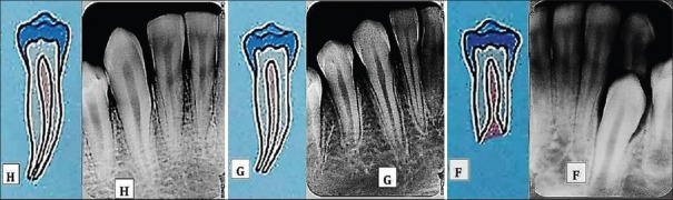
Schematic representation and radiographs of stages H, G and F of Demirjian system for mandibular canine
Figure 4.
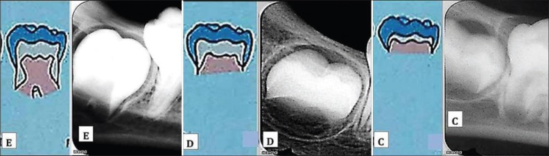
Schematic representation and radiographs of stages E, D and C of Demirjian system for mandibular molar
Table 1.
Dental calcification stages (adapted from demirjian et al. (1973))
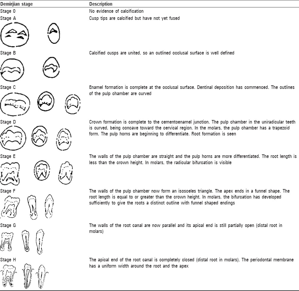
Table 2.
Demirjian's score tables based on developmental stage of tooth for boys and girls
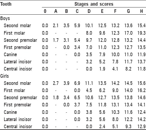
Table 3.
Demirjian's conversion chart from maturity score to final age for boys and girls
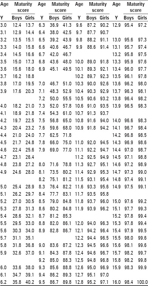
Table 4.
Willem's direct age scores for developmental tooth stages based on Demirjian's technique for boys and girls
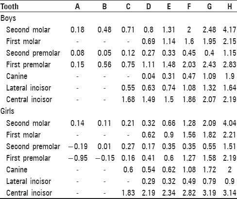
Figure 2.
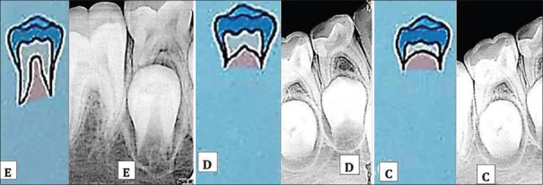
Schematic representation and radiographs of stages E, D and C of Demirjian system for mandibular premolar
Figure 3.
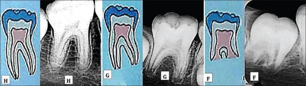
Schematic representation and radiographs of stages H, G and F of Demirjian system for mandibular molar
Skeletal age estimation
The right hand-wrist radiograph was taken for each subject at exposure parameters 60 kVp, 4 mA and 0.5 seconds with target to film distance of 5 feet and under strict radiation protection measures
The extraoral film was processed under standard processing conditions based on time-temperature method and dried for 10 minutes
The landmarks within the radiograph were closely examined and compared with the standard skeletal age plates of Radiographic Atlas of Skeletal Development of the Hand and Wrist Greulich and Pyle.[26]
Observers
The digital dental radiographs and hand-wrist radiograph of each subject were evaluated separately by observers X, Y and Z
The findings were recorded by them individually in the proforma of each patient while unaware of others findings
In case of variation in findings among the three observers, the findings that are common between any two out of three observers were considered as final
If the findings of all three observers are different, the radiographs were re-evaluated by all three observers together and findings were reconsidered.
Statistical analysis
SPSS (Statistical Package for Social Sciences) 12.0 for Windows (SPSS, Inc., Chicago, IL) was used for all analysis
The differences between the chronological age and the estimated dental and skeletal ages were statistically tested using paired ‘t’ test (P value)
The correlation between chronological age, dental and skeletal age estimation methods was confirmed statistically by Pearson's correlation (r value). In all these tests, r value closest to 1 was considered to indicate the strongest relation between the comparisons
The inter-observer variability (r value) was tested using the Spearman's Correlation Coefficient. The relation was considered strongest between the pair whose value was closest to 1
The intra-observer variability was evaluated by recording the tooth calcification stages of 20 randomly selected patients who were evaluated individually by each of the three observers with an interval of 30 days between the two estimations. The reproducibility of the estimations was statistically tested using the Pearson's Chi-square test (P value)
Probability of predicting the ages accurately was evaluated for each method by multiple regression analysis.
Results
A total of 180 patients (90 males and 90 females) ranging in age from 6 to 16 years were selected for the study.
Table 5 demonstrates the comparison between chronological age, Dental age estimated by Demirjian's method and Dental age estimated by Willem's method amongst male subjects. Demirjian's method of dental age estimation overestimated the age of male subjects from 6 to 10.99 years and 15-15.99 year age group, while it underestimated the age of male subjects from 11 to 14.99 years, when compared to chronological age. The P value obtained from sampled ‘t’ test was greater than 0.05 for all age groups except 8-8.99, 9-9.99 and 15-15.99 year age groups. Willem's method of dental age estimation overestimated the age of male subjects from 6 to 10.99 years and 15-15.99 year age group, while it underestimated the age of male subjects from 11 to 14.99 years, when compared to chronological age. The P value obtained from sampled t test was greater than 0.05 for all age groups except the 8-8.99 and 13-13.99 year age groups. The probability of predicting age accurately by Demirjian's method was 87.12%, while it was 89.06% by Willem's methods for male subjects.
Table 5.
Comparison between chronological age and dental age by Demirjian's and Willem's methods in males (age in years)
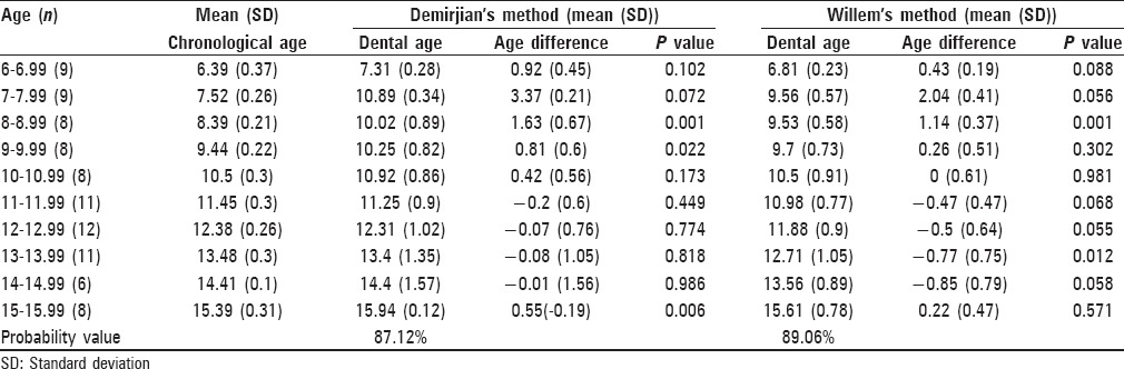
Table 6 demonstrates the comparison between chronological age, Dental age estimated by Demirjian's method and Dental age estimated by Willem's method amongst female subjects. Demirjian's method of dental age estimation consistently overestimated the age of female subjects for all age groups except 16-16.99 year age group when compared to chronological age. The P value obtained from sampled t test was greater than 0.05 for all age groups except 7-7.99, 11-11.99 and 12-12.99 year age groups. Willem's method of dental age estimation overestimated the age of female subjects belonging to all age groups except 6-6.99, 10-10.99, 13-13.99, 15-15.99 and 16-16.99 year age groups when compared to chronological age. The P value obtained from sampled t test was greater than 0.05 for all age groups. The probability of predicting age accurately by Demirjian's method was 86.73% while it was 89.08% by Willem's methods for female subjects.
Table 6.
Comparison between chronological age and dental age by Demirjian's and Willem's methods in females (age in years)
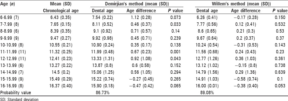
Table 7 demonstrates the comparison between chronologic age and skeletal age (Greulich and Pyle) amongst male subjects. Greulich and Pyle skeletal age estimation method consistently underestimated the age of male subjects belonging to all age groups. The P value obtained from sampled t test was greater than 0.05 for all age groups except 9-9.99, 10-10.99, 12-12.99, 13-13.99 and 15-15.99 year age groups, when compared to chronological age. The probability of predicting age accurately by Greulich and Pyle method was 90.65% for male subjects.
Table 7.
Comparison between Chronological age and Skeletal age (Greulich and Pyle method) in males (age in years)
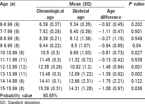
Table 8 demonstrates the comparison between chronological age and skeletal age amongst female subjects. Greulich and Pyle skeletal age estimation method consistently underestimated the age of female subjects belonging to all age groups when compared to chronological age. The P value obtained from sampled t test was greater than 0.05 for all age groups except the 11-11.99 and 12-12.99 year age groups. The probability of predicting age accurately by Greulich and Pyle method was 89.04% for female subjects.
Table 8.
Comparison between Chronological age and Skeletal age (Greulich and Pyle method) in females (age in years)
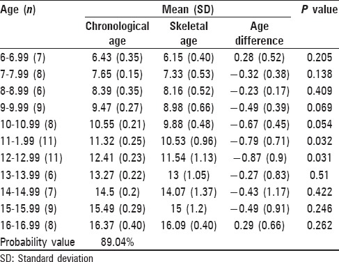
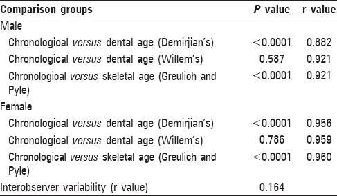
Table 9 demonstrates the comparison of dental ages estimated by Demirjian's and Willem's methods and skeletal age estimated by Greulich and Pyle method with the standard of chronological age of male and female subjects. The ‘r’ value representative of Pearson correlation coefficient between chronological age (CA) and dental age by Demirjian's method (DAD) was 0.882 for male subjects and 0.956 for female subjects. The ‘r’ value representative of Pearson correlation coefficient between CA and Dental age by Willem's method (DAW) was 0.921 for male subjects and 0.959 for female subjects. The ‘r’ value representative of Pearson correlation coefficient between CA and skeletal age by Greulich and Pyle method (SAGP) was 0.921 for male subjects and 0.960 for female subjects. The P value of paired sample t test for comparison between CA and DAD was <0.0001 for both male and female subjects. The P value of paired sample t test for comparison between CA and DAW was 0.587 for male subjects and 0.786 for female subjects. The P value of paired sample t test for comparison between CA and SAGP was <0.0001 for both male and female subjects. The ‘r’ value for the Pearson's Chi-square test employed to evaluate intra-observer variability was 0.164 (>0.05).
Table 9.
Comparison of dental ages estimated by Demirjian's and Willem's methods and skeletal age estimated by Greulich and Pyle method with the standard of chronological age of male and female subjects
Discussion
Age estimation should be as accurate as possible since it narrows down the search of a person of unknown age, enabling a more efficient and time saving approach.[4] Although various methods for the age determination do exist, a universal system has not been achieved due to the varying differences in different ethnic populations.[20,24] Hence, each method requires to be tested in different populations. The group under study was selected to ensure ethnic uniformity of the study sample. The present study consisted of 180 subjects; 90 males and 90 females residing in Gandhinagar district. Prabhakar et al., and Reshma et al., conducted studies with similar sample size but on different populations.[15,20]
The completion of crown calcification and root formation of all mandibular teeth (excluding third molars) occurs from 6 to 16 years of age, respectively.[27] The 6 to 16 year age group was selected for this study based on other maturation studies and the fact that orthodontic treatment is frequently performed in this age group which critically requires skeletal age assessment.[15]
In the present study, dental age estimation was conducted on seven teeth of left quadrant of mandible since these teeth represent the age range of commencement to completion of root calcification close to the age range of the patients selected for the study.[18] The maxillary posterior teeth were omitted from the study because superimposition of calcified structures in this area resulting in inaccurate assessment of the stage of development.[11]
The main disadvantage of the panoramic radiographs is that the image does not display the fine anatomic detail of periapical region available on intraoral periapical radiographs. Thus, it is not as useful as periapical radiography for studying root-apex calcification. Periapical radiographs require less radiation than panoramic radiographs. Further, digital intraoral receptors require less radiation than film, thus lowering patient exposure.[28] Hence, RVG with paralleling technique was utilized in the present study to acquire periapical images of the teeth under study.
The method described by Demirjian et al., (1973) was chosen in the present study because its criteria consists of distinct details based on shape and proportion of root length which are precise and simple, using the relative value to crown height rather than on absolute length.[15,24] Hence, it is best suited for forensic purposes.[23] However, there is consistent overestimation of age by Demirjian's method of dental age estimation in certain populations.[22] Hence, Willem's dental age estimation method[22] was also tested in this study.
The skeletal age for each hand-wrist radiograph was assigned according to the method outlined in the Greulich and Pyle atlas, because it is relatively easy to learn, less time consuming and shows greater reproducibility between observers.[11] Moreover, no norm for skeletal age assessment has been established for Indian population, hence Greulich and Pyle atlas was used in this study.[29]
The P value of paired sample t test for comparison between chronological age and dental age by Demirjian's method in the current study was greater than 0.05 for almost all age groups among male and females [Tables 5 and 6]. This suggested that there was no statistically significant difference between chronological age and dental age by Demirjian's method for most age groups and that this method was applicable to the population under study. This finding was consistent with the results of Hegde et al., Garamendi et al., and Bagherpour et al.[30,31,32]
The P value of paired sample t test for comparison between chronological age and dental age by Willem's method was greater than 0.05 for almost all age groups among males and all age groups in females [Tables 5 and 6]. This suggested that there was no statistically significant difference between the chronological age and dental age by Willem's method for almost all age groups and that this method was applicable to the population under study. Willems et al., Maber et al., Mani et al., and Amal et al., also had similar conclusions from their studies.[9,12,22,33]
The P value of paired sample t test for comparison between chronological age and skeletal age by Greulich and Pyle method was greater than 0.05 in almost all age groups among males and females [Tables 7 and 8]. This suggested that there was no statistically significant difference between the chronological age and skeletal age by Greulich and Pyle method and that this method was applicable to the population under study, more so among females than males. These findings were in agreement with those of Kraillasiri et al., and Garamendi et al.[11,31]
The ‘r’ value representative of Pearson correlation coefficient was close to 1 for all the pairs such as chronological age – dental age by Demirjian's method, chronological age – dental age by Willem's method as well as chronological age – skeletal age by Greulich and Pyle method for male and female subjects. This signified that there was a strongly positive correlation between the age estimation methods of all pairs. The ‘r’ value for all three age estimation methods between observers X and Y, Y and Z and X and Z was close to 1. This indicated that there was a strong agreement among the ratings of all observers. The ‘r’ value for inter-observer variability was greater than 0.05 indicating no statistically significant difference between the estimations of same patients by different observers at different points of time. The P value for paired sample t test was greater than 0.05 for the chronological age and dental age by Willem's method while it was less than 0.05 for the comparison of chronological age with both dental age by Demirjian method and skeletal age by Greulich and Pyle method. This indicates that there is no statistically significant difference between the chronological age of the sample and dental age estimated by Willem's method [Table 9].
There was overestimation and underestimation of age in almost equal number of groups with Willem's method [Tables 5 and 6]. However, these differences of chronological and dental age by Willem's method were consistently smaller than those between chronological age and dental age by Demirjian's method. This suggested that Willem's method is more accurate than the Demirjian's method for the population under study. This finding is supported by Maber et al., Mani et al. and Willems et al.[12,22,33]
The differences in certain age groups may be due to environmental factors such as the socio- economic status, nutrition and dietary habits that vary in different population groups.[20]
Conclusion
Digital dental radiography can be conveniently utilized to determine tooth calcification stages among 6-16-year-old children residing in Gandhinagar district
Demirjian's and Willem's dental age estimation methods are applicable for estimating the age of the population under study
Greulich and Pyle skeletal age estimation method can also be applied to estimate the age of the population under study
Although various age estimation methods do exist, the results are varied in different populations due to ethnic differences. Also, there is a lack of age estimation studies in Gujarati population. Hence, further studies are needed to formulate new tables for this population
Amongst the age estimation methods used in this study, the Willem's dental age estimation method was the most accurate and consistent for the 6-16 year old children residing in and around Gandhinagar district
The Willem's method (Modified Demirjian method) should be applied to estimate accurately chronological age for the population ranging from 6-16 years in age and residing in Gandhinagar district
The probability of predicting the age accurately is greater in males than in females by Demirjian's method. However, the probability of predicting the age accurately is greater in females than in males by Willem's method.
Footnotes
Source of Support: Nil
Conflict of Interest: None declared
References
- 1.Wikipedia. The online Encyclopedia. [Last accessed on 2011 Jul 21]. Available from: http://en.wikipedia.org .
- 2.William B, Fremingston M, Singh M, Blona K. Age determination in girls of North Eastern region of India. J Indian Acad Forensic Med. 2007;29:102–8. [Google Scholar]
- 3.Balwant R, Jasdeep K, Cameriere R. Radiological dental age estimation on third molars in South Indian population: Correlation between five tooth staging methods. Indian J Forensic Odontol. 2009;2:91–5. [Google Scholar]
- 4.Guy W. A review of the most commonly used dental age estimation techniques. J Forensic Odontostomatol. 2001;19:9–17. [PubMed] [Google Scholar]
- 5.Pradhuman V, Jatindra S, Kanika V, Som G, Guruprasad R. Age estimation of adolescents and young adults based on development of mandibular third molars – a panoramic study. J Indian Acad Oral Med Radiol. 2011;23:9–13. [Google Scholar]
- 6.Bosmans N, Ann P, Aly M, Willems G. The application of Kvaal's dental age calculation technique on panoramic dental radiographs. Forensic Sci Int. 2005;153:208–12. doi: 10.1016/j.forsciint.2004.08.017. [DOI] [PubMed] [Google Scholar]
- 7.Sasidhar S, Sharada P. Age estimation using pulp/tooth area ratio: A digital image analysis. J Forensic Sci. 2009;1:37–41. [Google Scholar]
- 8.Bala M, Pathak A, Jain RL. Assessment of skeletal age using MP3 and hand wrist radiographs and its correlation with dental and chronological ages in children. J Indian Soc Pedod Prev Dent. 2010;28:95–9. doi: 10.4103/0970-4388.66746. [DOI] [PubMed] [Google Scholar]
- 9.Amal B, Shara H, Fatma I. Comparison between two methods of dental age estimation among Egyptian children. Indian J Forensic Odontol. 2009;2:83–9. [Google Scholar]
- 10.Chertkow S. Tooth mineralization as an indicator of the pubertal growth spurt. Am J Orthod Dentofacial Orthop. 1980;77:79–91. doi: 10.1016/0002-9416(80)90226-2. [DOI] [PubMed] [Google Scholar]
- 11.Krailassiri S, Anuwongnukroh N, Dechkunakorn S. Relationships between dental calcification stages and skeletal maturity indicators in Thai individuals. Angle Orthod. 2002;72:155–66. doi: 10.1043/0003-3219(2002)072<0155:RBDCSA>2.0.CO;2. [DOI] [PubMed] [Google Scholar]
- 12.Mani SA, Naing L, John J, Samsudin AR. Comparison of two methods of dental age estimation in 7-15-year old Malays. Int J Paediatr Dent. 2008;18:380–8. doi: 10.1111/j.1365-263X.2007.00890.x. [DOI] [PubMed] [Google Scholar]
- 13.Baºaran G, Ozer T, Hamamci N. Cervical vertebral and dental maturity in Turkish subjects. Am J Orthod Dentofacial Orthop. 2007;131:447. doi: 10.1016/j.ajodo.2006.08.016. [DOI] [PubMed] [Google Scholar]
- 14.Flores-Mir C, Nebbe B, Major PW. Use of skeletal maturation based on hand wrist radiographic analysis as a predictor of facial growth: A systematic review. Angle Orthod. 2004;74:118–24. doi: 10.1043/0003-3219(2004)074<0118:UOSMBO>2.0.CO;2. [DOI] [PubMed] [Google Scholar]
- 15.Reshma N, US Krishna N, Gautam H. Assessment of growth using mandibular canine calcification stages and its correlation with modified MP3 stages. Int J Clin Paediatr Dent. 2010;3:27–33. doi: 10.5005/jp-journals-10005-1050. [DOI] [PMC free article] [PubMed] [Google Scholar]
- 16.Adel H, Hayder H, Mohammed D, Ali H. Interrelationship between dental maturity, skeletal maturity and chronological age in Saudi male children. J Egypt Dent Assoc. 2008;54:55–9. [Google Scholar]
- 17.Haiter-Neto F, Kurita LM, Menezes AV, Casanova MS. Skeletal age assessment: A comparison of 3 methods. Am J Orthod Dentofacial Orthop. 2006;130:435. doi: 10.1016/j.ajodo.2006.03.023. [DOI] [PubMed] [Google Scholar]
- 18.Soegiharto BM, Cunningham SJ, Moles DR. Skeletal maturation in Indonesian and White children assessed with hand wrist and cervical vertebrae methods. Am J Orthod Dentofacial Orthop. 2008;134:217–26. doi: 10.1016/j.ajodo.2006.07.037. [DOI] [PubMed] [Google Scholar]
- 19.Phillips VM, van Wyk Kotze TJ. Testing standard methods of dental age estimation by Moorrees, Fanning and Hunt and Demirjian, Goldstein and Tanner on three South African children samples. J Forensic Odontostomatol. 2009;27:20–8. [PubMed] [Google Scholar]
- 20.Prabhakar AR, Pande AK, Raju OS. Applicability of Demirjian's method of age assessment in children of Davangere. J Indian Soc Pedod Prev Dent. 2002;20:54–62. [PubMed] [Google Scholar]
- 21.Kuldeep S, Gorea K, Bharti V. Age estimation from eruption of permanent teeth. J Indian Acad Forensic Med. 2005;27:231–5. [Google Scholar]
- 22.Willems G, Van Olmen A, Spiessens B, Carels C. Dental age estimation in Belgian children: Demirjian's technique revisited. J Forensic Sci. 2001;46:893–5. [PubMed] [Google Scholar]
- 23.Rai B, Kaur J, Anand C, Rajnish J, Anil S, Sushil M. Accuracy of the Demirjian method for the Haryana Population. Internet J Dent Sci. 2008;6 [Google Scholar]
- 24.Ashish W, Panjab W, Tushar P. Correlation of radiographic and chronological age in human by using Demirjian's method: A radiographic study. J Indian Acad Oral Med Radiol. 2011;23:1–4. [Google Scholar]
- 25.Madhu S, Hegde AM, Munshi AK. The developmental stages of the middle phalanx of the third finger (MP3): A sole indicator in assessing the skeletal maturity? J Clin Paediatr Dent. 2003;27:149–56. doi: 10.17796/jcpd.27.2.qtj75rg3714l5543. [DOI] [PubMed] [Google Scholar]
- 26.Greulich W, Pyle S. Radiographic Atlas of skeletal development of hand wrist. Palo Alto, California: Stanford University Press; 1959. [Google Scholar]
- 27.Asher M. Wheeler's Dental Anatomy, Physiology and Occlusion. 6th ed. Philadelphia: W. B. Saunders Company; 2002. [Google Scholar]
- 28.Stuart W, Michael P. Oral Radiology: Principles and Interpretation. 6th ed. Noida: Reed Elsevier India Private Limited; 2009. pp. 37–53. [Google Scholar]
- 29.Koshy S, Tandon S. Dental age assessment: The applicability of Demirjian's method in South Indian children. Forensic Sci Int. 1998;94:73–85. doi: 10.1016/s0379-0738(98)00034-6. [DOI] [PubMed] [Google Scholar]
- 30.Hegde RJ, Sood PB. Dental maturity as an indicator of chronological age: Radiographic evaluation of dental age in 6 to 13 years children of Belgaum using Demirjian's Method. J Indian Soc Pedod Prev Dent. 2002;20:132–8. [PubMed] [Google Scholar]
- 31.Garamendi PM, Landa MT, Ballesteros J, Solano MA. Reliability of the methods applied to assess age minority in living subjects around 18 years old: A survey on a Moroccan origin population. Forensic Sci Int. 2005;154:3–12. doi: 10.1016/j.forsciint.2004.08.018. [DOI] [PubMed] [Google Scholar]
- 32.Bagherpour A, Imanimoghaddam M, Bagherpour M, Einolghozati M. Dental age assessment among Iranian children aged 6 - 13 years using the Demirjian method. Forensic Sci Int. 2010;197:121.e1–4. doi: 10.1016/j.forsciint.2009.12.051. [DOI] [PubMed] [Google Scholar]
- 33.Maber M, Liversidge H, Hector M. Accuracy of age estimation of radiographic methods using developing teeth. Forensic Sci Int. 2006;159:S68–73. doi: 10.1016/j.forsciint.2006.02.019. [DOI] [PubMed] [Google Scholar]