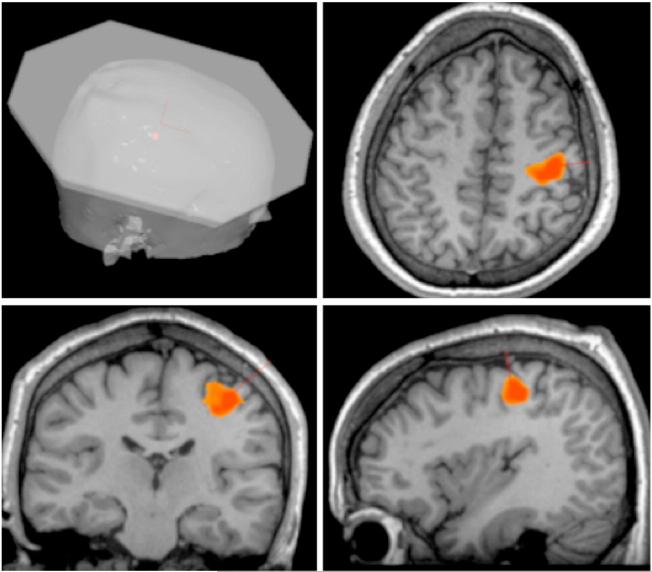
Fig. 1.
Individualized image guided TMS. TMS was applied to right M1hand area identified by a left index finger adduction and abduction task during BOLD fMRI acquisition overlaid on the individual's anatomical MRI. The TMS treatment-planning tool determined the scalp location and the orientation of the TMS coil.

