Abstract
Myiasis is a pathologic condition in humans occurring because of parasitic infestation. Parasites causing myiasis belong to the order Diptera. Oral myiasis is seen secondary to oral wounds, suppurative lesions, and extraction wounds, especially in individuals with neurological deficit. In such cases, neglected oral hygiene and halitosis attracts the flies to lay eggs in oral wounds resulting in oral myiasis. We present a case of oral myiasis in 40-year-old male patient with mental disability and history of epilepsy.
Keywords: Dipterous larvae, Maggots, Myiasis
INTRODUCTION
The term myiasis is derived from Greek words “muia” and “iasis”, which means “fly” and “disease”, respectively. German entomologist Fritz Zumpt defined myiasis as “the infestation of live human and vertebrate animals with dipterous larvae, which at least for a period, feed on the host's dead or living tissue, liquid body substances, or ingested food”.[1] Human or animal tissue acts as an intermediate host for the larvae in its life cycle. Myiasis can be seen worldwide, with a higher incidence being observed in tropical and subtropical regions of Africa and America due to the favorable climatic conditions of heat and humidity. In humans, the sites most commonly affected are skin, nose, ears, eyes, anus, vagina, and oral cavity.[2,3] Oral myiasis is associated with nosocomial infections, dental extractions, visits to tropical countries, alcoholism, and mouth breathing, and is commonly seen in mentally disabled individuals and people from low socioeconomic status. Oral myiasis was first described by Laurence in 1909.[4]
CASE REPORT
A 40-year-old male patient reported to our hospital with a chief complaint of facial wound on the left cheek region for past one month, which was initially smaller and increased progressively to the present large size, with appearance of worms in the wound. The patient was mentally challenged and revealed a medical history of epilepsy from childhood.
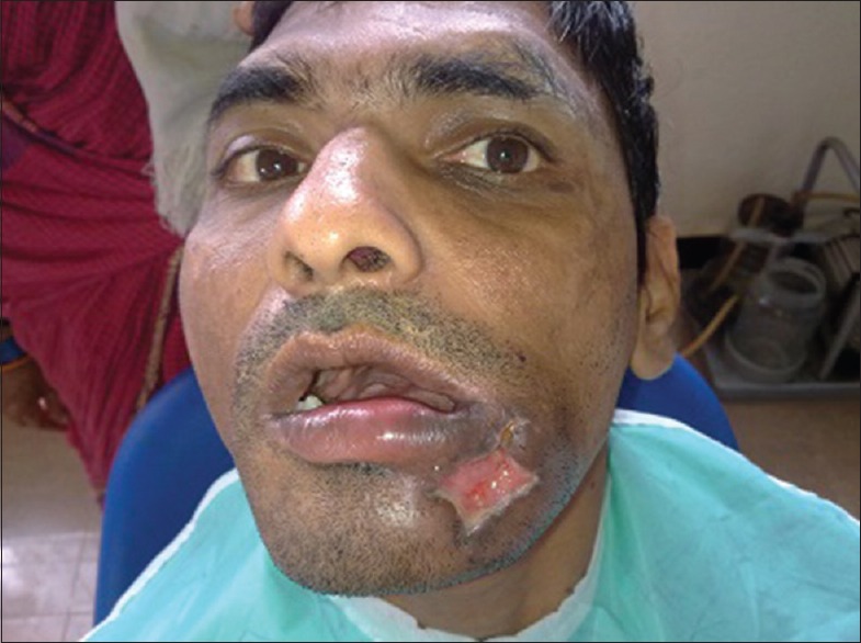
On extraoral examination, the ulcer was 5 cm × 4 cm in size on the left chin region, including lower lip and infiltrating the underlying tissues with an everted and erythematous border. Orocutaneous fistula was present at a point in the base of the ulcer. The wound was tender and firm. Maggots were seen burrowed deep in the wound [Figure 1]. The affected region was swollen, and the swelling extended to the middle of the upper lip. On the left side of the neck, two submandibular lymph nodes were palpable. On palpation, the ulcer was tender and indurate.
Figure 1.
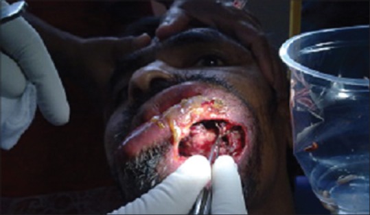
Maggots seen buried deep in the wound in the left cheek region (preoperative)
On intraoral examination, hard tissue revealed partially edentulous space and poor oral hygiene with severe deposits of calculus and stains. On soft tissue examination, a small fistula was seen in the left lower vestibule at the corner of the mouth.
Considering the patient's mental status, history of epilepsy, and poor oral hygiene, it was provisionally diagnosed as an ulcer infested with maggots (oral myiasis).
About 15–20 maggots were removed with a tweezer following the application of turpentine oil [Figures 2 and 3]. The area was irrigated with saline and betadine solution. The removed maggots were placed in a container, sealed tightly, and disposed off. Surgical debridement of the wound was carried out under local anesthesia [Figure 4]. The patient was prescribed antibiotic (cefotaxime 200 mg bd), and analgesic (ibuprofen) to prevent further infection and to control pain. When the patient was reviewed after a week, the swelling had subsided and wound healing was observed to be satisfactory [Figure 5].
Figure 2.
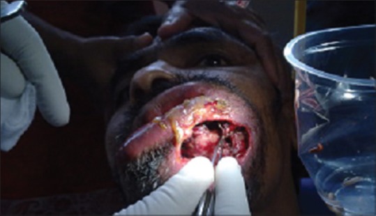
Removal of maggots using tweezers following turpentine oil application
Figure 3.
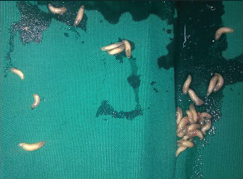
Removed maggots
Figure 4.
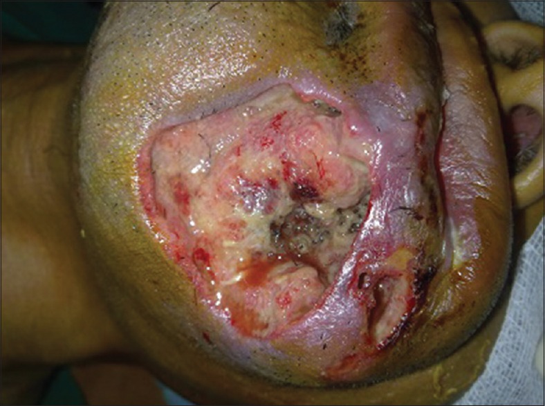
Immediate post-operative
Figure 5.
Wound healing satisfactory after a week
DISCUSSION
Clinically, myiasis is classified as primary and secondary. In the primary type, larvae feed on living tissue, and in the secondary type, larvae feed on dead tissue.[5,6] Depending upon the condition of involved tissue, myiasis is again classified as accidental myiasis (larvae ingested along with food), semi-specific (larvae laid on necrotic tissue in wounds), and obligatory myiasis (larvae affects the undamaged skin).[7,8]
Based on the tissue involved, cutaneous myiasis is subdivided into creeping and furuncle. In creeping type, larvae burrow through or under the skin, and in furuncle type, larvae remain in one spot, causing a boil-like lesion.[9]
Larvae of the common housefly Musca domestica (Indian housefly) have also been identified in neglected wounds. The adult female flies lay eggs or larvae on food, necrotic tissue, or open wounds. Warm humid climate and non-healing wound with halitosis attract the flies to lay eggs. Eggs hatch within 24 hours, and the larvae formed release toxins to destroy the host tissue. Larvae complete their development in 5-7 days, and they then wriggle out of the wound and fall to the ground to pupate.[10]
The treatment is primarily manual removal of larvae after the topical application of turpentine oil, mineral oil, chloroform, ethyl chloride, or mercuric chloride. These substances called asphyxiation drugs creates anaerobic atmosphere within the wound causing aerobic parasitic larvae to come to the surface making its removal easier.[11] Following the removal of the maggots, surgical wound debridement should be performed. A systemic treatment with ivermectin a semi-synthetic macrolide antibiotic isolated from Streptomyces avermitlis is another choice, which is given orally in one dose of 150-200 mg/kg of body weight.[12] It activates the release of gamma amino butyric acid, which induces the death of the larvae and their spontaneous elimination. Complete removal of larvae is very important to treat myiais successfully. Antibiotics can also be prescribed to prevent secondary bacterial infection.
Prevention of oral myiasis can be achieved by health awareness, enhancing oral and personal hygiene, and providing proper care to individuals with neurological deficit.
Footnotes
Source of Support: Nil.
Conflict of Interest: None declared.
REFERENCES
- 1.Zumpt F. Myiasis in man and animals in the old world. In: Zumpt F, editor. A Textbook for Physicians, Veterinarians and Zoologists. London: Butterworth and Co. Ltd; 1965. p. 109. [Google Scholar]
- 2.Droma EB, Wilamowsky A, Schnur H, Yarom N, Scheuer E, Schwartz E. Oral myiasis: A case report and literature rewview. Oral Surg Oral Med Oral Pathol Oral Radiol Endod. 2007;103:92–6. doi: 10.1016/j.tripleo.2005.10.075. [DOI] [PubMed] [Google Scholar]
- 3.Gunbay S, Bicacki N, Canda T, Canda S. A case of myiasis gingiva. J Periodontol. 1995;66:892–5. doi: 10.1902/jop.1995.66.10.892. [DOI] [PubMed] [Google Scholar]
- 4.Laurence SM. Dipterous larvae infection. Br Med J. 1909;9:88. [Google Scholar]
- 5.Poon TS. Oral myiasis in Hong Kong- A case report. Hong Kong Pract. 2006;28:388–93. [Google Scholar]
- 6.Bhoyar SC, Mishra YC. Oral myiasis caused by dipteral in epileptic patient. J Indian Dent Assoc. 1986;58:535–6. [PubMed] [Google Scholar]
- 7.Shinohara EH, Martini MZ, de Oliveira Neto HG, Takahashi A. Oral myiasis treated with ivermectin: Case report. Braz Dent J. 2004;15:79–81. doi: 10.1590/s0103-64402004000100015. [DOI] [PubMed] [Google Scholar]
- 8.Bhat AP, Jayakrishnan A. Oral myiasis: A case report. Int J Paediatr Dent. 2000;10:67–70. doi: 10.1046/j.1365-263x.2000.00162.x. [DOI] [PubMed] [Google Scholar]
- 9.Sowani A, Joglekar D, Kulkarni P. Maggots: A neglected problem in palliative care. Indian J Palliat Care. 2004;10:27–9. [Google Scholar]
- 10.Baskaran M, Jagankumar B, Geeverghese A. Cutaneous myiasis of face. J Oral Maxillofac Pathol. 2007;11:70–2. [Google Scholar]
- 11.Sharma H, Dayal D, Agarwal SP. Nasal myiasis: Review of 10 years experience. J Laryngol Otol. 1989;103:489–91. doi: 10.1017/s0022215100156695. [DOI] [PubMed] [Google Scholar]
- 12.Gealh WC, Ferreira GM, Farah GJ, Teodoro U, Camarini ET. Treatment of oral myiasis caused by Cochliomyia homnivorax: Two cases treated with ivermectin. Br J Oral Maxillofac Surg. 2009;47:23–6. doi: 10.1016/j.bjoms.2008.04.009. [DOI] [PubMed] [Google Scholar]


