Abstract
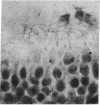
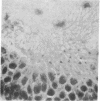
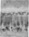
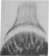
A study of the macula and crista organs of the frog's labyrinth with the use of an improved method of histological preparation has shown these endings to be more complex than heretofore believed. The structure lying over the layer of sensory and supporting cells is not a single “gelatinous” body as commonly described, but consists of two distinct layers with separate functions.
In all these endings—macula sacculi, macula utriculi, macula lagenae, and the cristae of the three semicircular canals—there is a special tectorial structure that lies over the cellular surface and makes the connections to the ciliary tufts of the hair cells. It has the general form of a reticulum, though in the saccule it is somewhat elaborated.
A preliminary study of other vertebrates indicates that this tectorial reticulum is present in all the labyrinthine endings throughout the series from fishes to mammals.
Consideration is given to the possible advantages of this special tectorial structure in the stimulation process.
Keywords: otic labyrinth, ear, amphibia
Full text
PDF


Images in this article
Selected References
These references are in PubMed. This may not be the complete list of references from this article.
- Hama K. A study on the fine structure of the saccular macula of the gold fish. Z Zellforsch Mikrosk Anat. 1969;94(2):155–171. doi: 10.1007/BF00339353. [DOI] [PubMed] [Google Scholar]
- Hillman D. E., Lewis E. R. Morphological basis for a mechanical linkage in otolithic receptor transduction in the frog. Science. 1971 Oct 22;174(4007):416–419. doi: 10.1126/science.174.4007.416. [DOI] [PubMed] [Google Scholar]
- Lewis E. R., Nemanic P. Scanning electron microscope observations of saccular ultrastructure in the mudpuppy (Necturus maculosus). Z Zellforsch Mikrosk Anat. 1972;123(4):441–457. doi: 10.1007/BF00335541. [DOI] [PubMed] [Google Scholar]


