Figure 3.
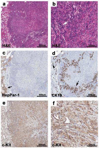
Poorly differentiated liver tumors. (a) Poorly differentiated liver tumor from a PtenLKO;Tgfbr2LKO mouse composed of poorly formed cords and trabeculae, with multifocal irregular glands, 4X. (b) Higher magnification of “a”. Poorly formed cords and trabeculae of anaplastic cells with multifocal, irregular glands (white arrow), 20X. (c) Absence of HepPar-1 staining in the tumor is shown. Note the adjacent positive staining in normal hepatocytes, arrow, 4X. (d) Normal bile ducts in adjacent parenchyma (arrowhead) and glandular structures within the tumor (arrow) stain positive for the cholangiocyte marker CK19, 4X. (e) Within the poorly differentiated carcinoma, anaplastic cells and glands are positive for c-Kit, 4X. (f) Higher magnification of “e” showing c-Kit staining, 20X.

