

Abstract
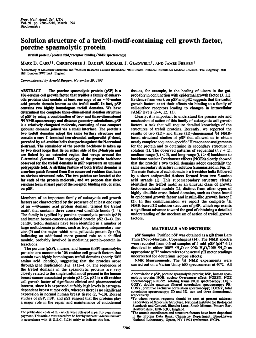
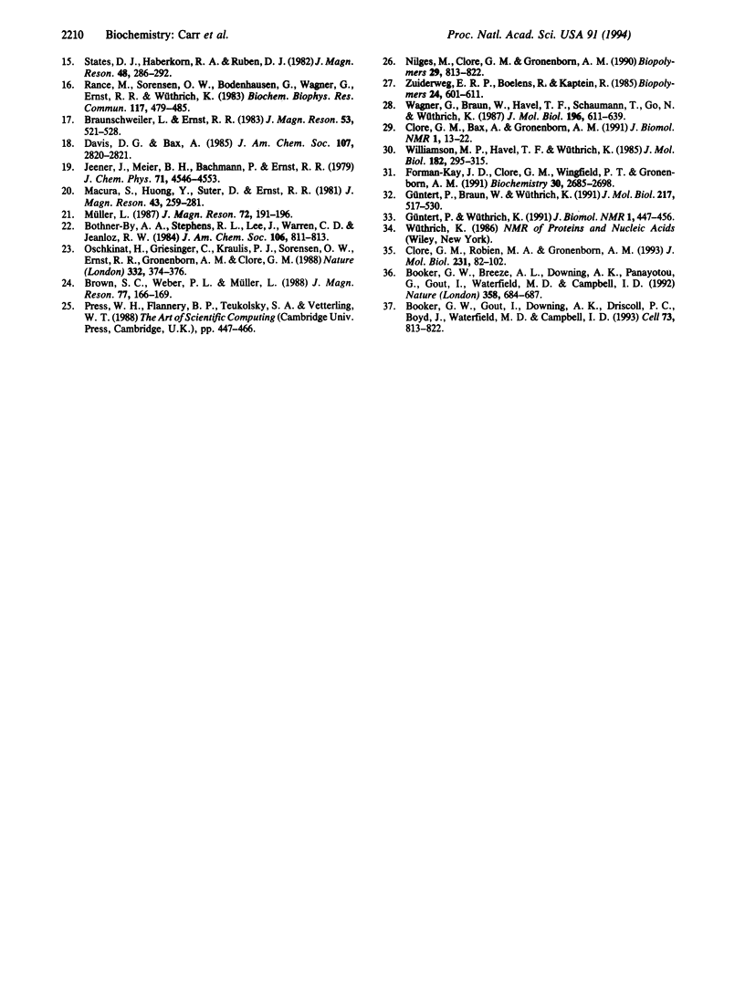
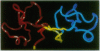
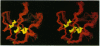
The porcine spasmolytic protein (pSP) is a 106-residue cell growth factor that typifies a family of eukaryotic proteins that contain at least one copy of an approximately 40-amino acid protein domain known as the trefoil motif. In fact, pSP contains two highly homologous trefoil domains. We have determined the complete three-dimensional solution structure of pSP by using a combination of two- and three-dimensional 1H NMR spectroscopy and distance geometry calculations. pSP is a relatively elongated molecule, consisting of two compact globular domains joined via a small interface. The protein's two trefoil domains adopt the same tertiary structure and contain a core C-terminal two-stranded antiparallel beta-sheet, preceded by a 6-residue helix that packs against the N-terminal beta-strand. The remainder of the protein backbone is taken up by two short loops that lie on either side of the beta-hairpin and are linked by an extended region that wraps around the C-terminal beta-strand. The topology of the protein backbone observed for the trefoil domains in pSP represents an unusual polypeptide fold. A striking feature of both trefoil domains is a surface patch formed from five conserved residues that have no obvious structural role. The two patches are located at the far ends of the protein molecule, and we propose that these residues form at least part of the receptor binding site, or sites, on pSP.
Full text
PDF
Images in this article
Selected References
These references are in PubMed. This may not be the complete list of references from this article.
- Booker G. W., Breeze A. L., Downing A. K., Panayotou G., Gout I., Waterfield M. D., Campbell I. D. Structure of an SH2 domain of the p85 alpha subunit of phosphatidylinositol-3-OH kinase. Nature. 1992 Aug 20;358(6388):684–687. doi: 10.1038/358684a0. [DOI] [PubMed] [Google Scholar]
- Booker G. W., Gout I., Downing A. K., Driscoll P. C., Boyd J., Waterfield M. D., Campbell I. D. Solution structure and ligand-binding site of the SH3 domain of the p85 alpha subunit of phosphatidylinositol 3-kinase. Cell. 1993 May 21;73(4):813–822. doi: 10.1016/0092-8674(93)90259-s. [DOI] [PubMed] [Google Scholar]
- Bork P. A trefoil domain in the major rabbit zona pellucida protein. Protein Sci. 1993 Apr;2(4):669–670. doi: 10.1002/pro.5560020417. [DOI] [PMC free article] [PubMed] [Google Scholar]
- Brown A. M., Jeltsch J. M., Roberts M., Chambon P. Activation of pS2 gene transcription is a primary response to estrogen in the human breast cancer cell line MCF-7. Proc Natl Acad Sci U S A. 1984 Oct;81(20):6344–6348. doi: 10.1073/pnas.81.20.6344. [DOI] [PMC free article] [PubMed] [Google Scholar]
- Carr M. D. 1H NMR-based determination of the secondary structure of porcine pancreatic spasmolytic polypeptide: one of a new family of "trefoil" motif containing cell growth factors. Biochemistry. 1992 Feb 25;31(7):1998–2004. doi: 10.1021/bi00122a015. [DOI] [PubMed] [Google Scholar]
- Clore G. M., Bax A., Gronenborn A. M. Stereospecific assignment of beta-methylene protons in larger proteins using 3D 15N-separated Hartmann-Hahn and 13C-separated rotating frame Overhauser spectroscopy. J Biomol NMR. 1991 May;1(1):13–22. doi: 10.1007/BF01874566. [DOI] [PubMed] [Google Scholar]
- Clore G. M., Robien M. A., Gronenborn A. M. Exploring the limits of precision and accuracy of protein structures determined by nuclear magnetic resonance spectroscopy. J Mol Biol. 1993 May 5;231(1):82–102. doi: 10.1006/jmbi.1993.1259. [DOI] [PubMed] [Google Scholar]
- Foekens J. A., Rio M. C., Seguin P., van Putten W. L., Fauque J., Nap M., Klijn J. G., Chambon P. Prediction of relapse and survival in breast cancer patients by pS2 protein status. Cancer Res. 1990 Jul 1;50(13):3832–3837. [PubMed] [Google Scholar]
- Forman-Kay J. D., Clore G. M., Wingfield P. T., Gronenborn A. M. High-resolution three-dimensional structure of reduced recombinant human thioredoxin in solution. Biochemistry. 1991 Mar 12;30(10):2685–2698. doi: 10.1021/bi00224a017. [DOI] [PubMed] [Google Scholar]
- Frandsen E. K., Jørgensen K. H., Thim L. Receptor binding of pancreatic spasmolytic polypeptide (PSP) in rat intestinal mucosal cell membranes inhibits the adenylate cyclase activity. Regul Pept. 1986 Dec 30;16(3-4):291–297. doi: 10.1016/0167-0115(86)90028-5. [DOI] [PubMed] [Google Scholar]
- Frandsen E. K. Receptor binding of pancreatic spasmolytic polypeptide in intestinal mucosal cells and membranes. Regul Pept. 1988 Jan;20(1):45–52. doi: 10.1016/0167-0115(88)90056-0. [DOI] [PubMed] [Google Scholar]
- Güntert P., Braun W., Wüthrich K. Efficient computation of three-dimensional protein structures in solution from nuclear magnetic resonance data using the program DIANA and the supporting programs CALIBA, HABAS and GLOMSA. J Mol Biol. 1991 Feb 5;217(3):517–530. doi: 10.1016/0022-2836(91)90754-t. [DOI] [PubMed] [Google Scholar]
- Güntert P., Wüthrich K. Improved efficiency of protein structure calculations from NMR data using the program DIANA with redundant dihedral angle constraints. J Biomol NMR. 1991 Nov;1(4):447–456. doi: 10.1007/BF02192866. [DOI] [PubMed] [Google Scholar]
- Hauser F., Hoffmann W. P-domains as shuffled cysteine-rich modules in integumentary mucin C.1 (FIM-C.1) from Xenopus laevis. Polydispersity and genetic polymorphism. J Biol Chem. 1992 Dec 5;267(34):24620–24624. [PubMed] [Google Scholar]
- Hoffmann W., Hauser F. The P-domain or trefoil motif: a role in renewal and pathology of mucous epithelia? Trends Biochem Sci. 1993 Jul;18(7):239–243. doi: 10.1016/0968-0004(93)90170-r. [DOI] [PubMed] [Google Scholar]
- Jørgensen K. H., Thim L., Jacobsen H. E. Pancreatic spasmolytic polypeptide (PSP): I. Preparation and initial chemical characterization of a new polypeptide from porcine pancreas. Regul Pept. 1982 Mar;3(3-4):207–219. doi: 10.1016/0167-0115(82)90126-4. [DOI] [PubMed] [Google Scholar]
- Nilges M., Clore G. M., Gronenborn A. M. 1H-NMR stereospecific assignments by conformational data-base searches. Biopolymers. 1990 Mar-Apr;29(4-5):813–822. doi: 10.1002/bip.360290415. [DOI] [PubMed] [Google Scholar]
- Oschkinat H., Griesinger C., Kraulis P. J., Sørensen O. W., Ernst R. R., Gronenborn A. M., Clore G. M. Three-dimensional NMR spectroscopy of a protein in solution. Nature. 1988 Mar 24;332(6162):374–376. doi: 10.1038/332374a0. [DOI] [PubMed] [Google Scholar]
- Rance M., Sørensen O. W., Bodenhausen G., Wagner G., Ernst R. R., Wüthrich K. Improved spectral resolution in cosy 1H NMR spectra of proteins via double quantum filtering. Biochem Biophys Res Commun. 1983 Dec 16;117(2):479–485. doi: 10.1016/0006-291x(83)91225-1. [DOI] [PubMed] [Google Scholar]
- Rio M. C., Bellocq J. P., Daniel J. Y., Tomasetto C., Lathe R., Chenard M. P., Batzenschlager A., Chambon P. Breast cancer-associated pS2 protein: synthesis and secretion by normal stomach mucosa. Science. 1988 Aug 5;241(4866):705–708. doi: 10.1126/science.3041593. [DOI] [PubMed] [Google Scholar]
- Rio M. C., Bellocq J. P., Gairard B., Rasmussen U. B., Krust A., Koehl C., Calderoli H., Schiff V., Renaud R., Chambon P. Specific expression of the pS2 gene in subclasses of breast cancers in comparison with expression of the estrogen and progesterone receptors and the oncogene ERBB2. Proc Natl Acad Sci U S A. 1987 Dec;84(24):9243–9247. doi: 10.1073/pnas.84.24.9243. [DOI] [PMC free article] [PubMed] [Google Scholar]
- Thim L. A new family of growth factor-like peptides. 'Trefoil' disulphide loop structures as a common feature in breast cancer associated peptide (pS2), pancreatic spasmolytic polypeptide (PSP), and frog skin peptides (spasmolysins). FEBS Lett. 1989 Jun 19;250(1):85–90. doi: 10.1016/0014-5793(89)80690-8. [DOI] [PubMed] [Google Scholar]
- Wagner G., Braun W., Havel T. F., Schaumann T., Go N., Wüthrich K. Protein structures in solution by nuclear magnetic resonance and distance geometry. The polypeptide fold of the basic pancreatic trypsin inhibitor determined using two different algorithms, DISGEO and DISMAN. J Mol Biol. 1987 Aug 5;196(3):611–639. doi: 10.1016/0022-2836(87)90037-4. [DOI] [PubMed] [Google Scholar]
- Williamson M. P., Havel T. F., Wüthrich K. Solution conformation of proteinase inhibitor IIA from bull seminal plasma by 1H nuclear magnetic resonance and distance geometry. J Mol Biol. 1985 Mar 20;182(2):295–315. doi: 10.1016/0022-2836(85)90347-x. [DOI] [PubMed] [Google Scholar]
- Wright N. A., Poulsom R., Stamp G. W., Hall P. A., Jeffery R. E., Longcroft J. M., Rio M. C., Tomasetto C., Chambon P. Epidermal growth factor (EGF/URO) induces expression of regulatory peptides in damaged human gastrointestinal tissues. J Pathol. 1990 Dec;162(4):279–284. doi: 10.1002/path.1711620402. [DOI] [PubMed] [Google Scholar]
- Wright N. A. Trefoil peptides and the gut. Gut. 1993 May;34(5):577–579. doi: 10.1136/gut.34.5.577. [DOI] [PMC free article] [PubMed] [Google Scholar]