Abstract
Bone marrow mesenchymal cells have been identified as a source of pluripotent stem cells with multipotential potential and differentiation in to the different cells types such as are osteoblast, chondroblast, adipoblast. In this research we describe pioneering experiment of tissue engineering in Bosnia and Herzegovina, of the isolation and differentiation rat bone marrow stromal cells in to the osteoblast cells lineages. Rat bone marrow stromal cells were isolated by method described by Maniatopulos using their plastic adherence capatibility. The cells obtained by plastic adherence were cultured and serially passaged in the osteoinductive medium to differentiate into the osteocytes. Bone marrow samples from rats long bones used for isolation of stromal cells (BMSCs). Under determinate culture conditions BMSCs were differentiated in osteogenic cell lines detected by Alizarin red staining three weeks after isolation. BMSCs as autologue cells model showed high osteogenetic potential and calcification capatibility in vitro. In future should be used as alternative method for bone transplantation in Regenerative Medicine.
KEY WORDS: Adult stem cells, bone marrow, bone marrow stem cells
INTRODUCTION
Bone marrow (BM) contains two different type of the progenitor cells, hematopoietic stem cells which are responsible for the production of the blood cells, and other cell type called Mesenchymal Stem Cells (MSCs), which have capability to differentiate in to the various cell lineages including osteoblast, adipose lineages and chondrocytes [1, 2]. Friedenstein et al. [3] was first who described MSC specifics to adhere for tissue culture plates, their characteristic fibroblast morphology and capacity to form colonies. Hunt et al. [4] also describe similar heterogeneous and fibroblastic appearance and a colony forming characteristic cells in to the bone marrow. Because of that, initially these cells were named as a plastic-adherent or colony-forming unit fibroblast (CFU-F) which today are named as marrow stromal cells, mesenchymal stem cells (MSC) or inducible Pluripotent Stem Cells (iPSC) due their heterogenous character and potential to differentiate into the various cells phenotypes [5, 6]. Those capabilities make MSCs valuable tools in Regenerative Medicine and Tissue Engineering fields. Bone Marrow Stem Cells (BMSCs) culture system derived from the rats, as a bone formative experimental system was first described by the Maniatopuoulas et al. and characterized morphologically, immunohistologically and biochemically [7]. This method of isolation is based on previously described characteristic of adhere to plastic surfaces, and then other cells such are hematopoetic don’t have such characteristic and removed with media changing [8]. In this article we will described our technical point of this culture system and rat BMSC isolation and differentiation in to the osteoblast-like cells what present pioneering biotechnology research in Bosnia and Herzegovina and first experiment of isolation stem cells from bone marrow. Aim of this study was to isolate Rat Bone Marrow Stem Cells and differentiate in to the osteoblast cell line, to show their osteoinductivity and potential for application in regenerative medicine.
MATERIALS AND METHODS
Animals
The 12 weeks old rats (n=4) were purchased from the Animal Laboratory Medical Faculty, University of Sarajevo. Rats were maintained under laboratory environment condition with ad libitum access to tap water and food. The experimental procedures handling and care of the animals, were approved by the ethical vote of the Institutional Ethical Committee, Medical Faculty, University of Sarajevo, Bosnia and Herzegovina.
Chemicals
The α- MEM medium, penicillin, streptomycin, L-glutamine, fetal calf serum (FCS), phosphate buffered saline (PBS), gentamicin sulphate, fungison, dexamethasone, β glycerophosphate (β-GP), ascorbic acid (AA), Alzarin red staining reagents.
Cell harvesting and culture
Primary osteoblasts were derived in culture from rat’s bone marrow. Briefly, 12 weeks old animals were sacrificed with chloroform anaesthesia. Both sites, femora and tibia were aseptically removed after sacrifice under chloroform anesthesia, asphyxiation, according to Institutional Animal Care and Use Committee (IACUC) guidelines. The cancellous bone was removed from the femur and tibia, and washed 3-5 times with 1x PBS. Thereafter, the bone marrows were flushed out using α-MEM culture medium, supplemented with 10% (v/v) FBS, freshly prepared AA (50 μg/ml), 50 IU L-1 penicillin, 2mM L-glutamine, 50 μg mL-1 streptomycin, 0.3 μg mL-1 fungizone, 50 μg mL-1 gentamycin sulphate, 10 mM β-GP, 10-8 M dexamethasone. The released cells were collected in to 75 cm2 culture flasks with 15 ml α-MEM culture medium. The cells cultures were maintained at 37° C in a humidified atmosphere of 95% air and 5% CO2. Cells were allowed to attach for 4-5 days, when after the non-adherent cell population was removed and the culture medium was replaced with fresh culture α-MEM medium. The culture medium was changed twice a week. Within 3-4 weeks cells start to growth.
Alizarin Red staining
The adherent cells were detached by 0.25% trypsin containing 50mM EDTA and seeded to petri dishes. After 24 hours, the culture medium was replaced with a fresh a-MEM medium supplemented with FCS (10%), dexamethasone (10-8 M), β-glycerophosphate (10 μM) and freshly prepared AA (50 μg/ ml). Alizarin Red staining was used to detect the calcified tissue in cultures.
Statistical analysis
The staining of Alizarine red and ALP activity was evaluated by three different persons independently of each other by assessing the staining intensity as follows: + weak staining, ++ modrate staining, +++ strong staining.
RESULTS
Isolation and culturing of MSCs in vitro
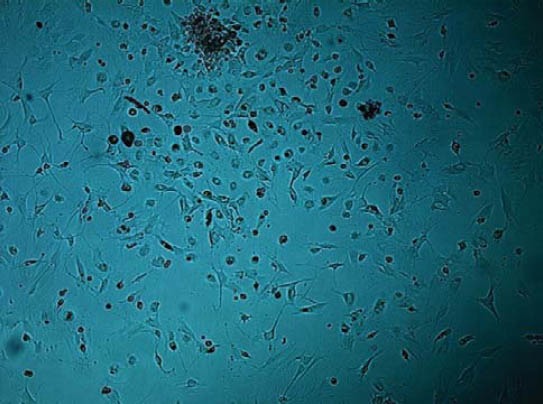
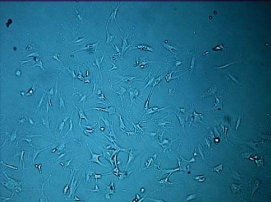
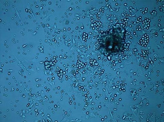
By plastic adherence capability of the bone marrow stem cells, the cells suspension containing both stromal and hematopoietic cells was seeded in tissue culture flasks using a-MEM 10% (FCS). After 48 hours, many of the rounded as well as spindle shaped cells had attached to the base of the tissue culture flask. The spindle shaped cells were attached to the bottom of the flask while the round cells remained suspended in the medium and were mostly eliminated from the culture with subsequent media changes. These cells began to proliferate at about day 4 (Figure 2) and gradually grow to form small colonies at day 10 (Figure 3).
FIGURE 2.

4th day of isolation. The spindle like cells were attached to the bottom of flask start to proliferate.
FIGURE 3.

10th day of isolation. Cells continued to proliferate.
FIGURE 1.

2nd day after isolation. The spindle shaped stromal cells of the bone marrow attached to the floor of cell flask, surrounded with rounded hematopoetic cells.
Alizarin red staining
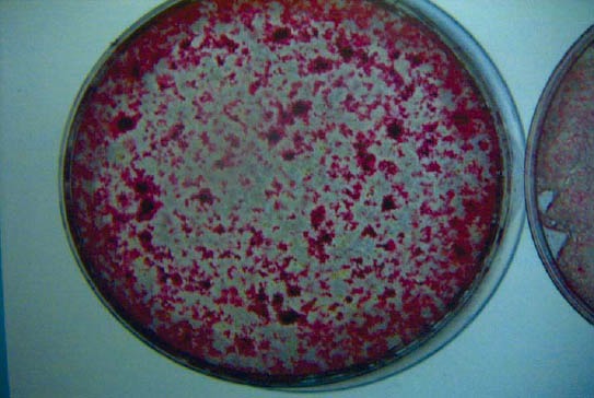
BMSCs were differentiated in vitro using osteogenic induction media. Following three weeks after seeding, these cells showed osteogensis upon staining with Alizarin Red S for calcium deposits.
FIGURE 4.

3rd weeks after isolation, the Alizarin red staining showed calcification of the culture showing Bone Marrow Stem osteogenic capability to form bone tissue.
DISCUSSION
In this study we successfully isolated MSCs from bone marrow of the mature rats, and showed their ostegenic potential and forming calcification tissue detected by Alizarin Red staining. That result indicated that MSCs play vital role in bone regeneration when were exposed to growth factors. That characteristic making possibility that in future MSCs as single cells or attached on some supplements could be successfully replacement for today gold standard of autologue bone transplant in bone reconstruction [9]. Also single MSCs could be use in combination with bone transplants or cancelous bone enrichment with growth factors as human Bone Morphogenetic Proteins-7 (rhBMP-7) to improve their osteoinductive and regenerative potential. Smajilagic et al. [10], showed successfully bridged non critical size defects on New Zealand Rabbits treated with autologue bone marrow enrichment with rhBMP-7. 3D CT reconstruction showed new formed bone Density (BMD) of 530 mg/cm3. In other research, authors showed synergistic effect of the rhBMP7 and osteopromotive membranes in naturally environment enriched with fibroplast from local maseter muscle flap transositioned in defect, in healing non critical size defects rabbit’s mandible [11]. Osteoinductive media growth factors we used in research were contest from Ascrobic acid (AA), Beta-Glycerophosphate and Dexametasone. Ascorbic acid (AA) is substance which is necessary for hydroxylation of the proline residue in the procollagen molecule, and AA become necessary for forming bone like matrix in culture [12]. It is unstable and always requires being prepared freshly for culture medium supplementation. Concentration of the 50 μg/ml AA was used in our study. Beta-glycerophosphate is organic phosphate and does not modulate any of biosynthesis activities cells in mineralized culture [13]. We used concentration of 10mM. Dexametasone is synthetic glucocorticoids prerequisite for mineralized tissue formation and key factor for controlling metabolism of bone tissue [14]. Concentration 10-8 was used in our study. The important technical point for these substances is that should be prepared just before changing the medium. This experimental system is very useful for investigation of the mechanism bone regeneration, impairment bone healing and a treatment of many bone metabolic diseases such as osteoporosis. Researcher could precisely and easily control the physicochemical environment in their experiments to determinate factors responsible for some of above diseases.
CONCLUSIONS
In conclusion, our study indicates that using simple principle of adhesion it is possible to isolate rats BMSCs and growth. Exposing to the osteogenic factors we were able to differentiate them into the osteoblast like cells in vitro and get calcification of the culture 3 weeks after isolation, detected with Alizarin Red Staininig. This experimental protocol of the rat BMSC culture system can be used for investigation of the wide variety of metabolic or regenerative bone diseases.
DECLARATION OF INTEREST
There is no conflict of interest.
REFERENCES
- 1.Prockop DJ. Marrow stromal cells as stem cells for nonhematopoietic tissues. Science. 1997;276:71–74. doi: 10.1126/science.276.5309.71. [DOI] [PubMed] [Google Scholar]
- 2.Aubin JE. Osteoprogenitor cell frequency in rat bone marrow stromal populations: role of heterotypic cell-cell interactions in osteoblast differentiation. J Cell Biochem. 1999;72:396–410. [PubMed] [Google Scholar]
- 3.Friedenstein AJ, Deriglasova UF, Kulagina NN, Panasuk AF, Ruda-kowa SF, Luriá EA, et al. Precursors for fibroblasts in different populations of hematopoietic cells as detected by the in vitro colony assay method. Exp Hematol. 1974;2:83–92. [PubMed] [Google Scholar]
- 4.Hunt P, Robertson D, Weiss D, Rennick D, Lee F, Witte ON. A single bone marrow-derived stromal cell type supports the in vitro growth of early lymphoid and myeloid cells Cell. 1987;48(6):997–1007. doi: 10.1016/0092-8674(87)90708-2. [DOI] [PubMed] [Google Scholar]
- 5.Jiang Y, Jahagirdar BN, Reinhardt RL, Schwartz RE, Keene CD, Ortiz-Gonzalez XR, et al. Pluripotency of mesenchymal stem cells derived from adult marrow. Nature. 2002;418:41–49. doi: 10.1038/nature00870. [DOI] [PubMed] [Google Scholar]
- 6.Pittenger MF, Mackay AM, Beck SC, Jaiswal RK, Douglas R, Mosca JD, et al. Multilineage potential of adult human mesenchymal stem cells. Science. 1999;284:143–147. doi: 10.1126/science.284.5411.143. [DOI] [PubMed] [Google Scholar]
- 7.Maniatopoulos C, Sodek J, Melcher AH. Bone formation in vitro by stromal cells obtained from bone marrow of young adult rats. Cell Tissue Res. 1988;254 (2):317–330. doi: 10.1007/BF00225804. [DOI] [PubMed] [Google Scholar]
- 8.Polisetti N, Chaitanya VG, Babu PP, Vemuganti GK. Isolation, characterization and differentiation potential of rat bone marrow stromal cells. Neurol India. 2010;58:201–208. doi: 10.4103/0028-3886.63789. [DOI] [PubMed] [Google Scholar]
- 9.Foster RD, Anthony Jp, Sharma A, Pogrel MA. Vascularized bone flap versus non vascularized bone grafts for mandibular reconstruction: an outcome analysis of primary bone union and endos- seous implant success. Head Neck. 1999;21:66. doi: 10.1002/(sici)1097-0347(199901)21:1<66::aid-hed9>3.0.co;2-z. [DOI] [PubMed] [Google Scholar]
- 10.Smajilagic A, Al-Khalil M, Redzic A, Filipovic S, Hadjihasanovic B, Lappalainen S. Recombinant human bone morphogenetic protein-7 and bone marrow as a substitute for bone graft in reconstruction defect of rabbit mandible. Saudi Med J. 2005;26(9):1398–1402. [PubMed] [Google Scholar]
- 11.Redzic A, Smajilagic A, Aljicevic M, Berberovic Lj. In vivo osteoin-ductive effect and in vitro isolation and cultivation bone marrow mesenchymal stem cells. Coll Antropol. 2010;34(4):1405–1409. [PubMed] [Google Scholar]
- 12.Prockop DJ, Kivirikko KI, Tuderman L, Guzman NA. The biosynthesis of collagen and its disorders. N Engl JMed. 1979;301(1):13–23. doi: 10.1056/NEJM197907053010104. [DOI] [PubMed] [Google Scholar]
- 13.Lee K, Aubin JE, Heersche JNM. Beta-glycerophosphate-induced mineralization of osteoid does not alter expression of extracellular matrixcomponents in fetal rat calvarian cell culture. J Bone Miner Res. 1992;7:1211–1219. doi: 10.1002/jbmr.5650071013. [DOI] [PubMed] [Google Scholar]
- 14.Kasugai S, Todescan R, Jr, Nagata T, Yao KL, Butler WT, Sodek J. Expression of bone matrix proteins associated with mineralized tissue formation by adult rat bone marrow cells in vitro: inductive effects of dexamethasone on the osteoblastic phenotype. J Cell Physiol. 1991;147(1):111–120. doi: 10.1002/jcp.1041470115. [DOI] [PubMed] [Google Scholar]


