

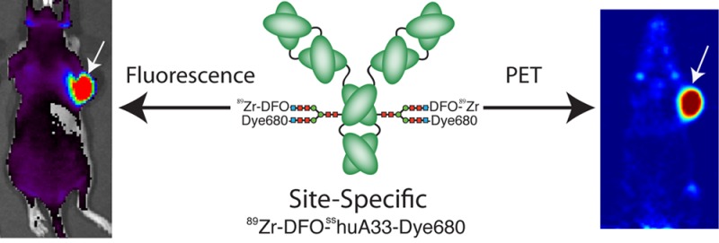
Abstract
The complementary nature of positron emission tomography (PET) and optical imaging (OI) has fueled increasing interest in the development of multimodal PET/OI probes that can be employed during the diagnosis, staging, and surgical treatment of cancer. Due to their high selectivity and affinity, antibodies have emerged as promising platforms for the development of hybrid PET/OI agents. However, the lack of specificity of many bioconjugation reactions can threaten immunoreactivity and lead to poorly defined constructs. To circumvent this issue, we have developed a chemoenzymatic strategy for the construction of multimodal PET/OI immunoconjugates that have been site-specifically labeled on the heavy chain glycans. The methodology consists of four steps: (1) the enzymatic removal of the terminal galactose residues on the heavy chain glycans; (2) the enzymatic incorporation of azide-bearing galactose (GalNAz) residues into the heavy chain glycans; (3) the strain-promoted click conjugation of chelator- and fluorophore-modified dibenzocyclooctynes to the azide-modified sugars; and (4) the radiolabeling of the immunoconjugate. For proof-of-concept, a model system was created using the colorectal cancer-targeting antibody huA33, the chelator desferrioxamine (DFO), the positron-emitting radiometal 89Zr, and the near-infrared fluorescent dye Alexa Fluor 680. The bioconjugation strategy is robust and reproducible, reliably producing well-defined and immunoreactive conjugates labeled with 89Zr, Alexa Fluor 680, or an easily and precisely tuned mixture of the two reporters. In in vivo PET and fluorescence imaging experiments, a hybrid 89Zr- and Alexa Fluor 680-labeled huA33 conjugate displayed high levels of specific uptake (>45% ID/g) in athymic nude mice bearing A33 antigen-expressing SW1222 colorectal cancer xenografts.
Introduction
Over the past 30 years, molecular imaging has transformed cancer care. The ability to noninvasively acquire anatomical and functional information about tumors has aided clinicians in all stages of cancer management, from diagnosis to staging to treatment.1−3 While the vast majority of imaging agents are specific to a single modality, recent years have played witness to a surge in the development of multimodal probes.4−7 In this regard, the complementary nature of positron emission tomography (PET) and optical imaging (OI) have made hybrid PET/OI probes particularly promising tools. While PET radiopharmaceuticals enable noninvasive whole body imaging and provide functional and anatomical information about lesions, near-infrared fluorescence (NIRF) optical imaging probes facilitate the high resolution imaging of tumor margins during surgical resection.3,5,7,8
Given their exquisite selectivity and affinity for their molecular targets, antibodies have emerged as particularly exciting platforms for the development of hybrid PET/OI agents. In the past few years, preclinical investigations employing PET/OI immunoconjugates targeting HER2, CD20, VEGF, and CD105 have emerged in the literature.9−15 Yet, despite their promise, one critical obstacle to the development of PET/OI immunoconjugates is the lack of site-specificity in the bioconjugation of radionuclides and fluorophores. At present, the vast majority of bioconjugation techniques rely on reactions between bifunctional probes and amino acids, most often lysines.12 Antibodies possess varying numbers of lysine residues, and thus controlling the precise molecular location of conjugation reactions is impossible. This lack of site-specificity can impair immunoreactivity if the conjugation reaction inadvertently occurs in or around the antigen binding domain. Furthermore, random bioconjugation strategies yield inadequately chemically defined constructs and can complicate the reliable reproduction of results from one antibody to the next. Not surprisingly, the potential complications of random bioconjugation are magnified in dual-labeled PET/OI constructs, as two types of reporter moieties are being appended to the antibody in two separate conjugation reactions.
In order to circumvent these issues, considerable effort has been dedicated to the development of strategies for the site-specific conjugation of payloads to antibodies.16−24 While promising, many of these methodologies are limited by their use of expensive and complex work flows and/or result in poor reproducibility of labeling between different antibodies. For example, one emergent group of strategies for the site-specific modification of antibodies—and the creation of antibody–drug conjugates in particular—has relied on the reaction between bifunctional, maleimide-bearing constructs and free cysteines in antibodies.19,25,26 These methodologies have largely proven successful. However, this sulfhydryl-based chemistry requires either the reduction of existing, natural disulfide bonds within the antibody or the introduction of free cysteine resides via genetic engineering, processes which require significant optimization and limit the general applicability of these strategies.
As a more universal alternative, we have developed a chemoenzymatic method for the site-specific radiolabeling of antibodies.27 This methodology leverages a promiscuous galactosyltransferase, an azide-modified substrate, and strain-promoted click chemistry to specifically modify the heavy chain glycans, the conserved, N-linked bianntennary oligosaccharide chains positioned on the CH2 domain of the Fc region, far from the antigen binding regions.28 The foundations of the approach lie in the glycoengineering work of Qasba, Hsieh-Wilson, and others, as well as the strain-promoted click chemistry work of Bertozzi and Boons.29−35 Admittedly, bioconjugation approaches based on the chemical manipulation of the heavy chain glycans predate our strategy.36,37 However, these earlier methods often require the prolonged exposure of the antibody to harsh redox conditions and have inherently high variability of labeling between different antibodies. It is also important to note that a number of descriptions of glycan-based bioconjugation strategies have appeared in the literature since our initial report; however, to the best of our knowledge, none of these have been applied to the development of multimodal probes.20−22,24
Here, we report the development of site-specifically labeled immunoconjugates for the multimodal PET/OI imaging of colorectal cancer. The bioconjugation strategy has four steps: (1) the enzymatic removal of the terminal galactose residues on the heavy chain glycans with β-1,4-galactosidase; (2) the enzymatic incorporation of azide-bearing galactose (GalNAz) residues into the glycans using a substrate-permissive galactosyltransferase, Gal-T(Y289L); (3) the strain-promoted click conjugation of chelator- and fluorophore-modified dibenzocyclooctynes (DIBO) to the azide-modified sugars; and (4) the radiolabeling of the immunoconjugate (Figure 1). For proof-of-concept, a model system was created using the colorectal cancer-targeting antibody huA33, the positron-emitting radiometal 89Zr, and the near-infrared fluorophore Alexa Fluor 680 (Dye680). The bioconjugation strategy proved robust, creating huA33 immunoconjugates with an easily tunable ratio of reporter moieties. Further, the methodology yielded a site-specifically labeled, hybrid PET/OI probe—89Zr-DFO-sshuA33-Dye680—with high immunoreactivity in vitro and high efficacy in vivo in a murine model of colorectal cancer. Ultimately, one of the greatest strengths of this strategy is its modularity, as the enzymatic tagging of the glycans with GalNAz can be performed with any IgG antibody, and dibenzocyclooctynes can be conjugated easily to a wide variety of reporter molecules.
Figure 1.
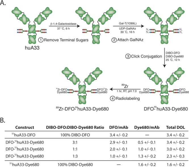
(A) Scheme of the site-specific methodology for the chemoenzymatic synthesis of multimodal imaging immunoconjugates. (B) Some properties of the site-specifically labeled huA33 immunoconjugates discussed in this work. DOL = degree of labeling [(DFO + Dye680)/mAb]. Alexa Fluor 680 is represented as Dye680 in the figure.
Results and Discussion
Design of the model system was the first step of the investigation. The huA33 antibody targets the A33 antigen, a transmembrane glycoprotein expressed on >95% of all colorectcal cancers, and has shown considerable promise in the clinic as a platform for radioimmunoconjugates.38 The ability to image colorectal cancer—particularly colorectal cancer metastatic to the liver—with PET during diagnosis and staging and with near-infrared fluorescence during surgical resection would certainly aid clinicians in the management of the disease. The positron-emitting radiometal 89Zr was chosen due to the advantageous match between the radiometal’s physical half-life (t1/2 ∼ 3.27 d) and the multiday biological half-life of the antibody.39 The selection of 89Zr in turn mandates the choice of its octandentate, siderophore-derived chelator desferrioxamine (DFO).40 Alexa Fluor 680 (Dye680) was chosen because of its near-infrared, tissue-penetrating 702 nm emission.
The site-specific modification process begins with the enzymatic manipulation of the heavy chain glycans. The antibody was first incubated with β-1,4-galactosidase at 37 °C for 6 h to expose the terminal GlcNAc sugars of the glycans. Subsequently, UDP-GalNAz and the promiscuous galactosyltrasnferase, Gal-T(Y289L), were added to the antibody solution, and the mixture was incubated at 30 °C for 16 h to append the azide-bearing galactose residues to the sugar chains. Since our initial report, these steps have been shortened from two 16 h incubations with an intermediate purification to a facile two enzyme, one pot reaction.27 Next, the DFO-, Alexa Fluor 680-, and dual-labeled conjugates were synthesized. While the singly labeled constructs were created by incubating the site-specifically labeled huA33-N3 construct (sshuA33-N3) with DIBO–DFO or DIBO–Alexa Fluor 680 (DIBO–Dye680), the dual labeled constructs were synthesized via incubation with 1:3, 1:1, or 3:1 mixtures of the two DIBOs. All of the immunoconjugates were purified using size exclusion chromatography, and a cumulative yield of 84.8 ± 8.4% was obtained over the course of the three step procedure (n = 20). Isotopic dilution experiments reveal that sshuA33-DFO had a degree of labeling of 3.4 ± 0.2 DFO/mAb, consistent with the near-quantitative labeling of four theoretically available terminal GlcNAc residues. In contrast, the degree of labeling of sshuA33-Dye680 was 1.6 ± 0.1 Dye680/mAb. Admittedly, this is below the typical degree of labeling previously observed with other chelators and dyes (∼3–3.5/mAb) and may be a result of the size and/or hydrophobicity of the fluorophore, though efforts to fully understand the cause are ongoing.27 For the hybrid probes, the ratio of conjugated DFO to Dye680 could be easily controlled by altering the initial reaction ratio of DIBO–DFO to DIBO–Dye680. For example, the degree of labeling of Dye680 increases from 0.5 ± 0.1 to 1.0 ± 0.1 to 1.3 ± 0.1 as the proportion of DIBO–Dye680 in the original reaction mixture is increased. Interestingly, the total degree of labeling ([DFO + Dye680]/mAb) decreases with increasing proportion of DIBO–Dye680 in the original reaction mixture, perhaps a result of the same phenomenon that leads to a lower degree of fluorescence labeling in the sshuA33-Dye680 construct.
SDS-PAGE electrophoresis experiments were run in order to characterize and confirm the site-specificity of the bioconjugation approach. In the resulting gels, a distinct increase in the molecular weight of the heavy chain of the DFO-, Dye680-, and dual-labeled sshuA33 constructs can be observed compared to unmodified huA33 and sshuA33-N3 (Figure 2). There is no corresponding shift in the molecular weight of the light chain, strongly suggesting site-specific labeling. In additional experiments, the immunoconjugates were treated with PNGaseF, an amidase that specifically cleaves between the innermost sugar of the glycans and the asparagine residues of the antibody (see Supporting Information). Upon PNGaseF treatment, a drop in the molecular weight of the heavy chain is apparent for all of the constructs, and, critically, the resulting bands are all shifted to the same molecular weight.
Figure 2.
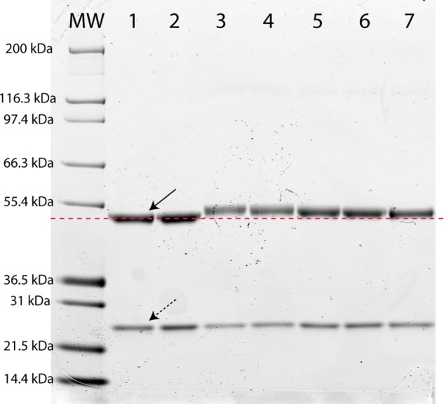
SDS-PAGE analysis of site-specifically labeled immunoconjugates. Lane 1: huA33. Lane 2: sshuA33-N3. Lane 3: sshuA33-DFO. Lanes 4, 5, and 6: DFO-sshuA33-Dye680 synthesized with 3:1, 1:1, and 1:3 mixtures of DIBO-DFO:DIBO-Dye680, respectively. Lane 7: sshuA33-Dye680. The solid and dotted arrows indicate the antibody heavy chain and light chain, respectively, and the red dotted line indicates the molecular weight of the unmodified heavy chain of huA33. MW = molecular weight ladder.
Ultimately, the DFO-sshuA33-Dye680 construct bearing 1.0 ± 0.1 Dye680/mAb and 2.0 ± 0.2 DFO/mAb was chosen for further in vitro and in vivo study. For the sake of comparison, a dual-labeled DFO-nshuA33-Dye680 construct bearing 1.0 ± 0.2 Dye680/mAb and 2.5 ± 0.4 DFO/mAb was also synthesized using a traditional, non-site-specific isothiocyanate-based coupling strategy (Figure 3A). Admittedly, the traditional bioconjugation method is more rapid (total time = 2 h) and results in slightly higher yields (89.9 ± 4.9%); however, the cumbersome optimization required to obtain a traditionally conjugated construct with degrees of labeling analogous to DFO-sshuA33-Dye680 undescores the robustness and reproducibility of the chemoenzymatic approach.
Figure 3.
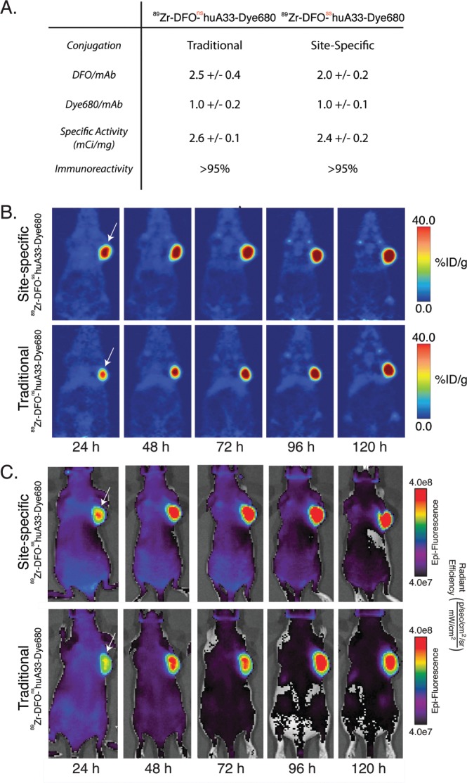
(A) Properties of site-specifically and traditionally labeled hybrid PET/OI immunoconjugates. PET (B) and fluorescence (C) images for 89Zr-DFO-sshuA33-Dye680 (top) and 89Zr-DFO-nshuA33-Dye680 (bottom) in mice bearing subcutaneous SW1222 xenografts. Mice were administered 89Zr-DFO-sshuA33-Dye680 or 89Zr-DFO-nshuA33-Dye680 [180–200 μCi (72–80 μg) in 200 μL 0.9% sterile saline] via lateral tail vein injection (t = 0) and imaged 24, 48, 72, 96, and 120 h postinjection. White arrows delineate the xenografts.
Both DFO-sshuA33-Dye680 and DFO-nshuA33-Dye680 (1000 μg) were subsequently labeled with 89Zr via incubation with 89Zr (3.0–3.2 mCi) in PBS buffer at pH 7.0–7.5 for 1 h at room temperature. After purification via size exclusion chromatography, both probes were isolated in >99% radiochemical purity. The specific activities of the site-specific and traditionally synthesized constructs were similar, 2.4 ± 0.2 mCi/mg and 2.6 ± 0.1 mCi/mg, respectively. Both DFO-sshuA33-Dye680 (0.95 ± 0.04) and DFO-nshuA33-Dye680 (0.93 ± 0.06) also displayed high immunoreactive fractions when assayed using A33 antigen-expressing SW1222 colorectal cancer cells. While a more significant increase in immunoreactivity might be expected for the site-specific methodology, huA33 is a well-optimized and robust antibody. We expect the immunoreactivity benefits of this approach to be more evident on immunoglobulins that are known to lose a significant amount of activity upon conjugation using standard conjugation chemistries. Finally, to assay the serum stability of the constructs, both 89Zr-DFO-sshuA33-Dye680 and 89Zr-DFO-nshuA33-Dye680 were incubated in human serum for 7 d at 37 °C. After 7 days, radioTLC analysis revealed both constructs to be >95% intact.
In order to investigate the in vivo behavior of the multimodal constructs, small animal PET and fluorescence imaging experiments were performed in a mouse model of colorectal cancer (Figure 3B,C). To this end, nude mice bearing subcutaneous A33 antigen-expressing SW1222 xenografts were intravenously injected with either 89Zr-DFO-sshuA33-Dye680 or 89Zr-DFO-nshuA33-Dye680 (180–200 μCi, 72–80 μg) and imaged with both PET and fluorescence at 24, 48, 72, 96, and 120 h postinjection. Both PET and fluorescence images clearly show that both immunoconjugates accumulate significantly in the tumors. At 24 h postinjection, uptake in the tumor is pronounced, but the constructs are also clearly visible—particularly via PET—in the blood and heart, reducing tumor-to-background contrast. Over the course of the experiment, however, the immunoconjugates clear from the blood and accumulate in the xenografts such that the tumors are prominently delineated from nontarget tissue with excellent tumor-to-background contrast. Complementary acute biodistribution experiments confirmed the imaging results (see Supporting Information). For 89Zr-DFO-sshuA33-Dye680, tumoral uptake values of 45.9 ± 7.9%ID/g and 45.7 ± 8.1%ID/g were observed at 48 and 120 h, respectively, resulting in tumor-to-muscle activity ratios of 30.1 ± 8.2 and 33.5 ± 15.5 at the two time-points. Critically, in the same biodistribution experiment, a blocking experiment in which an excess of 300 μg unlabeled DFO-sshuA33-Dye680 was co-injected with the radioimmunoconjugate reduced tumoral uptake at 48 h from 45.7 ± 13.1%ID/g to 15.7 ± 4.1%ID/g, demonstrating antigen specificity. Finally, ex vivo fluorescence microscopy and autoradiography were performed on xenografts excised from mice injected with 89Zr-DFO-sshuA33-Dye680. The fluorescence and radioactivity are clearly colocalized in the same regions of the tumor, the expected results for a dual labeled imaging agent. Taken together, this investigation clearly demonstrates that the in vitro and in vivo behavior of 89Zr-DFO-sshuA33-Dye680 is comparable to that of the traditionally constructed agent.
Conclusion
We have developed a methodology for the construction of site-specifically labeled multimodal PET/OI immunoconjugates. The synthetic strategy has proven to be robust and reliable, and we have used it to create a hybrid 89Zr-DFO- and Alexa Fluor 680-labeled probe shown to be as effective in vivo in a murine model of colorectal cancer as an analogous construct synthesized using traditional non-site-specific methods. Importantly, the ratio of chelator to fluorophore can be easily tuned according to the needs of the experiment. If high specific activity is a priority, the ratio of chelator-to-fluorophore can be increased. Alternatively, if fluorescent brightness is the goal, the relative number of fluorophores can be increased. Perhaps even more critically, the overall strategy is completely modular, as the chemoenzymatic GalNAz conjugation can be employed with any antibody, and myriad reporters can be appended to dibenzocyclooctynes. Ultimately, we believe that this methodology could have a transformational impact on the way multimodal immunoconjugates are developed.
Acknowledgments
Services provided by the MSKCC Small-Animal Imaging Core Facility were supported in part by NIH Grants R24 CA83084 and P30 CA08748. The authors also thank the NIH (Award 1K99CA178205-01A1), the DoD (Award W81XWH-12-1-0029, B.M.Z.), Mr. William H. Goodwin and Mrs. Alice Goodwin and the Commonwealth Foundation for Cancer Research, and the Experimental Therapeutics Center of Memorial Sloan Kettering Cancer Center for their generous funding.
Glossary
Abbreviations
- DIBO
dibenzocyclooctyne
- UDP
uridine-5′-diphosphate
- GalNAz
N-azidozacetylgalactosamine
- DFO
desferrioxamine
- Dye680
Alexa Fluor 680
- ss
site-specific
- ns
non-site-specific
Supporting Information Available
Reagents and general procedures; detailed experimental methods on chemical syntheses, bioconjugation procedures, in vitro, and in vivo experiments; diagram of the strain-promoted azide-cyclooctyne click reaction; synthesis of DIBO-Alexa Fluor 680; SDS-PAGE analysis of PNGase-F treated immunoconjugates; tables of biodistribution data, both absolute uptake values and tumor-to-tissue activity ratios. This material is available free of charge via the Internet at http://pubs.acs.org.
The authors declare no competing financial interest.
Funding Statement
National Institutes of Health, United States
Supplementary Material
References
- Wu A. M.; Olafsen T. (2008) Antibodies for molecular imaging of cancer. Cancer J. 14, 191–197. [DOI] [PubMed] [Google Scholar]
- Kircher M. F.; Hricak H.; Larson S. M. (2012) Molecular imaging for personalized cancer care. Mol. Oncol. 6, 182–195. [DOI] [PMC free article] [PubMed] [Google Scholar]
- Michalski M. H.; Chen X. (2011) Molecular imaging in cancer treatment. Eur. J. Nucl. Med. Mol. Imag. 38, 358–377. [DOI] [PMC free article] [PubMed] [Google Scholar]
- Cherry S. R. (2009) Multimodality imaging: beyond PET/CT and SPECT/CT. Sem. Nucl. Med. 39, 348–353. [DOI] [PMC free article] [PubMed] [Google Scholar]
- Louie A. Y. (2010) Multimodality imaging probes: Design and challenges. Chem. Rev. 110, 3146–3195. [DOI] [PMC free article] [PubMed] [Google Scholar]
- Seibold U.; Wangler B.; Schirrmacher R.; Wangler C. (2014) Bimodal imaging probes for combined PET and OI: Recent developments and future directions for hybrid agent development. Biomed. Res. Int. 1–13. [DOI] [PMC free article] [PubMed] [Google Scholar]
- te Velde E. A.; Veerman T.; Subramaniam V.; Ruers T. (2010) The use of fluorescent dyes and probes in surgical oncology. Eur. J. Surg. Oncol. 36, 6–15. [DOI] [PubMed] [Google Scholar]
- Frangioni J. V. (2003) In vivo near-infrared fluorescence imaging. Curr. Opin. Chem. Biol. 7, 626–634. [DOI] [PubMed] [Google Scholar]
- Xu H.; Baidoo K.; Gunn A. J.; Boswell C. A.; Milenic D. E.; Choyke P. L.; Brechbiel M. W. (2007) Design, synthesis, and characterization of a dual modality positron emission tomography and fluorescence Imaging agent for monoclonal antibody tumor-targeted imaging. J. Med. Chem. 50, 4759–4765. [DOI] [PMC free article] [PubMed] [Google Scholar]
- Paudyal P.; Paudyal B.; Iida Y.; Oriuchi N.; Hanaoka H.; Tominaga H.; Ishikita T.; Yoshioka H.; Higuchi T.; Endo K. (2009) Dual functional molecular imaging probe targeting CD20 with PET and optical imaging. Oncol. Rep. 22, 115–119. [DOI] [PubMed] [Google Scholar]
- Sampath L.; Kwon S.; Ke S.; Wang W.; Schiff R.; Mawad M. E.; Sevick-Muraca E. M. (2007) Dual-labeled trastuzumab-based imaging agent for the detection of human epidermal growth factor receptor 2 overexpression in breast cancer. J. Nucl. Med. 48, 1501–1510. [DOI] [PubMed] [Google Scholar]
- Cohen R.; Vugts D. J.; Stigter-van Walsum M.; Visser G. W. M.; van Dongen G. (2013) Inert coupling of IRDye800CW and zirconium-89 to monoclonal antibodies for single- or dual-mode fluorescence and PET imaging. Nat. Protoc. 8, 1010–1018. [DOI] [PubMed] [Google Scholar]
- Hong H.; Zhang Y.; Severin G. W.; Yang Y. N.; Engle J. W.; Niu G.; Nickles R. J.; Chen X. Y.; Leigh B. R.; Barnhart T. E.; Cai W. B. (2012) Multimodality imaging of breast cancer experimental lung metastasis with bioluminescence and a monoclonal antibody dual-labeled with Zr-89 and IRDye 800CW. Mol. Pharmaceutics 9, 2339–2349. [DOI] [PMC free article] [PubMed] [Google Scholar]
- Zhang Y.; Hong H.; Severin G. W.; Engle J. W.; Yang Y. A.; Goel S.; Nathanson A. J.; Liu G.; Nickles R. J.; Leigh B. R.; Barnhart T. E.; Cai W. B. (2005) ImmunoPET and near-infrared fluorescence imaging of CD105 expression using a monoclonal antibody dual-labeled with Zr-89 and IRDye 800CW. Am. J. Transl. Res. 4, 333–346. [PMC free article] [PubMed] [Google Scholar]
- Sampath L.; Kwon S.; Hall M. A.; Price R. E.; Sevick-Muraca E. M. (2010) Detection of cancer metastases with a dual-labeled near-infrared/positron emission tomography imaging agent. Trans. Oncol. 3, 307–U71. [DOI] [PMC free article] [PubMed] [Google Scholar]
- Hussain A. F.; Kampmeier F.; von Felbert V.; Merk H. F.; Tur M. K.; Barth S. (2011) SNAP-Tag technology mediates site-specific conjugation of antibody fragments with a photosensitizer and improves target specific phototoxicity in tumor cells. Bioconjugate Chem. 22, 2487–2495. [DOI] [PubMed] [Google Scholar]
- Jeong J. M.; Lee J.; Paik C. H.; Kim D. K.; Lee D. S.; Chung J. K.; Lee M. C. (2004) Site-specific 99mTc-labeling of antibody using dihydraziniophthalazine (DHZ) conjugation to Fc region of heavy chain. Arch. Pharm. Res. 27, 961–967. [DOI] [PubMed] [Google Scholar]
- Kampmeier F.; Ribbert M.; Nachreiner T.; Dembski S.; Beaufils F.; Brecht A.; Barth S. (2009) Site-specific, covalent labeling of recombinant antibody fragments via fusion to an engineered version of 6-O-alkylguanine DNA alkyltransferase. Bioconjugate Chem. 20, 1010–1015. [DOI] [PubMed] [Google Scholar]
- Tinianow J. N.; Gill H. S.; Ogasawara A.; Flores J. E.; Vanderbilt A. N.; Luis E.; Vandlen R.; Darwish M.; Junutula J. R.; Williams S. P.; Marik J. (2010) Site-specifically Zr-89-labeled monoclonal antibodies for ImmunoPET. Nucl. Med. Biol. 37, 289–297. [DOI] [PubMed] [Google Scholar]
- Dennler P.; Chiotellis A.; Fischer E.; Bregeon D.; Belmant C.; Gauthier L.; Lhospice F.; Romagne R.; Schibli R. (2014) Transglutaminase-based chemo-enzymatic conjugation approach yields homogeneous antibody–drug conjugates. Bioconjugate Chem. 25, 569–578. [DOI] [PubMed] [Google Scholar]
- Zhou Q.; Stefano J. E.; Manning C.; Kyazike J.; Chen B.; Gianolio D. A.; Park A.; Busch M.; Bird J.; Zheng X.; Simonds-Mannes H.; Kim J.; Gregory R. C.; Miller R. J.; Brondyk W. H.; Dhal P. K.; Pan C. Q. (2014) Site-specific antibody-drug conjugation through glycoengineering. Bioconjugate Chem. 25, 510–520. [DOI] [PubMed] [Google Scholar]
- Okeley N. M.; Toki B. E.; Zhang X.; Jeffrey S. C.; Burke P. J.; Alley S. C.; Senter P. D. (2013) Metabolic engineering of monoclonal antibody carbohydrates for antibody-drug conjugation. Bioconjugate Chem. 24, 1650–1655. [DOI] [PubMed] [Google Scholar]
- Boswell C. A.; Marik J.; Elowson M. J.; Reyes N. A.; Ulufatu S.; Bumbaca D.; Yip V.; Mundo E. E.; Majidy N.; Van Hoy M.; Goriparthi S. N.; Trias A.; Gill H. S.; Williams S. P.; Junutula J. R.; Fielder P. J.; Khawli L. A. (2013) Enhanced tumor retention of a radiohalogen label for site-specific modification of antibodies. J. Med. Chem. 56, 9418–9426. [DOI] [PubMed] [Google Scholar]
- Li Z.; Fang T.; Boons G. J. (2014) Preparation of well-defined antibody–drug conjugates through glycan remodeling and strain-promoted azide–alkyne cycloadditions. Angew. Chem., Int. Ed. 53, 7179–7182. [DOI] [PMC free article] [PubMed] [Google Scholar]
- Massa S.; Xavier C.; De Vos J.; Caveliers V.; Lahoutte T.; Muyldermans S.; Devoogdt N. (2014) Site-specific labeling of cysteine-tagged camelid single-domain antibody-fragments for use in molecular imaging. Bioconjugate Chem. 25, 979–988. [DOI] [PubMed] [Google Scholar]
- Pillow T. H.; Tien J.; Parsons-Reponte K. L.; Bhakta S.; Li H.; Staben L. R.; Li G.; Chuh J.; Fourie-O’Donohue A.; Darwish M.; Yip V.; Liu L.; Leipold D. D.; Su D.; Wu E.; Spencer S. D.; Shen B.-Q.; Xu K.; Kozak K. R.; Raab H.; Vandlen R.; Phillips G. D. L.; Scheller R. H.; Polakis P.; Sliwkowski M. X.; Flygare J. A.; Junutula J. R. (2014) Site-specific trastuzumab maytansinoid antibody-drug conjugates with improved therapeutic activity through linker and antibody engineering. J. Med. Chem. 57, 7890–7899. [DOI] [PubMed] [Google Scholar]
- Zeglis B. M.; Davis C. B.; Aggeler R.; Kang H. C.; Chen A.; Agnew B. J.; Lewis J. S. (2013) An enzyme-mediated methodology for the site-specific radiolabeling of antibodies based on catalyst-free click chemistry. Bioconjugate Chem. 24, 1057–1067. [DOI] [PMC free article] [PubMed] [Google Scholar]
- Raju T. S.; Briggs J. B.; Borge S. M.; Jones A. J. (2000) Species-specific variation in glycosylation of IgG: evidence for the species-specific sialylation and branch-specific galactosylation and importance for engineering recombinant glycoprotein therapeutics. Glycobiology 10, 477–486. [DOI] [PubMed] [Google Scholar]
- Ramakrishnan B.; Boeggeman E.; Qasba P. K. (2007) Novel method for in vitro O-glycosylation of proteins: Application for bioconjugation. Bioconjugate Chem. 18, 1912–1918. [DOI] [PubMed] [Google Scholar]
- Ramakrishnan B.; Qasba P. K. (2002) Structure-based design of beta 1,4-galactosyltransferase I (beta 4Gal-T1) with equally efficient N-acetylgalactosaminyltransferase activity: point mutation broadens beta 4Gal-T1 donor specificity. J. Biol. Chem. 7, 20833–20839. [DOI] [PubMed] [Google Scholar]
- Clark P. M.; Dweck J. F.; Mason D. E.; Hart C. R.; Buck S. B.; Peters E. C.; Agnew B. J.; Hsieh-Wilson L. C. (2008) Direct in-gel fluorescence detection and cellular imaging of O-GlcNAc-modified proteins. J. Am. Chem. Soc. 130, 11576–11582. [DOI] [PMC free article] [PubMed] [Google Scholar]
- Khidekel N.; Arndt S.; Lamarre-Vincent N.; Lippert A.; Poulin-Kerstien K. G.; Ramakrishnan B.; Qasba P. K.; Hsieh-Wilson L. C. (2003) A chemoenzymatic approach toward the rapid and sensitive detection of O-GlcNAc posttranslational modifications. J. Am. Chem. Soc. 125, 16162–16163. [DOI] [PubMed] [Google Scholar]
- Sletten E. M.; Bertozzi C. R. (2009) Bioorthogonal chemistry: fishing for selectivity in a sea of functionality. Angew. Chem., Int. Ed. 48, 6973–6998. [DOI] [PMC free article] [PubMed] [Google Scholar]
- Agard N. J.; Bertozzi C. R. (2009) Chemical approaches to perturb, profile, and perceive glycans. Acc. Chem. Res. 42, 788–797. [DOI] [PMC free article] [PubMed] [Google Scholar]
- Ning X.; Guo J.; Wolfert M. A.; Boons G. J. (2008) Visualizing metabolically labeled glycoconjugates of living cells by copper-free and fast huisgen cycloadditions. Angew. Chem., Int. Ed. 47, 2253–2255. [DOI] [PMC free article] [PubMed] [Google Scholar]
- Wolfe C. A.; Hage D. S. (1995) Studies on the rate and control of antibody oxidation by periodate. Anal. Biochem. 231, 123–130. [DOI] [PubMed] [Google Scholar]
- Clamp J. R.; Hough L. (1966) Some observations on the periodate oxidation of amino compounds. Biochem. J. 101, 120–126. [DOI] [PMC free article] [PubMed] [Google Scholar]
- Carrasquillo J. A.; Pandi-Taskar N.; O’Donoghue J. A.; Humm J. L.; Zanzonico P.; Smith-Jones P. M.; Divgi C. R.; Pryma D. A.; Ruan S.; Kemeny N. E.; Fong Y.; Wong D.; Jaggi J. S.; Scheinberg D. A.; Gonen M.; Panageas K. S.; Ritter G.; Jungbluth A. A.; Old L. J.; Larson S. M. (2011) 124I-huA33 antibody PET of colorectal cancer. J. Nucl. Med. 52, 1173–1180. [DOI] [PMC free article] [PubMed] [Google Scholar]
- Deri M. A.; Zeglis B. M.; Francesconi L. C.; Lewis J. S. (2013) PET imaging with 89Zr: From radiochemistry to the clinic. Nucl. Med. Biol. 40, 3–14. [DOI] [PMC free article] [PubMed] [Google Scholar]
- Zeglis B. M.; Houghton J. L.; Evans M. J.; Viola-Villegas N. T.; Lewis J. S. (2014) Underscoring the influence of inorganic chemistry on nuclear imaging with radiometals. Inorg. Chem. 53, 1890–1899. [DOI] [PMC free article] [PubMed] [Google Scholar]
Associated Data
This section collects any data citations, data availability statements, or supplementary materials included in this article.