
Figure 3.
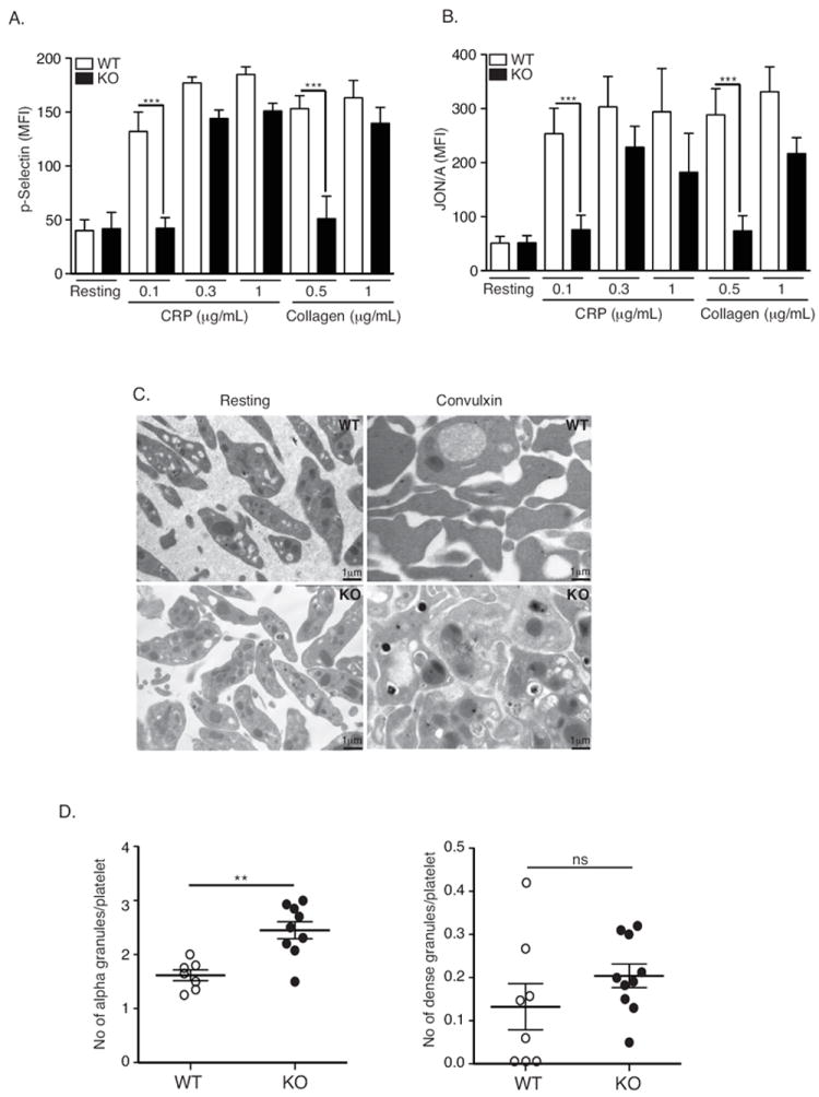
Impaired GPVI-mediated platelet activation in DUSP3-deficient platelets. (A-B) Washed platelets from WT or Dusp3-KO mice were stimulated with 0.1, 0.3, or 1 μg/mL CRP or 0.5 or 1 μg/mL collagen under non-stirring conditions, or left untreated. Surface expression of P-selectin and active integrin αIIbβ3 (JON/A) was quantified by flow cytometry. Mean fluorescence intensity histograms for P-selectin (A) and JON/A (B) are shown. Data were analyzed using ANOVA and the Bonferroni multiple comparison test and represent mean ± SEM of four independent experiments performed on platelet pools from three mice each; *p< 0.05, **p< 0.01. (C) Electron microscopy analysis of WT and Dusp3-KO washed platelets. Platelet ultrastructure was visualized in resting state or upon CVX stimulation (100 ng/mL). (D) Scatter plots of alpha and dense granules counted on five separated micrographs. Data were analyzed using unpaired Student t-test and represent mean ± SEM of three independent experiments performed on platelet pools from three mice; *p< 0.05, **p< 0.01.