



Summary
Background
Enucleation in young children often results in retarded orbital growth ipsilaterally. The need for an implant that will naturally grow with the child, like Dermis-fat Graft (DFG), for managing the anophthalmia has been of interest over the years.
Objective
To evaluate the use of DFG as an implant for volume replacement post-enucleation.
Study Design
A prospective non-comparative case series involving 18 consecutive children who had DFG either primarily or secondarily in conjunction with enucleation for intraocular pathologies, from December 2007 to September 2012, at the ophthalmology unit, Korle-Bu. Data from patients who had a minimum of three months follow up(FUP) were analysed.
Outcome measures
Complete covering of DFG with healthy conjunctiva, increase in volume of DFG, and presence or absence of complications.
Results
Fifteen patients were analysed, aged nine months to ten years (mean (SD), 3.7±2.7years). Eight (53.3%) were females. Thirteen (86.7%) DFGs were secondary and 2(13.3%) primary. Indications for enucleation were intraocular retinoblastoma (n=10, 66.7%), unexplained retinal detachment mimicking retinoblastoma (n=3,20.0%), anterior staphyloma (n=1,6.7%) and medulloepithelioma (n=1,6.7%). Fourteen (93.3%) patients showed increase in volume of DFG. Time for Conjunctival reepithelialisation of the dermal surface was four to fourteen weeks (mean/median=5.5/4.0). Complications encountered were infection (n=1,6.7%), infection with necrosis (n=1,6.7%), melanosis /keratinization (n=2, 13.3%) and cysts(n=2,13.3%). The patients were followed up for 3 to 54 months (mean/median 20.13 /16.00).
Conclusion
DFG for management of post-enucleation anophthalmia in Ghanaian children showed 93.3% success.
Keywords: Dermis- fat graft, enucleation, volume replacement, anophthalmia
Introduction
Enucleation in young children often results in retarded orbital growth ipsilaterally due to soft tissue volume loss in the anophthalmic socket.1–3 Management of anophthalmia consisted of fixed-diameter sphere within the muscle cone plus prosthetic shell which is often changed to keep up with the growing child.4
The need for an implant that will naturally grow with the child, like autogenous Dermis-fat Grafts (DFG), has been of interest over the years.5,6 A preliminary study in Ghanaian children which looked at the use of DFG for management of anophthalmia post enucleation had demonstrated initial success of 91%.7 This study is aimed at evaluating the use of DFG as an orbital implant for volume replacement post-enucleation in Ghanaian children and the associated complications over a five year period.
Methods
This was a prospective non-comparative case series involving 18 consecutive children who had DFG done at the Ophthalmology Unit of the Korle-Bu Teaching Hospital, Accra, Ghana, from December 2007 to December 2012. The study conformed to the Declaration of Helsinki and all parents gave consent for participation.
DFG was done either primarily or secondarily in conjunction with enucleation for intraocular tumours. Indication for enucleation was presence of intraocular tumour clinically diagnosed and confirmed by Computerised Tomography Scan (CT scan). Patients with pre- enucleation diagnosis of retinoblastoma had histopathological confirmation of no extra ocular extension before secondary DFG was performed.
Surgical Procedure
Enucleation was performed first, under general anaesthesia and the dimensions of the width and length of the socket were measured with callipers.
Harvesting of DFG
Donor site i.e. upper outer quadrant of the left gluteal region was prepared aseptically and an oval shaped margin marked with predetermined measurements from the eye socket after enucleation, making provision for an extra 2 millimetres for anticipated shrinkage when tissue is transplanted. The junction between the epidermis and dermis was infiltrated locally with normal saline mixed with Adrenalin 1 in 10000 injections.
The epidermis over the marked out site was shaved off with No. 22 surgical blade. The underlying dermis with fat was then excised using No. 11 blade, elliptically shaped to fit the orbital socket (18 –20mm in longest diameter, 14-16 mm in width and about 4–6mm in depth) and placed in saline solution. The wound at the donor site was then closed with interrupted stitches using 2/0 Vicryl suture and dressed with betadine ointment.
DFG implantation
The DFG was partially defatted with scissors and implanted into the posterior tenon's space. Conjunctiva was then sutured to the edge of DFG separated from anterior tenon's with 6/0 Vicryl suture in an interrupted fashion. The eye was padded with a well lubricated gauze (Chlorhexidine gauze with antibiotic ointment) to fit gently the rest of the socket for forty-eight hours in place of conformers which were not available in the country.
Post-operative management
Post operatively; the eyes were treated with combined antibiotic/corticosteroid ointment q.i.d for a month. Ocular prosthesis (thin shelled) was fitted when complete conjunctiva coverage was achieved, usually between 4 to 8 weeks post-operatively. Increase in volume or growth of the DFG and facial symmetry were measured by serial photographs post-operatively at 1 month then 3 monthly in year 1, 6 monthly in years 2 and 3, and yearly thereafter
Outcome measures
Outcome measures studied included a complete covering of DFG with healthy conjunctiva, an increase in volume of the DFG, good prosthetic fit with good facial symmetry and presence or absence of complications.
Data Analysis
Data from patients who had a minimum of 3 months follow up (FUP) were included in the analysis. Summary of results of the study were presented as proportions and percentages of outcomes for categorical variables. Continuous data were summarized as means, standard deviation and median values.
Results
Fifteen patients were analysed, aged 9 months to 10 years (mean (SD), 3.7±2.7years). Eight (53.3%) were females. Thirteen (86.7%) DFGs were secondary and 2(13.3%) primary. Indications for enucleation were intraocular retinoblastoma (n=10, 66.6%), unexplained retinal detachment mimicking retinoblastoma (n=3, 20.0%), anterior staphyloma (n=1, 6.7%) and medulloepithelioma (n=1, 6.7%).
There was increase in volume of DFG in 14 (93.3%) patients (Figure 1a). Good prosthetic fitting with good facial symmetry were achieved in the 14(93.3%) children who showed growth in the DFG.(Figure 1b).
Figure 1a.

Growth of secondary dermis-fat Graft - 3 years post-operatively.
Figure 1b.

Growth of secondary dermis-fat Graft with prosthesis - 3 years post-operatively.
Time for Conjunctival re-epithelialization of the dermal surface was 4-14 weeks, mean=5.5, median=4.0. Complications encountered were infection (n=1,6.7%), infection with necrosis (n=1, 6.7%), melanosis /keratinization(n=2,13.3%) and cysts(n=2,13.3%) (Figure 2).
Figure 2.
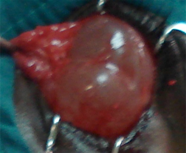
Macrocyst in dermis fat graft seen 5 months post-operatively.
Microscopy, culture and sensitivity tests done from wound swabs, showed negative results for the infection with necrosis but Staph. epidermidis was isolated in one case.
However, complete resolution of the infection with or without necrosis was achieved with antibiotic therapy, a combination of Guttae Ciprofloxacin 0.3% and Oc. Tetracycline.
The patient with infection and necrosis demonstrated no increase in volume of DFG and was lost to FUP after 13 weeks.
The microcyst is being monitored for progression, but the macrocyst (Figure 2) was treated by excision with residual mild ptosis. The patients were followed up for 3 to 54 months, mean 20.13±16.13, median and 16.00.
Discussion
Autologous dermis fat graft (DFG), composed of dermis and an attached subcutaneous fat, is an acceptable volume replacement implant for primary enucleation in children7–12 The dermal component, in orbit reconstruction provides structural support for the ingrowth of conjunctiva over the graft and its eventual vascularization. This minimizes reabsorption of graft fat with resultant replacement of lost orbital volume.9
It also preserves conjunctival surface area and deepens conjunctival fornix depth to enhance prosthesis fitting.9 Being autologous, it has neither the risk of rejection nor transfer of infection from cadaveric homologous tissue.10,12 However, DFG may be associated with disadvantages such as lack of predictability in determining the adequate volume required of the harvested graft, sometimes resulting in underestimation.10
In this series, 14 (93.3%) patients had good maintenance of orbital volume and good prosthetic fit with good facial symmetry, as evidenced by serial photographs. These findings corroborates findings from previous study in same population7 and other studies.11,13 Although some studies had longer follow up periods11,14, and were larger in series14 than our study, our finding is significant because of the paucity of data from the West African sub-region including Ghana. As part of the monitoring of patients in this study into adulthood, the prosthetic shell will be changed when indicated to keep up with the growing child
Complications encountered are similar to known complications of DFG as a volume replacement procedure for anophthalmia reported from other studies12, 13, 15 complete resolution of the infection and necrosis was achieved with antibiotic therapy, but the patient with the infection and necrosis demonstrated no increase in volume of DFG.
The complication of melanosis/ keratinization seen in two patients may be as a result of poor dissection of graft leaving epithelial islands, 12these two patients being part of the first three patients operated at the beginning of this series and therefore representing a learning curve. Cysts are recognised complications of DFG, and may be of epithelial origin from epithelial islands left on DFG from poor harvesting.15 Macrocysts, as occurred in one of our patients, can be treated with excision with good results.15
Our patient had residual mild ptosis post-excision. Other complications described in literature include graft overgrowth requiring re-operation or debulking11 and secondary revision of prosthesis.16 Some of these complications are sometimes seen years after implantation.16 None of these complications were encountered probably because of the relatively shorter follow-up period and smaller numbers studied.
The graft failure rate of 7% in this study compares with that by Lee MJ et al.13 The patient in this study who had graft failure had antecedent graft infection with necrosis of the graft. Probably from subsequent atrophy but this could not be confirmed because patient was lost to follow up after 13 weeks post-operatively. Friction and mechanical irritation between prosthesis and anterior covering tissue of DFG has been implicated as cause of failure in other studies.13
Significant atrophy of primary grafts does not occur very frequently, but may account for graft failure in some patients years following an apparently successful primary graft.14 The two patients who had primary DFG with enucleation; one for medulloepithelioma and the other for anterior staphyloma, the former with the longest follow up period of fifty-four months post operatively, all had good successes in growth of the DFG with no complications. In view of possible long term complications such as graft atrophy and graft failure, the patients in this study will need long term follow up into adulthood.
Strengths and limitations
The main strength of this study is that, as follow up to the maiden use of DFG as an implant for orbital volume replacement post- enucleation in Ghanaian children in same population, there is still a high success rate of graft growth.
However, the use of serial photographs for demonstrating graft growth rather than neuroimaging studies (such as MRI or CT Scan), the limited number of patients studied as well as the short follow up period were the possible limitations of this study.
Recommendation
There is the need for a larger series with long term follow up and a randomized controlled study to compare the use of DFG with synthetic implants for better outcome for volume replacement in Ghanaian children with anophthalmia.
Conclusion
Ninety-three percent of the dermis fat graft implanted post-enucleation in our paediatric patients showed success in volume increase, good prosthetic fitting and good facial symmetry. Minor complications were encountered except a macrocyst and graft necrosis.
Acknowledgement
We acknowledge the contributions of Mr. Benjamin Abaidoo the senior research assistant and nurses of the paediatric eye centre of the Ophthalmology Unit, Korle-Bu Teaching Hospital, Ghana. We also acknowledge Dr George Obeng Adjei of the University of Ghana Medical School, for reading through the manuscript.
References
- 1.Kennedy R. Growth retardation and volume determinations of the anophthalmic socket. Am J Ophthalmol. 1973;76:294–302. doi: 10.1016/0002-9394(73)90177-3. [DOI] [PubMed] [Google Scholar]
- 2.Apt L, Isenberg S. Changes in orbital dimensions following enucleation. Arch Ophthalmol. 1973;90(5):393–395. doi: 10.1001/archopht.1973.01000050395013. [DOI] [PubMed] [Google Scholar]
- 3.Osborne D, Hadden OB, Deeming LW. Orbital growth after childhood enucleation. Am J Ophthalmol. 1974;77(5):756–759. doi: 10.1016/0002-9394(74)90545-5. [DOI] [PubMed] [Google Scholar]
- 4.Price E, Simon JW, Calhoun JH. Prosthetic treatment of severe microphthalmos in infancy. J Pediatr Ophthalmol Strabismus. 1986;23(1):22–24. doi: 10.3928/0191-3913-19860101-06. [DOI] [PubMed] [Google Scholar]
- 5.Abel A D, Meyer DR. Enucleation with primary implant insertion for treatment of recalcitrant endophthalmitis and panophthalmitis. Ophthal Plast Reconstr Surg. 2005;21(3):220–226. doi: 10.1097/01.iop.0000159174.80985.e2. [DOI] [PubMed] [Google Scholar]
- 6.Bosniak SL, Nesi F, Smith BC, Schechter B, Cote R. A comparison of motility: autogenous dermis-fat vs synthetic spherical implants. Ophthalmic Surg. 1989;20(12):889–891. [PubMed] [Google Scholar]
- 7.Essuman VA, Tagoe N N, Ndanu T A, Ntim-Amponsah C T. Dermis-Fat Grafts and Enucleation in Children in a West African Population. Plast Reconstr Surg. 2013;131(3):457e–458e. doi: 10.1097/PRS.0b013e31827c7365. [DOI] [PubMed] [Google Scholar]
- 8.Sami D, Young S, Petersen R. Perspective on Orbital Enucleation Implants. Surv Ophthalmol. 2007;52(3):244–265. doi: 10.1016/j.survophthal.2007.02.007. [DOI] [PubMed] [Google Scholar]
- 9.Martin P, Rogers P, Billson F. Dermis-fat: evolution of a living prosthesis. Aust and N Z J Ophthalmol. 1986;14(2):161–165. doi: 10.1111/j.1442-9071.1986.tb00028.x. [DOI] [PubMed] [Google Scholar]
- 10.Al-Mujaini Abdullah, Ganesh Anuradha, Al-Zuhaibi Sana. Autogenous Dermis-Fat Orbital Implant for Anophthalmic Socket. Sultan Qaboos Univ Med J. 2007;7(2):145–148. [PMC free article] [PubMed] [Google Scholar]
- 11.Mitchell KT, Hollsten DA, White WL, O'Hara MA. The Autogenous Dermis-Fat Orbital Implant in Children. J AAPOS. 2001;5(6):367–369. doi: 10.1067/mpa.2001.118870. [DOI] [PubMed] [Google Scholar]
- 12.Reza V M, McMullan TFW, Burroughs JR, Isaacs DK, Tsirbas A, White GL, Jr, Anderson RL, McCann JD. Autologous dermis graft at the time of evisceration or enucleation. Br J Ophthalmol. 2007;91(11):1528–1531. doi: 10.1136/bjo.2007.115543. [DOI] [PMC free article] [PubMed] [Google Scholar]
- 13.Lee MJ, Khwarg SI, Choung HK, Kim NJ, Yu YS. Dermis-fat graft for treatment of exposed porous polyethylene implants in pediatric postenucleation retinoblastoma patients. Am J Ophthalmol. 2011;152(2):244–250. doi: 10.1016/j.ajo.2011.01.048. [DOI] [PubMed] [Google Scholar]
- 14.Smith B, Bosniak S, Nesi F, Lisman R. Dermis-fat orbital implantation: 118 cases. Orbit. 2010;29(4):183–189. [PubMed] [Google Scholar]
- 15.Patrinely JR, Anderson RL, Shore JW. Macrocystic enlargement of orbital dermis-fat grafts. Ophthalmic Surg. 1987;18(7):498–502. [PubMed] [Google Scholar]
- 16.Nunery WR, Hetzler KJ. Dermal-fat graft as a primary enucleation technique. Opthalmology. 1985;92(9):1256–1261. doi: 10.1016/s0161-6420(85)33882-4. [DOI] [PubMed] [Google Scholar]