

Abstract
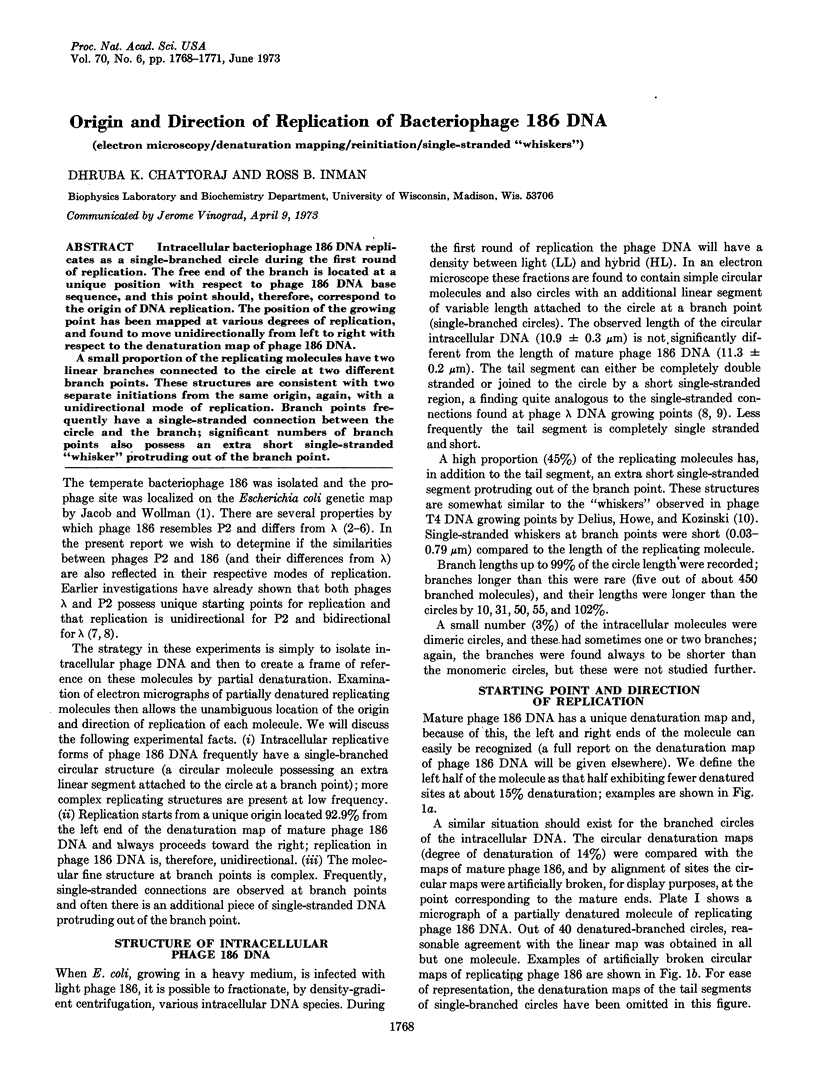
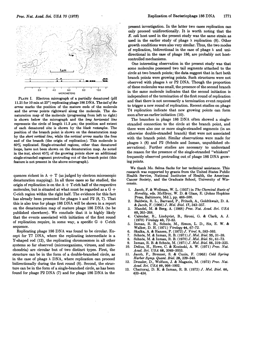
Intracellular bacteriophage 186 DNA replicates as a single-branched circle during the first round of replication. The free end of the branch is located at a unique position with respect to phage 186 DNA base sequence, and this point should, therefore, correspond to the origin of DNA replication. The position of the growing point has been mapped at various degrees of replication, and found to move unidirectionally from left to right with respect to the denaturation map of phage 186 DNA.
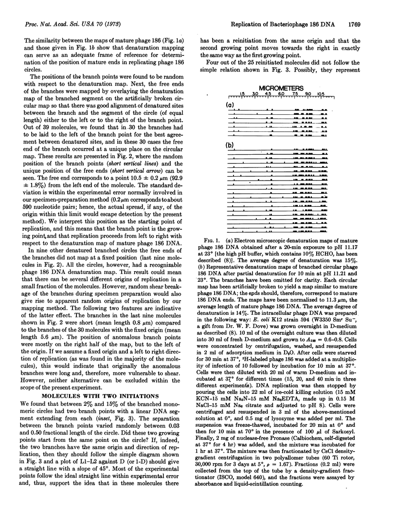
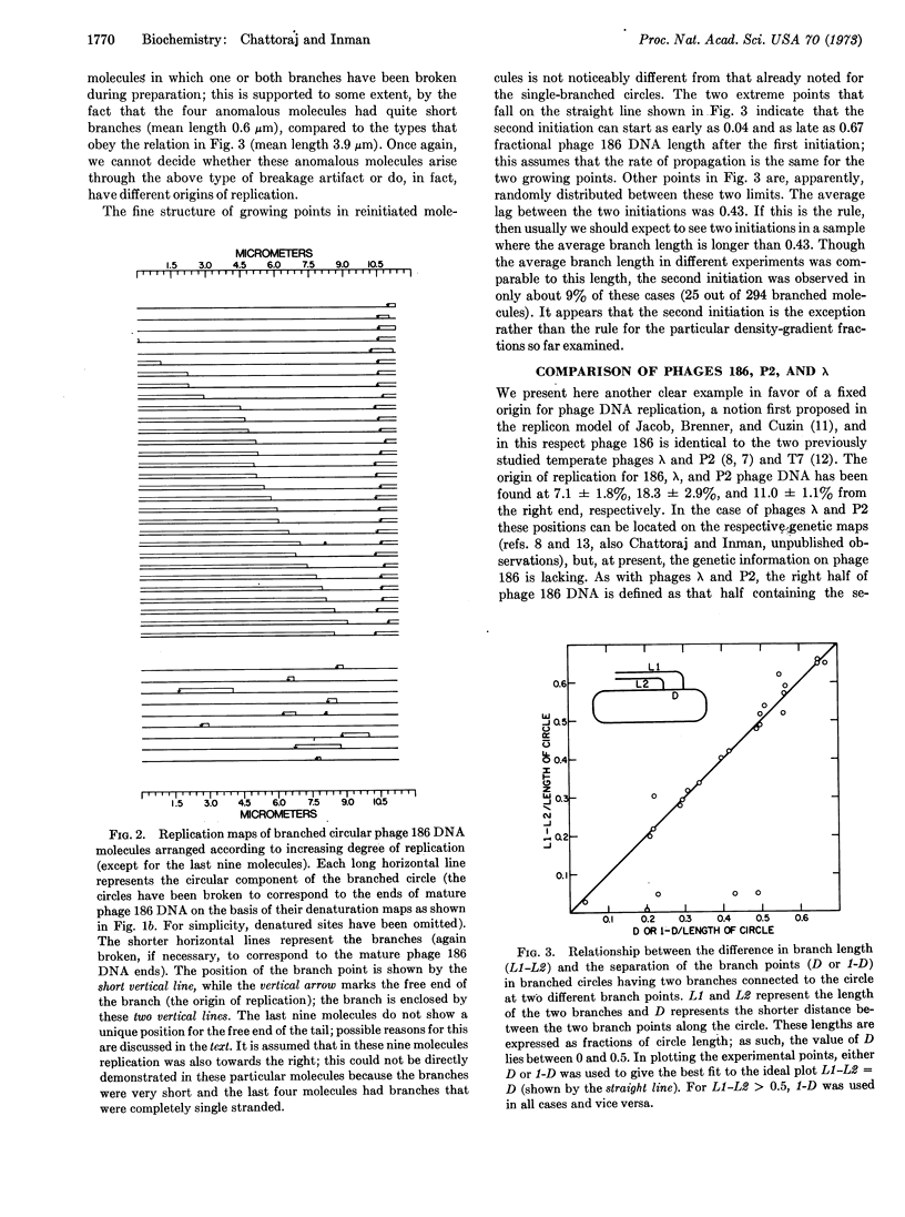
A small proportion of the replicating molecules have two linear branches connected to the circle at two different branch points. These structures are consistent with two separate initiations from the same origin, again, with a unidirectional mode of replication. Branch points frequently have a single-stranded connection between the circle and the branch; significant numbers of branch points also possess an extra short single-stranded “whisker” protruding out of the branch point.
Keywords: electron microscopy, denaturation mapping, reinitiation, single-stranded “whiskers”
Full text
PDF
Images in this article
Selected References
These references are in PubMed. This may not be the complete list of references from this article.
- Baldwin R. L., Barrand P., Fritsch A., Goldthwait D. A., Jacob F. Cohesive sites on the deoxyribonucleic acids from several temperate coliphages. J Mol Biol. 1966 Jun;17(2):343–357. doi: 10.1016/s0022-2836(66)80146-8. [DOI] [PubMed] [Google Scholar]
- Calendar R., Lindqvist B., Sironi G., Clark A. J. Characterization of REP- mutants and their interaction with P2 phage. Virology. 1970 Jan;40(1):72–83. doi: 10.1016/0042-6822(70)90380-6. [DOI] [PubMed] [Google Scholar]
- Chattoraj D. K., Inman R. B. Position of two deletion mutations on the physical map of bacteriophage P2. J Mol Biol. 1972 May 28;66(3):423–434. doi: 10.1016/0022-2836(72)90424-x. [DOI] [PubMed] [Google Scholar]
- Delius H., Howe C., Kozinski A. W. Structure of the replicating DNA from bacteriophage T4. Proc Natl Acad Sci U S A. 1971 Dec;68(12):3049–3053. doi: 10.1073/pnas.68.12.3049. [DOI] [PMC free article] [PubMed] [Google Scholar]
- Dressler D., Wolfson J., Magazin M. Initiation and reinitiation of DNA synthesis during replication of bacteriophage T7. Proc Natl Acad Sci U S A. 1972 Apr;69(4):998–1002. doi: 10.1073/pnas.69.4.998. [DOI] [PMC free article] [PubMed] [Google Scholar]
- Inman R. B., Schnös M., Simon L. D., Six E. W., Walker D. H., Jr Some morphological properties of P4 bacteriophage and P4 DNA. Virology. 1971 Apr;44(1):67–72. doi: 10.1016/0042-6822(71)90153-x. [DOI] [PubMed] [Google Scholar]
- Inman R. B., Schnös M. Structure of branch points in replicating DNA: presence of single-stranded connections in lambda DNA branch points. J Mol Biol. 1971 Mar 14;56(2):319–325. doi: 10.1016/0022-2836(71)90467-0. [DOI] [PubMed] [Google Scholar]
- Mandel M., Berg A. Cohesive sites and helper phage function of P2, lambda, and 186 DNA's. Proc Natl Acad Sci U S A. 1968 May;60(1):265–268. doi: 10.1073/pnas.60.1.265. [DOI] [PMC free article] [PubMed] [Google Scholar]
- Schnös M., Inman R. B. Position of branch points in replicating lambda DNA. J Mol Biol. 1970 Jul 14;51(1):61–73. doi: 10.1016/0022-2836(70)90270-6. [DOI] [PubMed] [Google Scholar]
- Skalka S. A., Hanson P. Comparisons of the distribution of nucleotides and common sequences in deoxyribonucleic acid from selected bacteriophages. J Virol. 1972 Apr;9(4):583–593. doi: 10.1128/jvi.9.4.583-593.1972. [DOI] [PMC free article] [PubMed] [Google Scholar]