
Figure 3. Tkv-containing cytonemes transport Dpp.
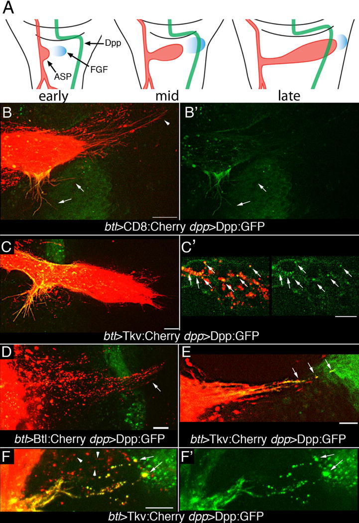
(A) Drawings of three 3rd instar stages depict growth and development of the ASP (red) relative to wing disc cells expressing Dpp (green) and FGF (blue). (B, B′) Expression of CD8:Cherry in the ASP and Dpp:GFP in the dpp domain of the disc (btl-Gal4 UAS-CD8:Cherry dpp-LHG/lexO-Dpp:GFP) marks the ASP and ASP cytonemes (red) and dpp-expressing disc cells (green). GFP fluorescence is in lateral ASP cytonemes (arrows) and in lower medial region of ASP, but not in tip ASP cytonemes (arrowhead). Left panel (merge), right panel (GFP). (C, C′) Expression of Tkv:Cherry in the ASP and Dpp:GFP in the in the dpp domain of the disc (btl-Gal4/UAS-Tkv:Cherry; dpp-LHG/lexO-Dpp:GFP) marks the ASP and lateral ASP cytonemes (red), but few tip cytonemes; lateral Tkv-containing ASP cytonemes and the medial region of the ASP have received Dpp:GFP (green) (C). Dpp:GFP and Tkv:Cherry colocalize in puncta in ASP cells (arrows, C′). (D) FGFR:Cherry expressed in ASP and Dpp:GFP in the dpp domain of the disc (btl-Gal4/UAS-Btl:Cherry dpp-LHG/lexO-Dpp:GFP) marks puncta in ASP tip cytonemes (arrow) that project beyond Dpp-expressing disc cells (green); no localization of FGFR:Cherry with Dpp:GFP was apparent in tip cytonemes. (E, F) Only cytonemes marked with Tkv:Cherry that appear to contact Dpp:GFP expressing disc cells (btl-Gal4 UAS-Tkv:Cherry; dpp-LHG/lexO-Dpp:GFP) have GFP fluorescence in puncta and at their tips (arrows). Cytonemes that do not appear to make contact do not have GFP fluorescence at their tips or in their Tkv-containing puncta (F, arrowheads) lack GFP fluorescence. (F) merge; (F′) Dpp:GFP. Animals were raised at 18°C to minimize transgene expression and were incubated at 22–25°C for 12–16 hrs prior to analysis. Scale bars, 10 μm.