

Highlights
-
•
This study investigated the aesthetic of the prosthesis rehabilitation.
-
•
Patient satisfaction after rehabilitation.
-
•
Importance of radiographic control case report.
Keywords: Dental implant, Esthetics, Mouth rehabilitation
Abstract
Introduction
The aim of this paper was to present a rehabilitation of a patient with a dynamic universal castable long abutment (UCLA) for a single tilted implant in the anterior maxillary area.
Presentation of case
A 57-year-old male patient attended the dentistry college clinic complaining of a vertical fracture of a residual root of the dental element 22. The tooth extraction was indicated for the implant installation. Due to the socket buccal wall thickness, the implant was installed with an inclination to the palate. It was done in a two-stage surgical protocol, and an external hexagon implant (3.75 × 11.5 mm) was placed. After a six-month healing period to correct the implant position, a dynamic UCLA was set in place, rectifying the implant emergence profile at 20°. The ceramic structure fitting was performed and, after the patient’s consent, the prosthesis was finalized and installed.
Discussion
After a follow-up period of twenty months, no complications were observed.
Conclusion
The installation of tilted implants with a dynamic UCLA may be a viable option, faster and less invasive than bone grafts.
1. Introduction
The osseointegrated implants are widely researched and safely applied in modern dentistry [1]. The main goal of this technique is to increase the stability of prosthesis during the masticatory function, improving the quality of life of the patient [2].
The upper jaw region possesses a trabecular bone (type 3 and 4) and some critical areas such as the nasal cavity and maxillary sinus, which could complicate the installation of parallel implants with adequate height [1,3–5]. Additionally, the anterior region has a thin bone cortical from both buccal and palatal sides [6].
When this occurs, tilted dental implants may be installed to guarantee proper retention, as well as the preservation of important anatomical structures [1,2,7]. The aim of the tilting is to improve the implant positioning, so that it is placed in the area that presents the greatest amount of bone [5,8], since more contact with trabecular bone allows a better implant anchorage and permits the utilization of longer implants [2,4,9,10]. Also, it is a simpler and less invasive option than bone grafting surgeries [1,3,4,7].
The dynamic universal castable long abutment (UCLA) is an alternative option of abutment used to rectify the implant tilting. That UCLA allows the correction of the implant emergence profile up to 20°, turning it to a favorable position [11].
Even though single tilted implants are frequently used at dental practice, few cases report their installation on anterior regions describing the whole process from the surgical procedure until the crown sets in [12]. The aim of this paper was to present a rehabilitation of a patient with a dynamic universal castable long abutment (UCLA) for a single tilted implant in the anterior maxillary area.
2. Case presentation
A 57-year-old patient came to the Department of Dental Material and Prosthodontics of the Aracatuba Dental School, complaining of a fracture of the dental element 22 (Fig. 1). This element was previously restored with a single partial fixed prosthesis with a metallic core. The patient presented good general health. The clinical exam indicated a lack of gingival smile, a class IV fracture at the incisal portion of the 11 and a vertical root fracture of the 22. Due to this fracture, tooth extraction was indicated. The patient assigned an informed consent for proposed oral rehabilitation.
Fig. 1.
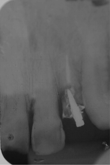
Initial radiograph.
The treatment plan established was to replace the fractured tooth with a single implant. After the extraction, it was verified that the socket buccal wall thickness was unsatisfactory, so the implant was installed with an inclination of 20° to the palate (Fig. 2). The surgical procedure was a two-stage protocol, and three months after the extraction, a dental implant (external hexagon 3.75 mm diameter and 11.5 mm length; Osteofit, Campo Largo, Paraná, Brazil) was placed and a 40 N torque was applied for primary stability. The top of the implant was at crestal bone level. A provisional adhesive prosthesis was manufactured prior to the surgery and installed after the implant placement. Two weeks after the surgery, the sutures were removed.
Fig. 2.

Comparison of the implant inclination to the dental arch by using a direction indicator.
After a six-month healing period, osseointegration was assessed through a radiographic analysis. No radiolucent line was observed around the implant. Then, the implant site was reopened and the healing cap was placed (Fig. 3). After the tissue remodeling, the squared transfer was positioned and had its setting to the implant confirmed through a radiograph. Then an open tray impression was made with addition silicon Express (3M ESPE, Sumaré, São Paulo, Brazil), alongside with the impression of the opposed arch.
Fig. 3.

Initial clinical aspect with the healing cap in position.
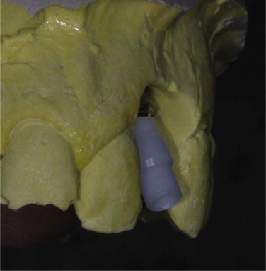
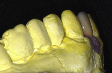
The depth and thickness of the gingival tissue was evaluated to confirm that the abutment metal collar would not be visible. The depth of the gingival sulcus was 2 mm. A chrome–cobalt 20° UCLA was screwed to the implant (Mangran Internacional, Curitiba, Parana, Brazil) to correct its tilting (Figs. 4 and 5).
Fig. 4.
UCLA at 0°, before the correction of implant inclination.
Fig. 5.

UCLA at 20° correcting the implant inclination on the stone cast.
After the metal casting (Figs. 6 and 7), the setting of the coping was assessed through radiographs so the ceramic cover could be applied (Fig. 8). On the next session, a prosthesis try-in was performed, the occlusal and proximal contacts were evaluated with a carbon paper (Accufilm II, Parkell, New York, USA) and, with the patient’s informed consent, the prosthesis was finalized.
Fig. 6.
Healthy gum margin around metal casting.
Fig. 7.
Metal casting of the prosthesis.
Fig. 8.
Prosthesis aspect after the ceramics application.
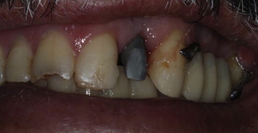
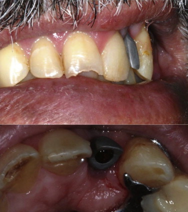
At the installation moment, the dental contacts were once again verified, as was the radiographic aspect (Fig. 9), a 20 N torque was applied to the abutment screw and, in a different session, the screw access hole was covered with a resin composite. The patient was pleased with the final clinical aspect (Figs. 10 and 11).
Fig. 9.
Radiographic image after the prosthesis installation.
Fig. 10.
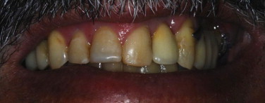
Frontal view of the clinical aspect after the case finalization.
Fig. 11.
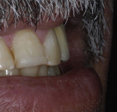
Right buccal view of the clinical aspect after the case finalization.
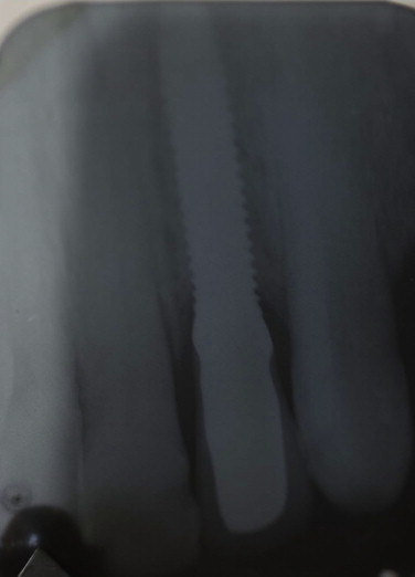
After a twenty-month follow-up (Fig. 12), complications such as loosening or fracture of the prosthetic screw and implant fracture were not observed [13]. Additionally, no bone loss was observed in the peri-implant bone area. The patient is pleased with the rehabilitation.
Fig. 12.

Radiographic image after 20-month follow up.
The 20° UCLA used in this case presents as an advantage the possibility of correction of the implant tilting in a screwed prosthesis whether a cemented one, in fewer clinical steps. Additionally, as the prosthesis is manufactured with the UCLA, only one screw is necessary in the assembly, between the abutment and the implant. However, there is a contraindication in cases of excessive load, especially in patients with parafunctional habits.
3. Discussion
Some clinical reports presented acceptable success rates with the installation of tilted implants. These implants minimize the need of bone grafting surgeries decreasing the procedure length and complexity thus increasing the amount of patients willing to undertake rehabilitations with such procedure [7].
Tilted implants are mostly used in posterior regions, in cases of atrophic maxillae where the proximity to the sinus may restrict the amount of bone available to the anchorage of dental implants [9]. However, some authors also report successful installation of tilted implants in anterior regions with no compromise of an aesthetic result [12].
There is no evidence that contraindicates the placement of tilted implants, no significant difference was noticed when evaluating the bone loss around axial and tilted implants [10,14,15]. In addition, the complications reported are very similar to the conventional implants [10]. Some of the complications reported were mucositis, sinus infection and periimplantitis [6,10]. In this clinical report no bone loss or complications were observed during the 20-month follow up period.
Bateli et al. [7] highlighted the importance of a tridimensional exam for the correct placement of angled implants. The authors affirmed that the inclination of dental implants prevent bone grafting procedures and should be used as often as possible.
The dynamic UCLA is a viable solution for cases in which the aesthetics is compromised during a prosthetic rehabilitation. The dynamic UCLA avoid possible discomfort that other systems may have, such as excessive buccal volume of the prosthesis infrastructure, depending on the planning and positioning of the implant. The dynamic UCLA handling is basically laboratorial. By means of an indexer, the dentist and dental technician select the best positioning and tilting of the system, to project a prosthesis that is both functional and aesthetic. This is possible because of the total freedom that the dynamic UCLA provides, with its characteristics of rotating on its own axis by 360° and tilting from 0° to 28° [11].
The clinical effect is: the dynamic UCLA provided movement freedom, pinpoint accuracy to the desired angle, gain in aesthetics and function, treatment agility and cost reduction.
4. Conclusions
The use of a dynamic UCLA to correct tilted implants is a viable treatment option for patients with atrophic maxillae, and may be faster and less invasive than bone grafts.
-
-
The dynamic UCLA allows dentists to rectify up to 20° of the implant emergence profile, which ensure an aesthetic result.
-
-
This treatment can result in high levels of patient satisfaction, with reestablishment of function and aesthetics and longevity of prosthetic rehabilitation.
Acknowledgment
None to declare.
References
- 1.Zampelis A., Rangert B., Heijl L. Tilting of splinted implants for improved prosthodontic support: a two-dimensional finite element analysis. J. Prosthet. Dent. 2007;97(Suppl. 6):S35–43. doi: 10.1016/S0022-3913(07)60006-7. [DOI] [PubMed] [Google Scholar]
- 2.De Vico G., Bonino M., Spinelli D., Schiavetti R., Sannino G., Pozzi A., Ottria L. Rationale for tilted implants: FEA considerations and clinical reports. Oral Implantol. 2011;4(3–4):23–33. [PMC free article] [PubMed] [Google Scholar]
- 3.Almeida E.O., Rocha E.P., Freitas Júnior A.C., Anchieta R.B., Poveda R., Gupta N., Coelho P.G. Tilted and short implants supporting fixed prosthesis in an atrophic maxilla: a 3D-FEA biomechanical evaluation. Clin. Implant Dent. Relat. Res. 2013:1–11. doi: 10.1111/cid.12129. [DOI] [PubMed] [Google Scholar]
- 4.Peñarrocha M., Carrillo C., Boronat A., Peñarrocha M. Maximum use of the anterior maxillary buttress in severe maxillary atrophy with tilted, palatally positioned implants: a preliminary study. Int. J. Oral Maxillofac. Implants. 2010;25(4):813–820. [PubMed] [Google Scholar]
- 5.Bevilacqua M., Tealdo T., Menini M., Pera F., Mossolov A., Drago C., Pera P. The influence of cantilever length and implant inclination on stress distribution in maxillary implant-supported fixed dentures. J. Prosthet. Dent. 2010;105(5–13):5–13. doi: 10.1016/S0022-3913(10)60182-5. [DOI] [PubMed] [Google Scholar]
- 6.Testori T., Mandelli F., Mantovani M., Taschieri S., Weinstein R.L., Del Fabbro M. Tilted trans-sinus implants for the treatment of maxillary atrophy: case series of 35 consecutive patients. J. Oral Maxillofac. Surg. 2013;71(7):1187–1194. doi: 10.1016/j.joms.2013.02.013. [DOI] [PubMed] [Google Scholar]
- 7.Bateli M., Woerner W., Att W. Tilted implants to support a maxillary removable dental prosthesis: a case report. Quintessence Int. 2012;43(3):191–195. [PubMed] [Google Scholar]
- 8.Pesqueira A.A., Goiato M.C., dos Santos D.M., Nobrega A.S., Haddad M.F., Andreotti A.M., Moreno A. Stress analysis in oral obturator prostheses over parallel and tilted implants: photoelastic imaging. J. Biomed. Opt. 2013;18(10):106009. doi: 10.1117/1.JBO.18.10.106009. [DOI] [PubMed] [Google Scholar]
- 9.Peñarrocha-Oltra D., Candel-Martí E., Ata-Ali J., Peñarrocha-Diago M. Rehabilitation of the atrophic maxilla with tilted implants: review of the literature. J. Oral Implantol. 2013;39(5):625–632. doi: 10.1563/AAID-JOI-D-11-00068. [DOI] [PubMed] [Google Scholar]
- 10.Pozzi A., Sannino G., Barlattani A. Minimally invasive treatment of the atrophic posterior maxilla: a proof-of-concept prospective study with a follow-up of between 36 and 54 months. J. Prosthet. Dent. 2012;108(5):286–297. doi: 10.1016/S0022-3913(12)60178-4. [DOI] [PubMed] [Google Scholar]
- 11.Mangran Internacional [Internet] 2014 [updated 2014 Aug 14, cited 2014 Aug 14]. Available from: www.mangraninternacional.com/index.php/ucla2.
- 12.Butler B.L., Suzuki C. Esthetic replacement of a maxillary central incisor with an ITI 15-degree angled implant: a case report. Int. J. Periodontics Restorative Dent. 1999;19(6):609–614. [PubMed] [Google Scholar]
- 13.Goiato M.C., Haddad M.F., Gennari Filho H., Villa L.M.R., dos Santos D.M., Pesqueira A.A. Dental implant fractures – aetiology, treatment and case report. J. Clin. Diagn. Res. 2014;8(3):300–304. doi: 10.7860/JCDR/2014/8074.4158. [DOI] [PMC free article] [PubMed] [Google Scholar]
- 14.Calandriello R., Tomatis M. Simplified treatment of the atrophic posterior maxilla via immediate/early function and tilted implants: a prospective 1-year clinical study. Clin. Implant Dent. Relat. Res. 2005;7(Suppl. 1):S1–S12. doi: 10.1111/j.1708-8208.2005.tb00069.x. [DOI] [PubMed] [Google Scholar]
- 15.Agliardi E.L., Francetti L., Romeo D., Del Fabbro M. Immediate rehabilitation of the edentulous maxilla: preliminary results of a single-cohort prospective study. Int. J. Oral Maxillofac. Implants. 2009;24(5):887–895. [PubMed] [Google Scholar]