Abstract
The year 2014 marked the 350th anniversary of the publication in London of Cerebri anatome, a ground-breaking work of neuroscience heavily influenced by the political and cultural context of Baroque Europe and mid-17th century England. This article aims to review the work of the English physician and anatomist Thomas Willis, specifically with regard to the contents of his Cerebri anatome. Willis's academic and professional career was influenced by the turbulent period of the English Civil War during which he studied medicine. Willis went from chemistry to dissection arguably because of his need to justify the body-brain-soul relationship. As a result, he became a fellow of a select club of eminent experimentalists, and afterward was a Fellow of the Royal Society. Later on, he went to London, leaving the academic life to dedicate himself fully to the profession of medicine. As a physician, Willis did not base his practice on aphorisms but on a ‘bench to bedside’ approach to medicine, while studying neuroanatomy – covering embryology, comparative anatomy and pathological anatomy – as a basis for the comprehension of neurological pathology. He developed innovative anatomical methods for the preservation and dissection of the brain, injection of coloured substances and illustration of his findings. In Cerebri anatome, Willis recognized the cerebral cortex as the substrate of cognition. He also claimed that the painful stimuli came from the meninges, but not from the brain itself. He explained for the first time the pathological and functional meaning of the brain's circular arterial anastomosis, which is named after him. He also specified some features of the cranial origin of the sympathetic nerves and coined the term ‘neurologie’. Cerebri anatome marked the transition between the mediaeval and modern notions of brain function, and thus it is considered a cornerstone of clinical and comparative anatomy of the nervous system. The new contributions and methods employed by Willis justify his place as a father of neurology and a pioneer of translational research.
Keywords: accessory nerve, circle of Willis, rete mirabile, 17th century anatomists, 17th century neuroscience, sympathetic nervous system, translational research
Introduction
In medical science, the eponym Willis refers to diverse anatomical structures, including the cerebral arterial circle (Dorland's Dictionary, 2005). The year 2014 marked the 350th anniversary of the publication in London of Thomas Willis's outstanding work: Cerebri anatome. Heavily influenced by the political and cultural context of Baroque Europe – and mid-17th century England in particular – this book was the result of what we now call translational research (Hörig et al. 2005) and marked the beginning of modern neurology and laid the foundations for future research in the fields of clinical and comparative anatomy of the nervous system.
Historical context
During the 17th century, the Baroque was an artistic movement that spread to the cultural and scientific world. This period was characterized by strong oppositions and disputes, not only religious (Protestants against Catholics) but also political (Parliamentarians against Absolutists) and epistemological (Experimental against Rationalistic methods).
The science of anatomy, the main source of medical research at that time, incorporated major technological innovations such as the microscope and the intravascular injection of substances as well as the experimental method, with which William Harvey proved his theory of blood circulation published in Motus cordis (1628). The interest in dynamics boosted the study of angiology and neuroanatomy, among others. In parallel with the geopolitical changes of that period, England, The Netherlands, France and Denmark displaced Italy as the major research powers in this field (Laín Entralgo, 1978).
Physiological processes were explained by two theories: as a balance among specific bodily fluids (Iatrochemistry) or through the laws of mechanics (Iatromechanics). Catholicism kept following Galen's theories or tried renewing them through Iatromechanics, whereas Protestantism shifted towards iatrochemistry, with Van Helmont and Franciscus de le Böe (Sylvius) being the major exponents of this theory in continental Europe. So was Thomas Willis in England; despite the fact that he was not a strong follower of Sylvius, he also wanted to merge the iatrochemical thinking with other areas of medical knowledge.
The apocalyptic vision of an imminent end of the world encouraged the study of god in the natural world to obtain a deeper knowledge of the creator. This fact accelerated the development of natural philosophy, deeply linked to religious beliefs. The nature of the soul was debated intensely during this period. Mediaeval and Renaissance physicians attempted to understand the brain by mixing Christian theology and Galen philosophy (Finger, 1994). The body was considered to be divided into three anatomical regions, each one designed to contain its own soul or spirit. The liver housed the ‘vegetative soul’ or ‘natural spirit’, which was responsible for desires and appetites. The heart housed the ‘soul’ or ‘vital spirit’, which fed passions and actions. The ‘rational soul’ or ‘animal spirit’ was immaterial and immortal, thus it could not live in any specific part of the body. Veins distributed the ‘natural spirit’ and arteries distributed the ‘vital spirit’ without any connection between the two systems. The ‘vital spirit’ reached the brain through the ‘great soporal’ (or carotid) arteries, which, having entered the cranial cavity, split into a ‘wonderful network’ of innumerable branches around the pituitary. This network was also called ‘rete mirabile’, the place where ‘vital’ spirits turned into ‘animal’ spirits (Eadie, 2003a).
On the other hand, reason, memory and imagination were carried out by invisible spirits located in different cerebral ventricles as described in the Doctrine of Ventricle Localisation of Mental Functioning by Nemesius, Bishop of Emesa, in the 4th century. This work explained that all sensory perceptions detected by the nerves – seen as channels – were sent to the two anterior ventricles (now, the lateral ventricles) so that fantasy and imagination could flow from this area. Later, this area was called ‘common sense’ (‘sensus communis’).
The middle ventricle (now the third ventricle) corresponded to the region of the intellectual abilities, responsible for approving, refuting or checking sensory perceptions collected by the anterior ventricles. Reason was placed in this area. Memory was located at the posterior ventricle (now the fourth ventricle) and stored all sensory perceptions after their evaluation by the intellectual abilities. Between the anterior and the middle ventricles there was a small lock that, when blocked, stopped all thinking skills.
During the 16th century, Nemesius's doctrine was being threatened by the works of Berengario da Carpi, Vesalius and Varolio. However, the doctrine was still being applied until the late 18th century. In relation to the role of the brain at this time, Henry More's words speak volumes: ‘a bowl of curds – hardly the sort of material that could handle the lofty work of the soul’ (Zimmer, 2004). In 1664, Willis published in London his outstanding work Cerebri anatome, considered a cornerstone of the history of neurology.
Life and work
Soon after the deaths of Shakespeare and Queen Elizabeth, while England was still basking in the artistic and cultural bloom of the Elizabethan era, Thomas Willis (Fig.1) was born on 27 January 1621 in Great Bedwyn, Wiltshire, UK, about 70 miles northwest of London (Symonds & Feindel, 1969). His father, having served several nobles before, settled as a farmer in a village near Oxford. Willis went to the School of Edward Sylvester and was accepted at the University of Oxford on 3 March 1637. There, he first studied at Christ Church College under the auspices of Dr Thomas Isles and later moved to All Saints College. Willis obtained a Bachelor of Arts (chemistry) on 12 June 1639 and a Master of Arts on 18 June 1642, the same year his father Thomas, bailiff of Sir Walter Smith at Bedwyn, lost his life defending the king at the siege of Oxford when he was ‘snatched away by the contagion of a camp-fever’ (Lo & Ellis, 2010).
Fig 1.
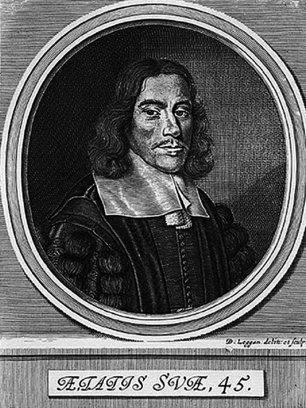
Portrait of Thomas Willis from the Title page of Pathologiae cerebri (1667).
Willis originally had a clerical vocation that led him to learn Latin, the language of both religious authority and political power. Although he disliked the inflexibility of theology, his great command of Latin was very useful in his later medical writings. He had to work as a servitor (a student from the lowest social class) for Dr Thomas Isles and Mrs Isles, who were the canon of Christ Church College and an unofficial medical practitioner, respectively. Willis's interest in Medicine awoke while helping Mrs Isles to make medical preparations (Rengachary et al. 2008) and so he was introduced into the growing art and the science of alchemy (Lo & Ellis, 2010).
Medical education in Oxford was characterized as traditional and low-quality. For 14 years, students listened and studied repeatedly outdated works by Aristotle, Hippocrates and Galen (Sinclair & Robb-Smith, 1950). In 1636, shortly before Willis started to study at Oxford, the University statutes were revised and dissection became part of the medical training. King Charles I granted the Tomlins Reader in Anatomy claim to the body of anyone who was executed within 21 miles around Oxford. Medical students had to participate in two public dissections, in addition to their anatomical lectures and other dissections, to obtain the Bachelor of Medicine degree (Molnár, 2004).
During the English Civil War (1642–51), Royalists severely disrupted academic life in Oxford. Willis started his medical studies in 1642. In November of the same year, King Charles I settled in Christ Church College in Oxford accompanied by the Royal Physician William Harvey. Willis learnt about the major developments that were then taking place in medicine from classes he attended under Dr Harvey. Willis, a Royalist, joined the auxiliary regiment of the Earl of Dover against the Parliamentarians. On 8 December 1646, his loyalty was rewarded with a Bachelor of Medicine after only 6 months of study; thus he could start his medical practice before the Parliamentarian forces took Oxford. The Protectorate prohibited Anglican worship but Willis, a pious man, began holding religious assemblies at his home, Beam Hall in Merton Street. Meanwhile, at the University of Oxford, the traditional elements of teaching were replaced by new trends that made Oxford the centre of current scientific knowledge, encouraged by Puritan thinking and inspired by the work of Francis Bacon a generation earlier.
In opposition to the Puritan influence, some members of the Invisible College of London built the Oxford Experimental Philosophy Club – a precursor of the Royal Society of London – which Willis became a Fellow of in 1649. Here Willis joined celebrities such as Robert Boyle, father of chemistry; William Petty, philosopher, physician, entrepreneur and economist; Christopher Wren, Savilian Professor of Astronomy at Oxford and architect of the Sheldonian Theatre, All Souls College and St Paul's Cathedral; Thomas Sydenham, known as the English Hippocrates; Robert Hooke, physicist and microscopist; and Thomas Millington, physician, Sedleian Professor of Natural Philosophy at Oxford, president of the Royal College of Physicians of London between 1696 and 1703, and Willis's close friend. They often gathered to discuss scientific topics or to carry out experiments which made a huge contribution to the development of independent translational research in Oxford. Aristotelian Natural Philosophy was replaced by the Atomism of Pierre Gassendi, whose guidelines had a great influence on Willis's work together with the iatrochemical concepts coming from Walter Charleton – Royal Physician of the Stuarts and main supporter of Jan Baptist van Helmont, and Théodore Turquet de Mayerne – responsible for the admission of the chemical medicines in the pharmacopoeia of the Royal College of Physicians of London.
Paradoxically, Willis's lack of a traditional medical education had a positive effect on him, as he became involved in experimental science, so despised by the authorities of the time. He gained a lot of medical experience by visiting markets of villages near Oxford. For instance, in Abingdon he performed a uroscopy (Hughes, 1991) in which he made genuine observations, such as that flies preferably flew over urine samples with a higher sugar content. The semiological value of this act is evident today, as two types of diabetes are still distinguished: diabetes mellitus (sugar diabetes) and diabetes insipidus (water diabetes) (Molnár, 2004). Willis also studied a range of miscellaneous conditions hitherto unexplained. Willis regarded asthma as a nervous condition. He proposed explanations for the basis of typhoid fever, whooping cough, meningitis and narcolepsy. In keeping with his belief that the brain was the centre of behaviour, and indeed the personality, Willis concluded that hysteria had a cerebral origin; prior to this discovery, physicians thought that the uterus was responsible for excessive emotionality (Pearce, 2003).
It was common at the time for physicians to consult with each other regarding their patients, and Willis did so with many prominent clinicians. The Library of St John's College, Oxford, holds letters that were exchanged between Willis and Richard Higges of Coventry. These letters are good sources for studying Willis's way of reasoning, which was rather different from that of many of his contemporaries. Letters depict Willis's original way of observing and treating patients, with pertinent observations and opinions (Dewhurst, 1972). Thus in a letter addressed to Higges dated 24 March 1665, Willis provides details of the appropriate treatment of the feet of a female patient. This letter also contains detailed descriptions of how the medication intended for her during night and day should be prepared (Molnár, 2004). Examples of the above indicate how he tried to make use of his experimental knowledge to assist patients at the bedside.
King James II consulted him about the health of his children born with ulcers, ‘originating in the amours of their father’. Willis's opinions (‘mala stamina vitae’) were too candid and he was not consulted again. Willis was widely held to be pious and ‘a man of no carriage, little discourse, complaisance, or society … yet for his deep insight, happy researches in natural and experimental philosophy, anatomy, and chemistry … none since hath equalled, much less outdone him’. (Anthony Wood [Athenae Oxon. ii, 402] as quoted verbatim in Pierce, 2000).
Tomlins, Reader in Anatomy at Oxford did private dissections at Petty's home. On 14 December 1650, Willis and Petty underwent the most unusual experience among anatomists: the resuscitation of a corpse. Anne Greene, a 22-year-old servant, was executed by hanging. Half an hour after the procedure, she was carried to Petty's home to carry out her dissection. When opening the coffin, a strange noise coming from her throat was heard, so they decided to start resuscitation. A month later, the young girl was completely recovered (Dalley, 2002). This awesome event changed their professional fortune.
On 7 April 1657, Willis married Mary Fell, John Fell's sister (John Fell was the Dean of Christ Church and vice-chancellor of Oxford University). In 1660, after the Restoration, Oxford was purged of Puritan elements. In the same year, Willis became Doctor of Medicine and Sedleian Professor of Natural Philosophy at Oxford (1660–75) (Pearce, 2000). This position required lectures, at least twice weekly, on the Aristotelian tradition. However, the education received from the members of the Invisible College placed him at the opposite side of the scholastic tradition that had recently been restored. Thanks to the protection of his friend, patient and Bishop of London Gilbert Sheldon, Willis could reveal personal theories and genuine observations to his students. Among them were John Locke, physician and philosopher, and Richard Lower, whose notes of Willis's conferences became the basis of Willis's Oxford Lectures (Molnár, 2004). Lower became a great anatomist while helping Willis in his neurological studies. He followed Willis to London, where Lower noted how the lungs changed dark red venous blood into bright red arterial blood, and conducted the first blood transfusion experiments transfusing blood from dog to dog and human to human, which was presented at the Royal Society (Dalley, 2002).
In 1662, King Charles II founded the Royal Society of London. A year later, Willis had been elected Fellow of this Society, being admitted to it on 24 October 1667. In 1664 he was elected Honorary Fellow of the Royal College of Physicians of London (Pearce, 2000). In 1665 Sheldon suggested that he went back to London where other scientists were gathering. Once again London was at the heart of the ‘New Science’, and Willis could not be absent. He moved to St Martin's Lane in 1666 and immediately was successful. There, his professional medical practice occupied most of his time and he became ‘the most famous doctor in Europe’, according to an engraved plaque commissioned by Willis's grandson Brawne Willis, which stands in St Martin's Chapel at Fenny Stratford, England. In 1674, he bought 3000 acres of land near Bletchley from the Duke of Buckingham. He died (supposedly ‘of drudgery in pursuit of lucre’) of tuberculosis and pneumonia on 11 November 1675 (Compston, 2011). His remains now rest next to those of his wife in the north transept of Westminster Abbey.
At the time, Willis's findings had only a moderate impact, being perceived as theoretically impressive but of little practical medical importance (Wilson, 2012). After Willis, there was a relative lull in activity concerning brain anatomy and function until the latter years of the 19th century, when physicians, mostly in France, Germany and the UK, started reporting case studies of patients.
Work and publications
Willis's writings were neglected by his contemporaries. Nonetheless, Willis's opus is well described by Hughes and Isler, and in Munk's roll (Pearce, 2000). Willis's scientific production comprises seven works and is divided into three stages (López-Piñero, 1973) (see Table1). It is the result of his intense interest in studying the human soul from an iatrochemical point of view. As the Reformation facilitated the use of local languages, most of his contemporaries (e.g. Boyle) published their works in English. Willis, however, wrote his works in Latin for several reasons: it was a neat and elegant language and it supported traditional medical education together with the University, the Monarchy and the Church (French, 1994). Exceptionally, Willis's Plain and Easie Method was originally published in English as it was a posthumous edition of a work written around 1666. The numerous collected editions of the works of Willis which appeared within 50 years of his death, nine in Latin and two in English, testify to his widespread influence.
Table 1.
| Stage | Title (year of publication) place of publication: publisher (size of book) |
|---|---|
| 1: Introduction to his iatrochemical concepts and applications to the doctrine of fever. | Diatribae duae medico–philosophicae quarum prior agit de fermentatione sive de motu intestino particularum in quovis corpore, altera de febribus sive de motuearundum in sanguine animalum (1659) London: T. Roycroft (8vo) |
| 2: Research on the functions and pathologies of the nervous system. | Cerebri anatome cui accessit nervorum descriptio et usus (1664) London: J. Flesher (4to)* |
| Pathologiae cerebri et nervosi generis specimen in quo agitur de morbis convulsivis et de scorbuto (1667) Oxford: J. Allestry (4to)* | |
| Affectionum quae dicuntur hystericae et hypochondriacae pathologia spasmodica vindicata. Accesserunt exercitationes medico-physicae duae de sanguinis accensione et de motu musculari (1670) London: J.Allestry (4to)* | |
| De anima brutorum quae hominis vitalis ac sensitiva est, excertitationes duæ; prior physiologica ejusdem naturam, partes, potentias et affectiones tradit; altera pathologica morbos qui ipsam, et sedem ejus primarium, nempe ceerebrum et nervosum genus atticiunt, explicat, eorumque therapeias instituit (1672) Oxford: Sheldonian Theatre (4to)* | |
| 3: Attempt to develop a pharmacology based on modern principles. | Pharmaceutice rationalis, sive diatriba de medicamentorum operationibus in humano corpore, Part 1 (1674) and Part 2 (1675) Oxford: Sheldonian Theatre for Ric. Davis (4to)* |
| A Plain And Easie Method Of Preserving Those That Are Well From the Plague (1691) London: W. Crook (8to) |
Willis's main contribution to medicine involved the investigation of the morphology of the nervous system, including embryology, comparative anatomy and pathological anatomy as a basis for the comprehension of neurological pathology. In 1664, Cerebri anatome (Willis, 1664a,b) was published, and Descartes released Traité de l'homme. Both authors in their effort to understand the relationship between soul and body, shared the goal of understanding the structure and function of the nervous system; however, their approaches were very different.
Descartes had previously proposed a description that did not rely on the ‘vegetative’ or ‘vital souls’ but on the particles obeying the laws of physics. His line of thought was influenced by the 17th century advances in engineering, e.g. the hydraulic automata installed in Versailles, France (Donaldson, 2009), and also by the theory of the ‘automatism of beasts’, as written by the Spanish physician Gómez-Pereira (1500–58) in his Antoniana Margarita (1554) (Martín-Araguz et al. 2001), who compared the body to a mechanical doll with no need of a soul to lead its movements. Descartes imagined the nerves as a system of cords and inflating tubes that produced mechanical involuntary movements. Despite the fact that he was awfully mistaken about the brain, Descartes took a crucial step towards a science of the nervous system (Zimmer, 2004). He accepted the mediaeval notion of ‘spirits’ flowing through the ventricles and used it to determine where the ‘rational soul’ was located. Besides, he was sure that the pineal gland, which dangled over the ventricles, was the place where the ‘rational soul’ influenced the ‘spirits’, steering them towards different nerves to produce voluntary movements (López-Muñoz et al. 2010). Significantly, he did not hesitate to justify his theory by changing or inventing anatomical structures (Donaldson, 2009).
On the other hand, Willis's ambitions were much more modest. He did not speculate about the relationship between soul and body. To him, the observation of the real structure must prevail and should not be modified to fit any theory. Willis, unlike Descartes, wanted to understand normal nervous system functions in order to comprehend neurological pathologies. In his opinion, ‘we can easily imagine that a defect or lack of nervous fluids damages the vital spirit’ (Donaldson, 2010) and this was reflected in his De anima brutorum in which Willis studied the ‘animal’ or ‘sensitive soul’ of man. Although it might appear a theoretical and speculative book, Willis made fairly mature contributions to the comparative anatomy of the nervous system because he based his theories on animal research (López-Piñero, 1973). He claimed that the ‘animal soul’ was the most subtle and igneous part of blood and all sensations, motion and impulses were dependent on ‘animal spirits’. Against the material nature of the ‘sensitive soul’, the ‘rational soul’ had an immaterial and immortal nature; it housed the faculties of wisdom and reason and thus should be exclusive to man. According to Gassendi, both ‘souls’ were closely related through imagination or fantasy (López-Piñero, 1973). Neuropsychiatric diseases occurred when both ‘sensitive and rational souls’ were altered. In this scheme, the influence of van Helmont seems obvious (López-Piñero, 1973). Willis made one of the first systematic descriptions of neuropsychiatric diseases such as cephalalgia, sleep disorders, coma, stroke, vertigo, paralysis, mania, delusion and melancholy. In De anima brutorum, Willis, he claimed that ‘Descartes is hot on the heels of Gómez-Pereira when discussing the animal mind’ (Martín-Araguz et al. 2001).
Cerebri anatome
Original publication in Latin
The original publication of Cerebri anatome consists of a cover (Fig.2), Dedication, Preface to Readers, Index, 29 chapters including the methodology employed as well as parts and uses of the brain and nerves, Conclusion and 23 individual figures in 15 printer's plates. The figures in Cerebri anatome were probably drawn by at least two different hands. Professor Alistair Compston (2011) has stated that the artist of the more artistically elaborated figures (i.e. figs I–VIII) was no doubt Christopher Wren, but the author of the schematic figures remains unknown. To achieve his opus, Willis did not work alone but in collaboration with a multidisciplinary team composed of Thomas Millington, Christopher Wren and Richard Lower. In the Preface of Cerebri anatome, Willis thanked them for their collaboration and the instruction received and he especially praised Lower's abilities in dissection and his capacity for work.
Fig 2.
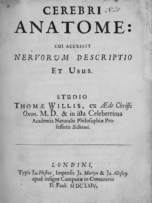
Title page of Cerebri anatome (1664). Available on: https://archive.org/stream/cerebrianatomecu00will#page/n8/mode/1up.
Early translation into English
Cerebri anatome was translated into English by Samuel Pordage, a cavalier, poet, playwright and translator. Pordage's translation of Cerebri anatome was published in 1681 under the title, ‘The Anatomy of the Brain and the Description and Use of the Nerves in the Remaining Medical Works of that Famous and Renowned Physician Dr Thomas Willis' (Willis, 1681). Pordage's translation was reprinted in two volumes on the occasion of its tercentenary (Willis, 1965). There, the First Volume concludes with a bibliographic chapter by HR Denham of the Wellcome Historical Library which provides a survey of the 22 previous editions (either separate or included in Opera Omnia) of Cerebri anatome (Hierons & Meyer, 1967). Pordage himself seems to have been aware of his medical ignorance and linguistic difficulties, for he added at the end of the book, ‘A table of all the hard words from the Greek and Latin for the benefit of the meer English Readers …’ where he ‘… fully explained them and rendered them intelligible to the meanest capacity’. Of these, the ‘hard word’ which arguably causes more controversy nowadays is the term ‘neurologie’. In Willis's Cerebri anatome, this term is written in Greek (νευρολογία, Willis, 1664a,b; p. 273). In Pordage's translation (Willis, 1965), the term used is ‘neurologie’. Νευρολογία (transliterated, ‘neurologia’) was used by Willis to include the cranial, spinal, peripheral and autonomic nerves as distinct from the brain and spinal cord, as when he states, ‘We have resolved to undertake the task of the Doctrine of the Nerves; and this rather because without the perfect knowledge of the Nerves, the Doctrine of the Brain and its Appendix would be left wholly lame and imperfect’ (Feindel, 1962). Willis's usage of the root ‘neuro’ – as transliterated from the Greek word νευρο and also meaning sinew, tendon or bowstring – predates its appearance in all other combinations in English by a century or more. Later, toward the end of the 18th century, the word ‘neurology’ gradually came to its present broader meaning as given in the Oxford English Dictionary: ‘the scientific study or knowledge of the anatomy, functions and diseases of the nerves and the nervous system’ (Feindel, 1962).
Body-brain-soul relationship as the starting point of Willis's work
Willis began his research on the comprehension of the ‘rational soul’ and its interaction with the brain because of his need for new material for his conferences in Oxford (O'Connor, 2003). In the Dedication of Cerebri anatome, addressed to Sheldon, then Archbishop of Canterbury, Willis was quite explicit in stating that ‘Natural Philosophy’ and ‘Christian Theology’ should not be seen as alternatives or rivals, while claiming that anatomy could ‘… unlock the secret places of Man's mind and look into the living and breathing Chapel of the Deity’. Willis ended his dedication claiming that ‘… after studying the brain, even the most perverse atheist shall acknowledge God or refuse both religion and reason’. Afterwards, in the Preface of Cerebri anatome, Willis's religiosity, combined with his scientific methods, led him to view the brain as a ‘harmonious, interconnecting system designed by God’ (Wilson, 2012). Later on in Cerebri anatome, while writing about the brain, Willis expressed his opinions about the role of the king and the church using a metaphor, a very frequent resource in lectures and philosophic works of the Restoration period (Harley, 1993; O'Connor, 2003). Then, the brain was ‘the Capital of the Empire’ or ‘the Chapel of the Deity’, the main organ of the body, so divided in ‘provinces’ and ‘villages’ separated by ‘water’ (the blood vessels).
Willis also believed that working on anatomy could demonstrate the structural resemblances between man and animals at the same time as they were different in their highest function. This meant that man had an ‘immortal soul’ in addition to the ‘one shared with animals’. For that reason, Cerebri anatome is a work of comparative anatomy with many references and drawings in which humans are compared with horses or sheep (Figs3 and 4). Willis realized that the functions of the nervous system were much more difficult to understand than those of other organs.
Fig 3.
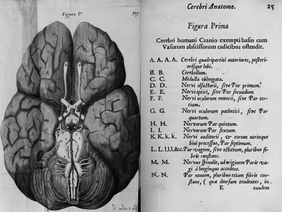
Figure 1 of Willis's Cerebri anatome (1664) with explanatory text showing the base of the human brain, particularly the arterial circle and the spinal roots of the accessory nerve accompanying the vagus nerve. Attributed to Christopher Wren (Compston, 2011). Available on: https://archive.org/stream/cerebrianatomecu00will#page/n71/mode/2up.
Fig 4.
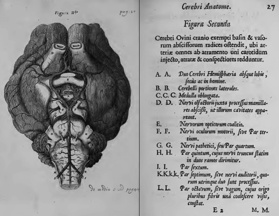
Figure 2 of Willis's Cerebri anatome (1664) with explanatory text showing the base of the sheep brain. Attributed to Christopher Wren (Compston, 2011). Available on: https://archive.org/stream/cerebrianatomecu00will#page/n75/mode/2up.
Willis replaced Nemesius's doctrine. He deduced that the ventricles contained cerebrospinal fluid which collected waste products from effluents. Willis recognized the cortex as the substrate of cognition and claimed that the gyrencephalia was related to a progressive increase in the complexity of cognition. In his functional scheme, the origin of voluntary movements was placed at the cerebral cortex while involuntary movements came from the cerebellum; memory was placed at the white matter; the gyri controlled memory, will and imagination (widely associated with ‘human soul’). The striatum had an essential role in sensation and movement. Willis was an enthusiastic advocate of the ‘spiritus animalis’ which were extracted from a cerebral distillation of blood and descended through the nerves towards the organic territories in order to cause sensations and movements (Eadie, 2003b). The nervous fluids carried by the ‘spiritus’ were located not inside the nerve fibres as commonly believed at that time, but on their interstitial space. Willis posed a hypothesis about sensation and movement which involved the nervous tubuli discovered at the brain by Malpighi with Baglivi's membranous fibres (Barcia Goyanes & Evans, 1995). Furthermore, he considered the chemical process to be fundamental: the substance must reach the muscles through the nerves to join the ‘spiritus animalis’ extracted from the blood. The resulting reaction triggered muscle contraction (Eadie, 2003b).
Willis's new technical approaches of study
The first and longest chapter of Cerebri anatome describes Willis's new approaches and technologies in the field of organ preservation and injection of coloured solidifiable substances. In Cerebri anatome,Willis set out explicitly his scientific methodology when writing, ‘… wherefore all delay being laid aside, I determined with my self seriously to enter presently upon a new corpse, and to rely on this one thing, not to pin my faith on the received Opinions of others, nor on the suspicious and guesses of my own mind, but for the future to believe Nature and ocular demonstrations. Therefore henceforward I betook myself wholly to the study of Anatomy … and so a firm and stable Basis might be laid, on which not only a more certain Physiologie than I had gained in the Schools, but what I had long thought upon, the Pathologie of the Brain and nervous stock, might be built’. Concerning the dissection method, Willis realized that the previous anatomists studied the brain by in situ dissection, that is to say, after opening the cranial vault they successively extracted brain slices. This method allowed them to carefully study the ventricles but made it more difficult to appreciate the delicate anatomy of the brain stem. In turn, Willis removed the entire brain intact which enabled him to study a much less deformed organ. When writing Cerebri anatome, Willis had a vast collection of anatomical charts and atlases that he could rely upon during his original investigations. Although Willis regarded Galen and Hippocrates as the founding fathers of medicine, he held the view that the Classical anatomists not only lacked sufficient anatomical detail but that they were also affected by a flawed, pagan-based belief system (Dewhurst, 1972). This lack of faith, Willis opined, rendered the Ancients incapable of ‘… recognizing the true beauty of God's ultimate creation, mankind’ (Wilson, 2012).
Willis was also an innovator in techniques for conserving the brain for dissection (Lo & Ellis, 2010). To delay the quick decomposition of the brain, Willis preserved it in pure alcohol, a body preservation method first proposed by Boyle. Boyle and Wren had previously studied the effect of snake venom on dogs from which Wren developed a preservation technique called ‘chiriguia infusoria’ (intravenous injection). Similarly, Willis and Lower used ‘… an inky liquor being injected into one of the Carotides’ after which ‘vessels got revealed in every secret place of the brain and cerebellum by filling with the same colour’. This technique not only helped to demarcate the arteries but also proved the purpose of vessel anastomosis. Since then, Lower remarked, ‘without syringes, Anatomy is as much deficient as a physician would be without laudanum’. A translational application of Willis and Lower's innovative staining technique by means of intravascular perfusion in cadavers is the current angiography/arteriography/venography. First developed by the Portuguese neurologist António Egas Moniz (1874–1944) at the University of Lisbon, angiography provides series of in vivo X-ray-images of blood vessels, which has been widely used in order to diagnose several classes of diseases, such as tumors, artery disease and arteriovenous malformations. Moniz performed the first cerebral angiogram in 1927. He also helped develop Thorotrast for use in the angiographic procedure; Thorotrast was extensively used after its introduction in 1931 (Tondreau, 1985). Moniz received the Nobel Prize for Physiology and Medicine in 1949, shared with Walter Rudolf Hess. In 1953 the angiographic technique was modified by Sven-Ivar Seldinger (1921–98).
Illustrations and engravings
Knowing how to illustrate his findings on dissection was one of Willis's greatest achievements in Cerebri anatome. Leonardo da Vinci urged the anatomist to ‘… dispel from your mind the thought that the comprehension of the human body in every aspect of its structure could be given in words … it is therefore necessary to be able to draw as well as to describe’ (Kenney, 1998). Since the publication of the ‘History of the Composition of the Human Body’ by Valverde de Amusco (1556), anatomical texts were gradually incorporating some kind of visual caption and higher quality illustrations engraved on sheets made through copper plates. Thus, of the 42 tables of Valverde's opus, Tauloa I and Tauloa II of Book V show a total of 20 figures of in situ brain dissection. Indeed, the 23 figures found in Cerebri anatome had no precedents in the history of anatomy (Lo & Ellis, 2010). The first figure (Fig.3) shows the cerebral arterial circle that carries Willis's eponym but which he never claimed as discovered by him or asked to be called after him. Conversely, he demanded the authorship of the names of the accessory nerve and the cranial origin of the sympathetic nerves (Willis referred to them as ‘intercostal nerves’).
Novel anatomical descriptions
Notably, in Cerebri anatome Willis described several structures, such as the striate nucleus, the internal capsule, the cerebellar peduncles, the anterior commissure, the claustrum, the inferior olivary nucleus, the pyramids, thalamus, the stria terminalis and some nerves. He claimed that brain tissue had no sensitivity to painful stimuli but that the meninges did. He also clarified some concepts about the vegetative nervous system, and made a classification of nine cranial nerves (Table2).
Table 2.
Classification of the cranial nerves based on Dalley (2002), Rengachary et al. (2008), Simon et al. (2011) and Davis et al. (2014)
| Willis | Sömmering (still used) | Galen |
|---|---|---|
| I – Olfactory nerves | I – Olfactory nerve | Not mentioned |
| II – Nerves of visual perception | II – Optic nerve | Par primum |
| III – Nerves moving the eyes | III – Oculomotor nerve | Par secundum |
| IV – Pathetic nerves of the eyes | IV – Trochlear nerve | Not mentioned |
| V – Trifacial nerve: | V – Trigeminal nerve | Par tercium |
| Sensory branch | Par quartum | |
| Motor branch | ||
| VI – A pair of small nerves moving the eyeball | VI – Abducens nerve | Probably attached to II |
| VII – Facial and auditory nerve, it has two divisions: | VII – Facial nerve | Par quintum |
| “ramus durus – facialis” | VIII – Vestibulocochlear nerve | Par quintum |
| “ramus mollis – acousticus” | ||
| VIII – Wandering or vagus nerve | IX – Glossopharyngeal nerve | Par sextum |
| X – Vagus nerve | Par sextum | |
| XI – Accessory nerve | Par sextum | |
| IX – Motor nerve of the tongue | XII – Hypoglossal nerve | Par septimum |
The sympathetic nerves and the vagus nerve
The division between the sympathetic nerves and the vagus nerve is often attributed to Charles Estienne (1504–64). However, Eustachius (1500 or 1514–74) and Thomas Willis were the only anatomists of this époque who gave clear examples of this separation (Hierons & Meyer, 1962). They also stated that there was an intracranial origin for the sympathetic subdivision of the autonomic nervous system. That is why some authors (e.g. Sheehan, 1936) think Willis knew about Eustachius's unpublished Tables, whereas other authors (Hierons & Meyer, 1962) consider that different lines of thought led to similar conclusions. Cerebri anatome includes two schematic, accurate and clear illustrations of this separation, which only seem to look like those of Eustachius. Although Willis's illustrations were of less artistic beauty, they had the advantage of showing the topographic relations with visceral organs (Fig.5). Willis also declared that the vagus nerve was more important for lower animals than the sympathetic nerves, a statement revised by Gaskell in 1916, although he did not mention Willis in his work (Hierons & Meyer, 1962).
Fig 5.
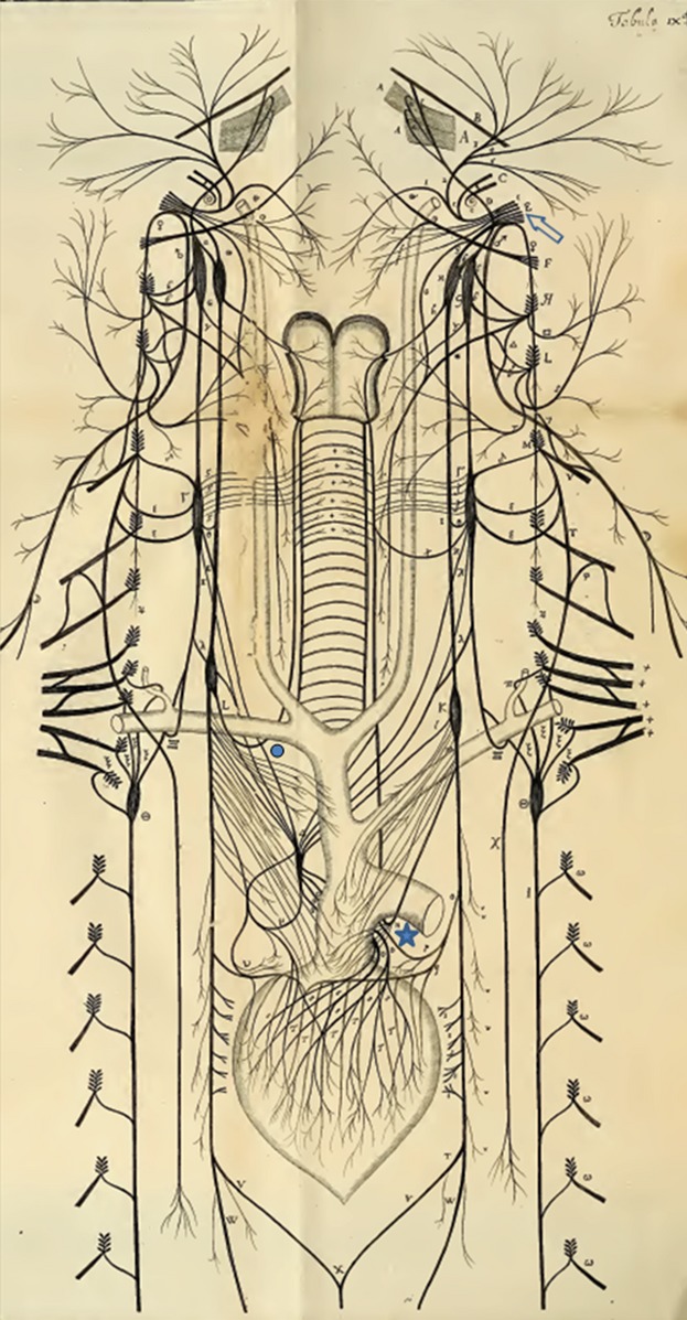
Diagram of the distribution of some cranial and spinal nerves and the ‘wandering pair’ (vagi; clear arrow) in Willis's Cerebri anatome (1664; p. 493 modified). Notice the recurrent laryngeal nerves which are looping round the subclavian artery on the right side (circle) and the aortic arch on the left side (star), giving off cardiac branches. Available on: https://archive.org/stream/cerebrianatomecu00will#page/n493/mode/1up.
One of Willis's major principles proposed that the brain influenced the cerebellum through the quadrigeminal plate and the superior cerebellar peduncles, and so the latter coordinated the vagus and ‘intercostal’ (sympathetic) nerves, which were involuntary. This influence was often evidenced in hysteria by difficulty in breathing, chest pain, abdominal distension and outbursts of weeping and laughter. Hence, Willis anticipated modern ideas of the physiology of emotion (Vinchon & Vie, 1928).
Willis's description of the intercostal nerves has not received much attention by historians of medicine. Yet the intercostal nerves are of paramount importance for his neurology. Willis explained that via these nerves, which connect the brain to the heart and lower viscera, the brain controls the passions and instincts of the lower body. In other words, Willis believed that the intercostal nerves mediate a kind of rationality and that therefore they make a human a rational being. Willis's theory must be seen in the context of the early modern mind–body problem. Oxford theologist Samuel Parker (1640–88) took up Willis's argument when stating that the intercostal nerves are the most important instruments (reins) of the soul. They control the bodily passions so that humans can transform themselves into more virtuous beings. The explanation of the intercostal nerves offered by Willis and Parker fits the Anglican optimism about the abilities of human reason as well as about the moral potential of humankind (Knoeff, 2004).
The accessory nerve
Willis was the first anatomist to describe and illustrate the accessory nerve as an independent nerve. He studied the accessory nerve in fish and poultry which supplied fins and wings, instead of neck muscles as it does in humans and other mammals. The temporary connection between the accessory nerve and the vagus drew his attention and led him to speculate about the response of the brain to fear and other passions linked to the neck and arm muscles (Hierons & Meyer, 1962).
The cranial nerves
Willis made important contributions to the naming and numbering of the cranial nerves (Simon et al. 2011; Davis et al. 2014). From Galen to the 16th century (Vesalius, Colombo, Estienne, Bahuin) these nerves were called ‘nervi cerebri’ (‘brain nerves’). Willis set the origin of these nerves inside the skull, claiming that ‘nervorum paria intra cranium oriunda’ (‘the pair of nerves are being born inside the skull’).
Willis's popularity helped to consolidate a new classification composed of nine cranial nerves (see Table2). His classification of the cranial nerves was a marked improvement over its predecessors. It included nine cranial nerves, CN I through VI being those we recognize today. In Willis's scheme, CN VII encompassed both the facial and vestibulocochlear nerves; CN VIII included the glossopharyngeal and vagus nerves, and the cranial root of the accessory nerve; and CN IX consisted of the hypoglossal nerve and the spinal root of the accessory nerve (Dalley, 2002). This classification remained in place for the next 114 years until in 1778 Sömmering introduced the current classification of 12 cranial nerves. In fact, Willis's work was so popular in England that some English works used his classification until the end of the 19th century.
The cerebral arterial circle
Willis was not the first anatomist to give a reasonable description or illustration of the cerebral arterial circle. However, the acknowledgement and comprehension of this structure had been hindered by several factors such as the preservation of Galen's concepts in physiology, its confusion with the rete mirabile, the higher frequency of anatomical variants within the ‘normal’ circle and the deficient understanding of blood circulation before the publication of Harvey's Motus cordis in 1628 (Hierons & Meyer, 1962).
In the mid-16th century, many important anatomists, such as Berengario da Carpi (1521) and Vesalius (1543), rejected the existence of the rete mirabile in humans. Then Fallopius, Vesalius's disciple, introduced the first acceptable description of the vessels involved at the circle, in his Observationes anatomicae (1561). Fallopius observed the ascent of the carotid arteries into the skull and division of these into inner branches (now anterior cerebral arteries). He described two-thirds of the posterior communicating arteries, although he was mistaken in claiming that the posterior communicating arteries branched into small ones before joining the carotid arteries. In 1627, Casserius, Harveys's teacher in Padua, provided the first illustration of the arterial circle showing it as complete on one side while adhering to Fallopius's mistaken viewpoint on the other. Twenty years later, Vesling fixed this paradox in his Syntagma anatomicum (1647), published in English in 1653, but still could not demonstrate how the junction of the anterior cerebral arteries occurred.
Finally, in 1658, Wepfer gave a more detailed and accurate description of the arterial circle in his Observationes anatomicae. Consequently, some authors consider him the true discoverer of this structure. Although Willis added nothing new to Wepfer's findings, Cerebri anatome contains the first complete illustration of the arterial anastomosis as a complete circle located at the cerebral base. Willis wrote these unequivocal words about it (p. 92): ‘… nempe arteriae carotides unius lateris, pluribus in locis, uniuntur cum carotidibus alterirus; infuper vertebrales utriusque lateris inter sese, & in posteriores carotidum ramos, prius unitos, inosculantur …’ [sic; italics as in the original] (to wit, ‘… the carotid arteries of one side, in many, are united with the carotides of the other side; besides the vertebrals of either side among themselves, and are also inoculated into the posterior branches of the carotides before united …’).
It is important to emphasize that Willis never claimed to be the first person to discover the arterial circle. Willis's immediate successors in the field, among them Vieussens (Neurographia universalis, 1684), Ridley (The Anatomy of the Brain, 1695), Leclerc and Manget (Bibliotheca anatomica, 1699), Manget (Theatricum anatomicum, 1717), and Boerhaave (Institutiones medicae, 1708) did not quote him, nor for that matter anybody else, as the discoverer of the arterial circle, although most of them make appreciative or critical comments on Cerebri anatome, and in particular on the description of the cerebral arteries. Haller (1708–77) was the first to associate Willis's name with the anastomosis in his Bibliotheca anatomica (1774) (Meyer & Hierons, 1962) through the expression ‘circulum Willisii’ (p. 290) and ‘circulum qui dicitur Willisii’ (p. 476). However, Willis really was the first to comprehend the functional and pathological significance of this arterial circle. As a true academic physician, and exemplary translational researcher as well, he performed experimental surgery to prove the brain's compensatory mechanisms (Lo & Ellis, 2010) and sensed the existence of the blood–brain barrier, about which he suggested that only the smallest particles that were essential to the swift performance of nerve function moved from blood to the brain (Rengachary et al. 2008).
A shadow of a doubt
Despite the excellence of Cerebri anatome, for almost 300 years many authors questioned Willis's authorship and honesty on the basis of an annotation written by Anthony Wood (1632–95), an English antiquary who wrote about the history of Oxford and its University, in which it is said: ‘Whatever is anatomical in that Book, the Glory thereof belongs to the said R Lower, whose indefatigable Industry at Oxon produced that Elaborate Piece’. This quotation was taken verbatim from Legends No Histories (1670) by Henry Stubbe who wrote it at the request of the physician Baldwin Hamey as he wanted to minimize the growing power of the Royal Society over the Royal College of Physicians of London. In another part of his book, Stubbe changed his opinion, saying that Willis first suggested the work to Lower and concluded by claiming that Willis ‘… was the grand occasion of the work and in much the author’ (Hierons & Meyer, 1962). Willis likewise showed his deep gratitude to Lower in the preface to Cerebri anatome, as mentioned above. Besides, Lower never suggested in any of his publications that his colleague had ever stolen his work. Willis's position may reasonably be taken as that of a senior member of what we might call today a medical research team (Feindel, 1962).
Epilogue
Both the history of humanity and the history of science repeat cyclically up to a point. At the present time, neuroscience is undergoing an important conceptual and technological revolution. Just as the biological psychiatry of the 20th century refuted Freud's principles, Thomas Willis overthrew the mediaeval concepts about brain function 350 years ago. Willis's works, particularly Cerebri anatome, had a great influence in Europe and contributed to the weakening of the prevailing Galenian movement.
During the Baroque era, the normal operation of the anatomical structures was explained by two lines of thought strongly influenced by religious beliefs: ‘Iatromechanics’ and ‘Iatrochemistry’. Authors such as Descartes (iatromechanics), Silvyus or Willis (iatrochemists) sought to describe the relationship between the soul and body to reach an understanding of the function of the nervous system. However, their approaches and methods were absolutely different, even between the latter two authors.
The historical review of anatomic studies of the ‘circle’ before Willis's own work shows that earlier writers recognized the existence of this anatomic structure, at least in some part. However, nobody recognized, before Willis, the functional relevance of the anastomotic arterial complex that is the ‘circle’.
Willis rejected Cartesian ideas and recognized the cerebral cortex, not the ventricles, as the substrate of cognition. He visualized the brain as an alembic in which brain disturbances are caused by distillation problems (chemical disorders). He performed several experiments to demonstrate that brain functions were located in different regions. His multidisciplinary research done for clinical purposes set a precedent for current translational research.
Acknowledgments
This study was conducted in the context of a stage of L.-A.A.-A at the Department of History of Science-CSIC. Thank you to Rafael Huertas from IH-CCHS-CSIC for his support. J.L.B.-L. gratefully acknowledges the support of UPV-EHU (Grants GIU 07/14 and UFI 11/41). The authors wish to express gratitude to Mrs Ana-Maria Álvarez-Castrosín for her technical assistance with manuscript preparation and to Nicolas Raio, MD, for his generous assistance in reviewing the English language style.
Conflict of interest
None.
Author contributions
Concept/design (L.-A.A.-A), acquisition of data (L.-A.A.-A), data analysis (L.-A.A.-A, P.N.-A), data interpretation (L.-A.A.-A, P.N.-A), drafting of the manuscript (L.-A.A.-A, P.N.-A, T.F.-R, J.L.B.-L.), critical revision of the manuscript (L.-A.A.-A, J.L.B.-L.) and approval of the article (L.-A.A.-A, P.N.-A, T.F.-R, J.L.B.-L.).
References
- Barcia Goyanes JJ, Evans NR. Notes on the historical vocabulary of neuroanatomy. Hist Psychiatry. 1995;6:471–482. doi: 10.1177/0957154X9500602404. [DOI] [PubMed] [Google Scholar]
- Compston A. 2011. Dr Thomas Willis' Works. Available: https://history.medsci.ox.ac.uk/seminars/prof-alastair-compston-dr-thomas-williss-works-the-most-learned-christopher-wren-and-the-inward-dens-of-the-brain/. Last accessed 10th November 2014.
- Dalley AF. Thomas Willis 1621–1675. Clin Anat. 2002;15:2–3. doi: 10.1002/ca.1083. [DOI] [PubMed] [Google Scholar]
- Davis MC, Griessenauer CJ, Bosmia AN, et al. The naming of the cranial nerves: a historical review. Clin Anat. 2014;27:14–19. doi: 10.1002/ca.22345. [DOI] [PubMed] [Google Scholar]
- Dewhurst K. Some letters of Dr. Thomas Willis (1621–1675) Med Hist. 1972;16:63–76. doi: 10.1017/s0025727300017269. [DOI] [PMC free article] [PubMed] [Google Scholar]
- Donaldson IM. The Treatise of man (De homine) by René Descartes. J R Coll Physicians Edinb. 2009;39:375–376. doi: 10.4997/jrcpe.2009.418. [DOI] [PubMed] [Google Scholar]
- Donaldson IM. Cerebri anatome: Thomas Willis and his circle. J R Coll Physicians Edinb. 2010;40:277–279. doi: 10.4997/jrcpe.2010.318. [DOI] [PubMed] [Google Scholar]
- Dorland. Dorland: Diccionario Enciclopédico Ilustrado De Medicina. Madrid: Elsevier; 2005. [Google Scholar]
- Eadie MJ. A pathology of the animal spirits – the clinical neurology of Thomas Willis (1621–1675) part I – background, and disorders of intrinsically normal animal spirits. J Clin Neurosci. 2003a;10:14–29. doi: 10.1016/s0967-5868(02)00165-0. [DOI] [PubMed] [Google Scholar]
- Eadie MJ. A pathology of the animal spirits – the clinical neurology of Thomas Willis (1621–1675). Part II – disorders of intrinsically abnormal animal spirits. J Clin Neurosci. 2003b;10:146–157. doi: 10.1016/s0967-5868(02)00164-9. [DOI] [PubMed] [Google Scholar]
- Feindel W. Thomas Willis (1621–1675) – the founder of neurology. Can Med Assoc J. 1962;87:289. [PMC free article] [PubMed] [Google Scholar]
- Finger S. Origins of Neuroscience: A History of Explorations into Brain Function. New York: Oxford University Press; 1994. [Google Scholar]
- French RK. The languages of William Harvey's natural philosophy. J Hist Med Allied Sci. 1994;49:24–51. doi: 10.1093/jhmas/49.1.24. [DOI] [PubMed] [Google Scholar]
- Harley DN. Medical metaphors in English moral theology, 1560–1660. J Hist Med Allied Sci. 1993;48:396–435. doi: 10.1093/jhmas/48.4.396. [DOI] [PubMed] [Google Scholar]
- Hierons R, Meyer A. Observations on the history of the ‘circle of Willis’. Med Hist. 1962;6:119–130. doi: 10.1017/s0025727300027095. [DOI] [PMC free article] [PubMed] [Google Scholar]
- Hierons R, Meyer A. The Anatomy of the Brain and Nerves. Proc R Soc Med. 1967;60:314. [Google Scholar]
- Hörig H, Marincola E, Marincola FM. Obstacles and opportunities in translational research. Nat Med. 2005;11:705–708. doi: 10.1038/nm0705-705. [DOI] [PubMed] [Google Scholar]
- Hughes JT. Thomas Willis 1621–1675: His Life and Work. London: Royal Society of Medicine Services; 1991. [Google Scholar]
- Kenney CA. A historical review of the illustrations of the circle of Willis from antiquity to 1664. J Biocommun. 1998;25:26–31. [PubMed] [Google Scholar]
- Knoeff R. The reins of the soul: the centrality of the intercostal nerves to the neurology of Thomas Willis and to Samuel Parker's theology. J Hist Med Allied Sci. 2004;59:413–440. doi: 10.1093/jhmas/jrh090. [DOI] [PubMed] [Google Scholar]
- Laín Entralgo P. Historia de la Medicina. Barcelona: Salvat; 1978. [Google Scholar]
- Lo WB, Ellis H. The circle before Willis: a historical account of the intracranial anastomosis. Neurosurgery. 2010;66:7–18. doi: 10.1227/01.NEU.0000362002.63241.A5. ; discussion 17–8. [DOI] [PubMed] [Google Scholar]
- López-Muñoz F, Marín F, Alamo C. The historical background of the pineal gland: I. From a spiritual valve to the seat of the soul. Rev Neurol. 2010;50:50–57. [PubMed] [Google Scholar]
- López-Piñero JM. La iatroquímica de la segunda mitad del siglo XVII. In: Laín-Entralgo P, editor. Historia Universal de la Medicina. Barcelona: Salvat; 1973. pp. 185–193. [Google Scholar]
- Martín-Araguz A, Bustamante-Martínez C, Fernández-Armayor Ajo V, et al. Antoniana Margarita: Gómez Pereira, Francisco Lobato and the antecedents of cerebral mechanicism during the Spanish renaissance. Rev Neurol. 2001;33:82–89. [PubMed] [Google Scholar]
- Meyer A, Hierons R. Observations on the history of the ‘circle of Willis’. Med Hist. 1962;6:119–130. doi: 10.1017/s0025727300027095. [DOI] [PMC free article] [PubMed] [Google Scholar]
- Molnár Z. Thomas Willis (1621–1675), the founder of clinical neuroscience. Nat Rev Neurosci. 2004;5:329–335. doi: 10.1038/nrn1369. [DOI] [PubMed] [Google Scholar]
- O'Connor JP. Thomas Willis and the background to Cerebri anatome. J R Soc Med. 2003;96:139–143. doi: 10.1258/jrsm.96.3.139. [DOI] [PMC free article] [PubMed] [Google Scholar]
- Pearce JM. The circle of Willis (1621–75) J Neurol Neurosurg Psychiatry. 2000;69:86. doi: 10.1136/jnnp.69.1.86. [DOI] [PMC free article] [PubMed] [Google Scholar]
- Pearce JM. Willis on narcolepsy. J Neurol Neurosurg Psychiatry. 2003;74:76. doi: 10.1136/jnnp.74.1.76. [DOI] [PMC free article] [PubMed] [Google Scholar]
- Rengachary SS, Xavier A, Manjila S, et al. The legendary contributions of Thomas Willis (1621–1675): the arterial circle and beyond. J Neurosurg. 2008;109:765–775. doi: 10.3171/JNS/2008/109/10/0765. [DOI] [PubMed] [Google Scholar]
- Sheehan D. Discovery of the autonomic nervous system. Arch Neurol Psychiatry. 1936;35:1081–1115. [Google Scholar]
- Simon F, Marečková-Štolcová E, Páč L. On the terminology of cranial nerves. Ann Anat. 2011;193:447–452. doi: 10.1016/j.aanat.2011.04.012. [DOI] [PubMed] [Google Scholar]
- Sinclair H, Robb-Smith A. A Short History of Anatomical Teaching in Oxford. Oxford: Oxford University Press; 1950. [Google Scholar]
- Symonds C, Feindel W. Birthplace of Thomas Willis. Br Med J. 1969;3:648–649. doi: 10.1136/bmj.3.5671.648. [DOI] [PMC free article] [PubMed] [Google Scholar]
- Tondreau RL. The retrospectoscope. Egas Moniz 1874–1955. Radiographics. 1985;5:994–997. doi: 10.1148/radiographics.5.6.3916824. [DOI] [PubMed] [Google Scholar]
- Vinchon J, Vie J. Un maître de la neuropsychiatrie au XVII siécle: Thomas Willis (1662–1675) Ann Med Psychol (Paris) 1928;2:109–144. [Google Scholar]
- Willis T. 1664a. Cerebri anatome. Londini: Typis Ja. Flesher, Impensis Jo. Martyn and Ja Allestry. https://archive.org/details/cerebrianatomecu00will.
- Willis T. 1664b. Cerebri anatome. On open library.org – https://archive.org/stream/cerebrianatomecu00will#page/44/mode/2up.
- Willis T. The anatomy of the brain and the description and uses of the nerves. In: Pordage S, editor. The Remaining Medical Works of that Famous and Renowned Physician Dr Thomas Willis. London: Dring, Harper, Leigh & Martyn; 1681. [Google Scholar]
- Willis T. Anatomy of the Brain and Nerves: Volumes 1 & 2. Montreal: McGill University Press; 1965. [Google Scholar]
- Wilson MD. The life and times of Thomas Willis and his impact on contemporary medicine. J Hist Neurosci. 2012;21:127–131. doi: 10.1080/0964704X.2011.595632. [DOI] [PubMed] [Google Scholar]
- Zimmer C. Beyond the ivory tower. A distant mirror for the brain. Science. 2004;303:43–44. doi: 10.1126/science.1090319. [DOI] [PubMed] [Google Scholar]


