Abstract
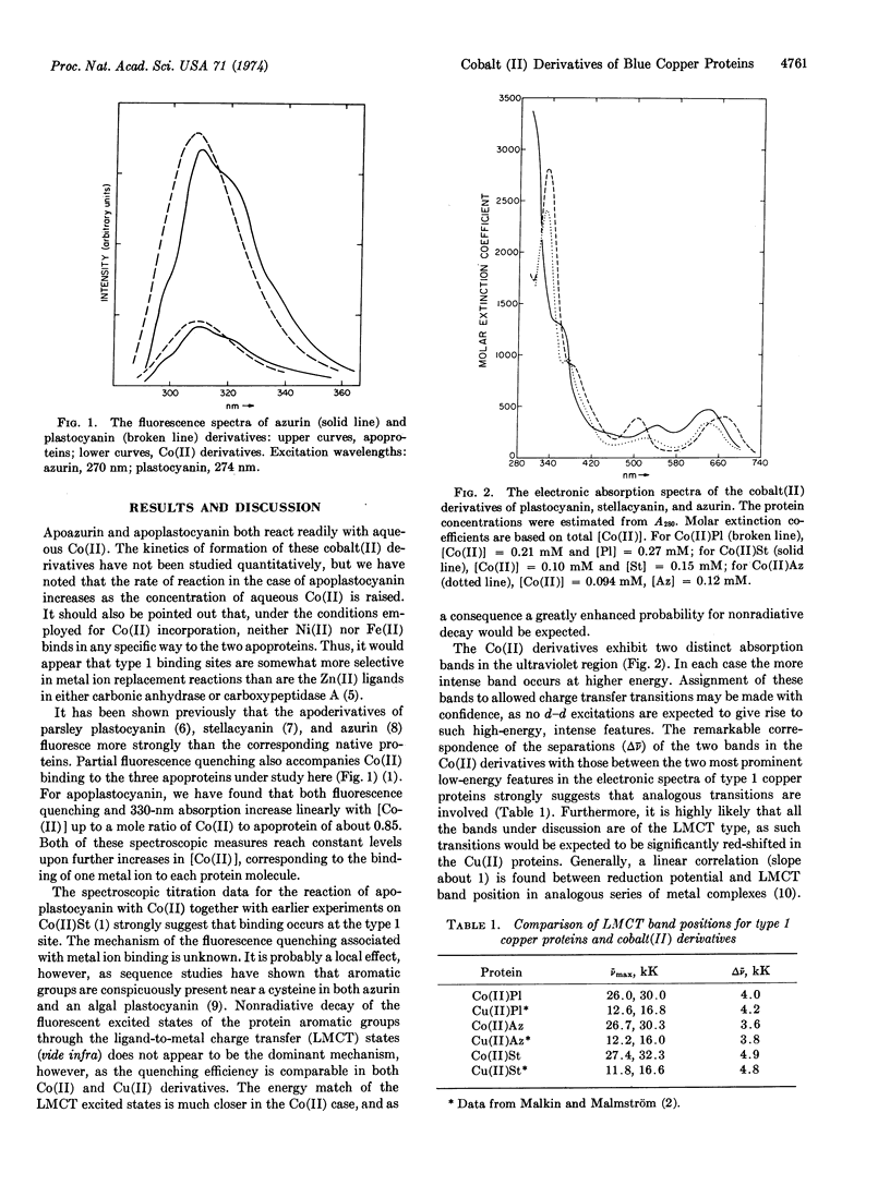
Preparation of cobalt(II) derivatives of type 1 copper proteins has been extended to include bean plastocyanin and azurin from Pseudomonas aeruginosa. Fluorescence quenching data suggest that Co(II) binds to the type 1 site of both apoplastocyanin and apoazurin. Electronic absorption spectral measurements have been made for cobalt(II)-stellacyanin, cobalt(II)-plastocyanin, and cobalt(II)-azurin. In each case two visible bands with moderate intensities and two rather strong ultraviolet absorption peaks are observed. The intensity pattern and the separation of the two ultraviolet bands in the Co(II) derivatives correspond closely to the two intense, low-lying absorption peaks in the analogous, type 1 copper proteins. The evidence suggests that the intense, two-band systems represent ligand-to-metal charge transfer transitions in both Cu(II) and Co(II) derivatives. It is proposed that the transitions in each case originate in the 3pπ and 3pσ orbitals of a cysteine sulfur ligand at the type 1 site. The visible absorption bands in the Co(II) derivatives are assigned to d-d transitions. The large 4P splitting observed indicates that the type 1 Co(II) site is of low symmetry. The d-d band pattern suggests that the high-spin Co(II) center is either distorted tetrahedral or five-coordinate. The observed band intensities are in much closer accord with the values for Co(II) complexes known to have distorted tetrahedral structures.
Keywords: metal replacement, electronic spectroscopy, charge transfer transitions
Full text
PDF


Selected References
These references are in PubMed. This may not be the complete list of references from this article.
- Ambler R. P., Brown L. H. The amino acid sequence of Pseudomonas fluorescens azurin. Biochem J. 1967 Sep;104(3):784–825. doi: 10.1042/bj1040784. [DOI] [PMC free article] [PubMed] [Google Scholar]
- Finazzi-Agrò A., Giovagnoli C., Avigliano L., Rotilio G., Mondovì B. Luminescence quenching in azurin. Eur J Biochem. 1973 Apr 2;34(1):20–24. doi: 10.1111/j.1432-1033.1973.tb02723.x. [DOI] [PubMed] [Google Scholar]
- Graziani M. T., Agrò A. F., Rotilio G., Barra D., Mondovi B. Parsley plastocyanin. The possible presence of sulfhydryl and tyrosine in the copper environment. Biochemistry. 1974 Feb 12;13(4):804–809. doi: 10.1021/bi00701a025. [DOI] [PubMed] [Google Scholar]
- KATOH S., TAKAMIYA A. NATURE OF COPPER-PROTEIN BINDING IN SPINACH PLASTOCYANIN. J Biochem. 1964 Apr;55:378–387. doi: 10.1093/oxfordjournals.jbchem.a127898. [DOI] [PubMed] [Google Scholar]
- Malkin R., Malmström B. G. The state and function of copper in biological systems. Adv Enzymol Relat Areas Mol Biol. 1970;33:177–244. doi: 10.1002/9780470122785.ch4. [DOI] [PubMed] [Google Scholar]
- McMillin D. R., Holwerda R. A., Gray H. B. Preparation and spectroscopic studies of cobalt(II)-stellacyanin. Proc Natl Acad Sci U S A. 1974 Apr;71(4):1339–1341. doi: 10.1073/pnas.71.4.1339. [DOI] [PMC free article] [PubMed] [Google Scholar]
- Morpurgo L., Finazzi-Agrò A., Rotilio G., Mondovì B. Studies of the metal sites of copper proteins. IV. Stellacyanin: preparation of apoprotein and involvement of sulfhydryl and tryptophan in the copper chromophore. Biochim Biophys Acta. 1972 Jul 21;271(2):292–299. doi: 10.1016/0005-2795(72)90203-6. [DOI] [PubMed] [Google Scholar]
- Wells J. R. Purification and properties of a proteolytic enzyme from French beans. Biochem J. 1965 Oct;97(1):228–235. doi: 10.1042/bj0970228. [DOI] [PMC free article] [PubMed] [Google Scholar]


