Abstract
Background
The orientation of the acetabular cup component of a total hip arthroplasty can be evaluated in a number of ways, utilizing a myriad of imaging techniques and measurement parameters, including intraoperative surgical estimates, postoperative radiographs, and cross-sectional imaging such as computed tomography (CT) and magnetic resonance imaging (MRI).
Questions/Purposes
How do traditional versus corrected measurements of acetabular version vary from one another based on the inclination of the cup? What is the reliability of the corrected acetabular version measurements based on interobserver and intraobserver consistency?
Patients and Methods
Two fellowship-trained musculoskeletal radiologists reviewed CT scans on 60 total hip arthroplasties. Acetabular inclination, traditional CT acetabular version, and CT acetabular version corrected for inclination (by utilizing multi-planar reformations to measure in the plane of the cup face) were each measured. The difference was then calculated between the “traditional” axial CT and “corrected” acetabular version measurements, and the association between this difference and the acetabular inclination was assessed.
Results
The “traditional” axial CT and “corrected” acetabular version measurements differed from one another in every case, with the traditional method yielding a version measurement that was on average 9.5° higher than the corrected technique. However, as the acetabular cup inclination angle decreased, the “traditional” measurement became more variable and increasingly discordant with the “corrected” version measurement.
Conclusions
There is inherent variability between the many methods utilized for defining and measuring acetabular version, with axial CT measurements often used as an accepted proxy for true cup anteversion. However, the variability between different measurement techniques is correlated with acetabular inclination, and this variability is most pronounced when acetabular inclination is low, ultimately leading to potential confusion in measurement terminology. The increasingly widespread availability of multi-planar CT reformations provides an opportunity to standardize methodology, eliminate the impact of inclination on acetabular version measurements, and potentially provide a more reliable comparison of the impact of cup orientation on surgical outcomes.
Electronic supplementary material
The online version of this article (doi:10.1007/s11420-014-9416-6) contains supplementary material, which is available to authorized users.
Keywords: acetabulum, arthroplasty, computed tomography, cross-table, CT, hip, imaging, inclination, measurement, multi-planar, pelvis, radiograph, reformations, version
Introduction
Accurate placement and orientation of the acetabular and femoral components of a total hip arthroplasty are crucial for the prevention of arthroplasty dislocation, premature component wear, and limited postoperative range of motion [5, 8, 10, 11]. Component orientation can be evaluated in a number of ways, utilizing a myriad of imaging techniques and measurement parameters. Intraoperative surgical estimates, postoperative radiographs, and cross-sectional imaging such as computed tomography (CT) and magnetic resonance imaging (MRI) have all been used to evaluate arthroplasty orientation [1, 3, 4, 12, 15]. To discuss the evaluation of arthroplasty orientation, it is thus important to first understand and define the terms utilized to describe acetabular cup position.
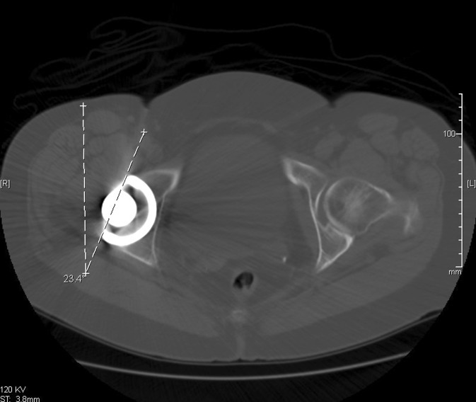
While there are numerous methods of defining acetabular version anatomically and radiographically [1, 6, 9, 11, 13], the most commonly used method for radiographic analysis is the one proposed by Woo and Morrey [19], namely the “angle formed by a line drawn tangential to the face of the acetabulum and a line perpendicular to the horizontal plane, as seen on a lateral view of the pelvis.” For evaluating the orientation of the acetabular cup component, CT has also demonstrated utility [1, 2, 12, 14, 16], and CT with multi-planar reformation has been demonstrated to be more accurate than intraoperative surgical estimates or radiographs [18]. Authors most frequently define CT version of the acetabulum by presenting an axial CT image of the pelvis (Fig. 1). The most anterior and posterior points of the prosthetic cup are marked and a straight line is drawn. The angle between this line and the sagittal plane of the body (defined as a line perpendicular to two identical points on either side of the pelvis) is described as the version. Authors thus define version as anterior (anteversion) if the angle opens to the front of the pelvis and posterior (retroversion) if it opens to the back of the pelvis [7, 18] (Fig. 1).
Fig. 1.
A line drawn tangential to the acetabular cup base is compared with the sagittal plane of the body as defined by a line perpendicular to the ischial spines.
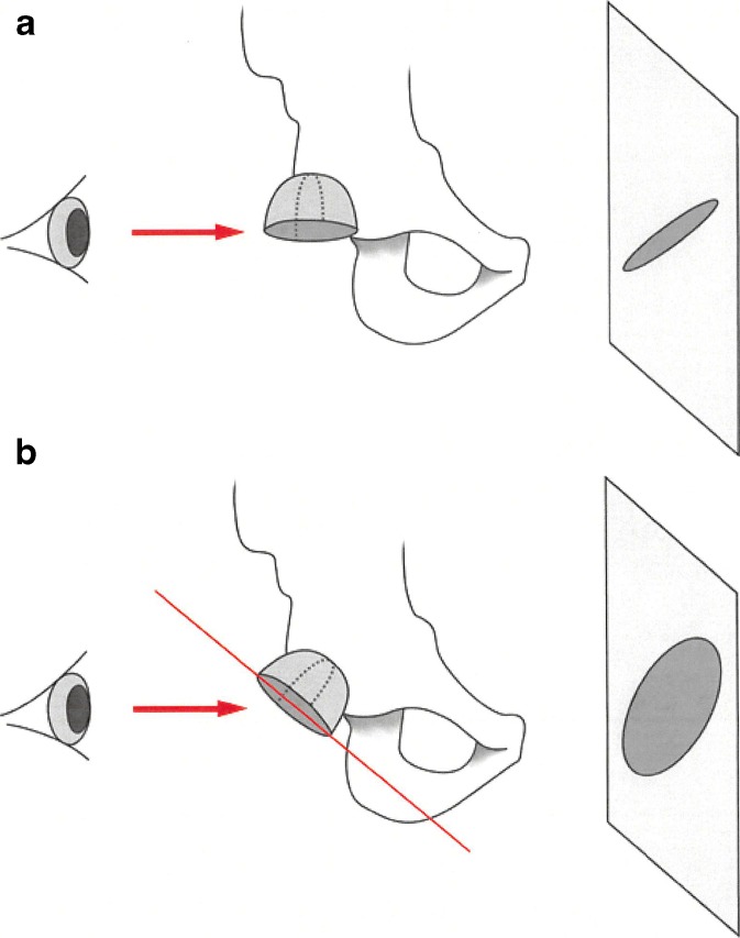
“Degrees of anteversion” as reported in research literature is thus an imprecise term if the specific measurement technique utilized is not defined and understood by the manuscript audience. Furthermore, there is a temptation to use “anteversion” as measured utilizing a cross-table lateral radiograph and “anteversion” as measured on an axial CT interchangeably, while these numbers in fact differ, and the discrepancy between these two methods is not independent of the inclination of the cup [17]. As the inclination of the cup changes, the axis of rotation that determines the version of the cup changes its position in the coronal plane, and observing or radiographing the cup along the right–left axis or top–bottom axis demonstrates progressive change in the contour of the base of the cup (Fig. 2a, b). If one considers the extreme example of an acetabular inclination angle of zero, a “traditional version” axial CT measurement obtained by drawing a line tangential to the acetabular face and along the sagittal plane would always yield a version measurement of 90° regardless of the true position of the acetabular axis. However, in the same situation (acetabular inclination angle of zero), a “reformatted/corrected” measurement obtained by drawing a line tangential to the acetabular face and along the transverse plane would reveal whether cup anteversion or retroversion was truly present. We therefore propose utilizing a “corrected” measurement of CT version that effectively creates a line tangential to the face of the acetabular cup (as would be seen with a cross-table lateral radiograph) and eliminates the impact of acetabular inclination on CT measurement of acetabular cup version. Preliminary work by the primary investigator [6] suggests this corrected method is more accurate when compared with an in vitro model as the gold standard.
Fig. 2.
a With the acetabular cup at nearly zero degrees of inclination, the base of the cup appears as a straight line when viewed along the right–left axis. b As the inclination angle of the acetabular cup increases, the shadow cast by the base of the cup becomes a changing ellipse.
We addressed two main questions: (1) How do traditional versus corrected CT measurements of acetabular version vary from one another based on the inclination of the acetabular cup? (2) What is the reliability of the corrected acetabular version measurement based on interobserver and intraobserver consistency?
Patients and Methods
Institutional IRB approval was sought and obtained for all aspects of this project. Two musculoskeletal radiologists with 51 years of combined radiology experience independently reviewed CT scans of the pelvis or hip in 48 sequential hip arthroplasty patients, yielding retrospective data on 60 total hip arthroplasties. CT images were obtained on a Philips Brilliance 64 slice CT scanner and Philips Brilliance 16 slice CT scanner (Philips Healthcare 3000 Minuteman Road, Andover MA 01810). The imaging data for each case was anonymized prior to interpretation and stored in an online database where patient identification was removed, and the interpreting radiologists were blinded to prior measurements.
The arthroplasty procedures reviewed were performed by a range of orthopedic surgeons both within and outside our institution, and patients from more than a dozen individual surgeons were reviewed. Patient ages ranged from 26 to 90 years old, and the most common indication for arthroplasty was osteoarthritis.
Acetabular inclination (as measured on the CT scout image), traditional CT acetabular version (as measured on axial CT images), and CT acetabular version corrected for inclination (via multi-planar reformation performed on a Sectra workstation) were each measured in a randomized order as assigned by a research coordinator. Each radiologist then made a second measurement of the inclination, version, and corrected version at a separate time point and in a different order, resulting in a total of four measurements per data point and 12 measurements per hip. Inter- and intraobserver reliability was analyzed for each measurement parameter.
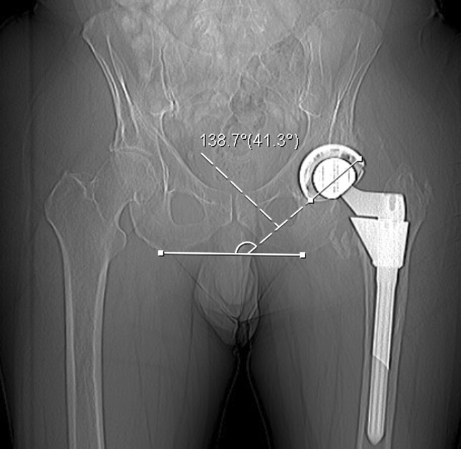
Acetabular inclination measurements were obtained by drawing a line tangential to the face of the acetabular cup on the anteroposterior (AP) CT scout image and calculating the angle relative to a line drawn tangential to the ischial tuberosities as described by Murray et al. (Fig. 3).
Fig. 3.
Acetabular inclination was calculated by drawing a line tangential to the face of the acetabular cup on the AP scout image and calculating the angle relative to a line drawn between the ischial tuberosities.
Version measurements were obtained utilizing two techniques. First, “traditional” CT version measurements were made on axial CT images by drawing a line tangential to the face of the acetabular cup on a single axial CT image obtained at the center of the cup (identified via cross-reference to the CT scout image) and calculating the angle relative to the sagittal plane of the body as defined by a line drawn perpendicular to a line traversing two corresponding points across the pelvis (see Fig. 1 above).
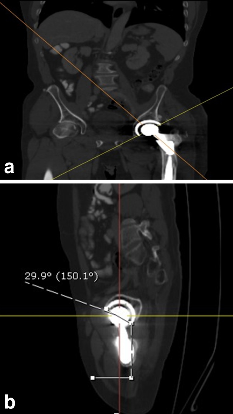
“Corrected” version measurements were obtained by utilizing multi-planar reformations performed on a Sectra IDS7 v12.5 PACS workstation (Sectra AB Teknikringen 20, SE-583 30 Linköping Sweden). The sagittal imaging plane was rotated along its axis to be perpendicular to the face of the acetabular cup, bringing it in line with the “acetabular axis” as defined by Murray (Fig. 4a) [13]. Acetabular version was then measured from the reformatted sagittal plane by drawing a line tangential to the face of the acetabular cup and calculating the angle relative to the transverse (axial) plane of the body (Fig. 4b).
Fig. 4.
a A CT reformation performed on an independent workstation demonstrates the acetabular axis (orange line). b An oblique CT reformation obtained in the plane of the acetabular base demonstrates corrected acetabular version.
This “corrected” imaging approach approximates an X-ray beam oriented along the face of the acetabular cup in a cross-table lateral radiograph (similar to the Woo and Morrey radiographic technique) and eliminates variations in version measurements that may be seen when measuring in the transverse or axial plane, resulting in a constant version measurement at all points along the face of the acetabular cup regardless of cup inclination.
Absolute difference was calculated between the “traditional” axial CT and “corrected” acetabular version measurements, and the association between this difference and the acetabular inclination angle (as measured on the CT scout image) was assessed by Pearson correlation coefficient (rho). Intraclass correlation coefficient (ICC) was used to measure intra- and interobserver reliability.
Results
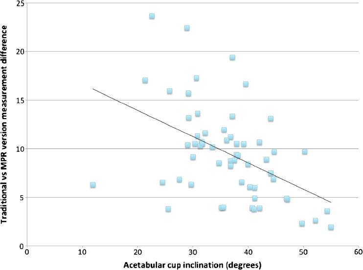
The “traditional” axial CT and “corrected” acetabular version measurements differed from one another in every case as expected, with the traditional measurement being on average 9.5° higher than the corrected version measurement. However, the degree of absolute discrepancy demonstrated a statistically significant increase as the acetabular inclination angle decreased (Pearson correlation coefficient (rho) = −0.47, p value = 0.0002) (Fig. 5).
Fig. 5.
A scatterplot of the data demonstrates the inverse relationship between acetabular cup inclination and the discrepancy between traditional and corrected version measurements. Differences were calculated based on an average of all four measurements per method used.
There was no decrease in reproducibility when utilizing multi-planar reformation techniques for CT version measurement. Intraobserver reliability as measured by ICC was 0.98 for both radiologists (0.96–0.98 bounds) for traditional methods and 0.95–0.97 (0.95–0.98 bounds) for corrected acetabular version measurements. Interobserver reliability as measured by ICC was 0.98 (0.95–0.99 bounds) for the traditional method, while interobserver reliability for corrected version measurements was 0.95 (0.92–0.98 bounds). Both traditional and corrected measurements of version were within the statistically acceptable range based on GLM statistical analysis.
Discussion
Proper understanding of the version and inclination of the acetabular component of a hip prosthesis is essential for the measurements of these angles. These two angles are paramount in the assessment of a hip prosthesis and have significant role in the ultimate clinical outcomes of these arthroplasty procedures. We demonstrate in this study that the most commonly utilized CT technique for measuring acetabular version has inherent variability that is correlated with acetabular inclination and that a “corrected” method of CT measurement which is independent of acetabular inclination is no less reproducible when compared with the axial measurement method.
There are several limitations to this study. First, the AP scout image is not actually an AP radiograph and is thus obtained with the patient supine rather than standing. This has the potential to lead to discrepancies in the measured inclination of the cup if the pelvis is in a different state of tilt. Second, while there is literature to support the higher accuracy of multi-planar CT reformations as compared to other methods, there is no consensus “gold standard” for which method is the most accurate reflection of the true orientation of the cup. Finally, not all institutions have access to software packages that allow easy multi-planar reformations. However, this software post-processing capability is rapidly becoming widely available, and we feel that increasing awareness of the potential limitations of axial anteversion/retroversion declarations still holds merit.
Only CT scans with multi-planar reformation capability allow retroactive manipulation of the imaging data to create a true measurement along the axis of rotation of the cup. All version measurements performed with the CT scan can be made independently of the position of the patient on the CT scanner table and can be determined based on reformatted CT images. This type of analysis allows for true comparison of “version” between research subjects, without the variability introduced by differing cup inclination or patient position. Previous work has demonstrated that CT measurements are as accurate or more accurate than radiographs when compared with intraoperative or in vitro model standards [6]; however, the obliquity or inclination of the acetabulum has been shown to have an impact on measured acetabular version [17].
Thus, it is our opinion that acetabular version measurements should be performed along the axis of rotation of the cup, regardless of its position in the coronal plane. Such measurement requires no correction for inclination since one is always imaging along the base of the cup. CT imaging with multi-planar reformation allows the imager to easily view the acetabular cup along its axis of rotation with a few simple manipulations of the images, allowing the version of the cup to be evaluated accurately. Furthermore, this method can be applied regardless of what type of computed navigation was used intraoperatively and can be created and interpreted without sacrificing the reproducibility which has been previously demonstrated by CT [2, 12, 18].
While there are many methods for defining and measuring acetabular version, accurate description is ultimately an important part of post-arthroplasty evaluation and outcomes research.
Our analysis demonstrates that the inherent variability between these measurement methods is further exacerbated by low acetabular inclination. We therefore feel that the most consistent and reproducible measurements should take advantage of the isotropic data obtained with modern multi-detector CT and eliminate the impact of inclination via multi-planar reformation. This method for calculating acetabular version is rapid, reproducible, and would allow more direct comparisons between arthroplasty techniques and the subsequent clinical and radiologic outcomes of those techniques.
The ability to perform multi-planar reformation on axially acquired CT data has already changed the way that many imaging queries are approached in other spheres of medicine. The evaluation of acetabular version following hip arthroplasty is an excellent opportunity to apply the technique in a context that will improve reproducibility and applicability when compared to the myriad of other potential techniques available for acetabular version evaluation.
Electronic Supplementary Material
(PDF 1,224 kb)
(PDF 1,224 kb)
(PDF 1,224 kb)
Acknowledgments
The authors wish to thank Cyndi Conklin—Hospital for Special Surgery Department of Digital Media.
Disclosures
ᅟ
Conflict of Interest
Michael Loftus, MD, MBA and Bernard Ghelman, MD have declared that they have no conflict of interest. Yan Ma, PhD reports grants from NIH during the conduct of the study.
Human/Animal Rights
All procedures followed were in accordance with the ethical standards of the responsible committee on human experimentation (institutional and national) and with the Helsinki Declaration of 1975, as revised in 2008 (5).
Informed Consent
Informed consent was waived from all patients for being included in the study.
Required Author Forms
Disclosure forms provided by the authors are available with the online version of this article.
Footnotes
Level of Evidence: Diagnostic Study, level III
References
- 1.Ackland MK, Bourne WB, Uhthoff HK. Anteversion of the acetabular cup. Measurement of angle after total hip replacement. J Bone Jt Surger. 1986;68(3):409–413. doi: 10.1302/0301-620X.68B3.3733807. [DOI] [PubMed] [Google Scholar]
- 2.Barmeir E, Dubowitz B, Roffman M. Computed tomography in the assessment and planning of complicated total hip replacement. Acta Orthop Scand. 1982;53:597–604. doi: 10.3109/17453678208992265. [DOI] [PubMed] [Google Scholar]
- 3.Blendea S, Eckman K, Jaramaz B, Levison TJ, Digioia AM. Measurements of acetabular cup position and pelvic spatial orientation after total hip arthroplasty using computed tomography/radiography matching. Comput aided Surg Off J Int Soc Comput Aided Surg. 2005;10(1):37–43. doi: 10.3109/10929080500178032. [DOI] [PubMed] [Google Scholar]
- 4.Chandler DR, Tarr RR, Gruen TA, Sarmiento A. Radiographic assessment of acetabular cup orientation. A new design concept. Clin Orthop Relat Res. 1984;186:60–64. [PubMed] [Google Scholar]
- 5.D’Lima DD, Urquhart AG, Buehler KO, Walker RH, Colwell CW. The effect of the orientation of the acetabular and femoral components on the range of motion of the hip at different head-neck ratios. J Bone Jt Surg. 2000;82(3):315–321. doi: 10.2106/00004623-200003000-00003. [DOI] [PubMed] [Google Scholar]
- 6.Ghelman B, Kepler CK, Lyman S, Della Valle AG. CT outperforms radiography for determination of acetabular cup version after THA. Clin Orthop Relat Res. 2009;467(9):2362–2370. doi: 10.1007/s11999-009-0774-1. [DOI] [PMC free article] [PubMed] [Google Scholar]
- 7.Hassan DM, Johnston GHF, Dust WNC, Watson G, Dolovich AT. Accuracy of intraoperative assessment of acetabular prosthesis placement. J Arthroplasty. 1998;13:80–84. doi: 10.1016/S0883-5403(98)90079-1. [DOI] [PubMed] [Google Scholar]
- 8.Herrlin K, Pettersson H, Selvik G. Comparison of two- and three-dimensional methods for assessment of orientation of the total hip prosthesis. Acta radiol. 1988;29(3):357–361. doi: 10.3109/02841858809171914. [DOI] [PubMed] [Google Scholar]
- 9.Kummer FJ, Shah S, Iyer S, DiCesare PE. The effect of acetabular cup orientations on limiting hip rotation. J Arthroplasty. 1999;14:509–513. doi: 10.1016/S0883-5403(99)90110-9. [DOI] [PubMed] [Google Scholar]
- 10.Lewinnek GE, Lewis JL, Tarr R, Compere CL, Zimmerman JR. Dislocations after total hip-replacement arthroplasties. J Bone Jt Surg. 1978;60(2):217–220. [PubMed] [Google Scholar]
- 11.Mian SW, Truchly G, Pflum FA. Computed tomography measurement of acetabular cup anteversion and retroversion in total hip arthroplasty. Clin Orthop Relat Res. 1992;276:206–209. [PubMed] [Google Scholar]
- 12.Murphy SB, Simon SR, Kijewski PK, Wilkinson RH, Griscom NT. Femoral anteversion. J Bone Joint Surg Am. 1987;69:1169–1176. [PubMed] [Google Scholar]
- 13.Murray MM. Current status and potential of primary ACL repair. Clin Sports Med. 2009;28(1):51–61. doi: 10.1016/j.csm.2008.08.005. [DOI] [PMC free article] [PubMed] [Google Scholar]
- 14.Murray D. The definition and measurement of acetabular orientation. J Bone Jt Surgery, Br Vol. 1993: 228–232. Available at: http://www.bjj.boneandjoint.org.uk/content/75-B/2/228.abstract. Accessed June 18, 2013. [DOI] [PubMed]
- 15.Najarian BC, Kilgore JE, Markel DC. Evaluation of component positioning in primary total hip arthroplasty using an imageless navigation device compared with traditional methods. J Arthroplasty. 2009;24:15–21. doi: 10.1016/j.arth.2008.01.004. [DOI] [PubMed] [Google Scholar]
- 16.Olivecrona H, Weidenhielm L, Olivecrona L, et al. Spatial component position in total hip arthroplasty. Accuracy and repeatability with a new CT method. Acta radiol. 2003;44(1):84–91. [PubMed] [Google Scholar]
- 17.Van Bosse HJP, Lee D, Henderson ER, Sala DA, Feldman DS. Pelvic positioning creates error in CT acetabular measurements. Clinical Orthopaedics and Related Research. 2011;469(6):1683–91. doi: 10.1007/s11999-011-1827-9. [DOI] [PMC free article] [PubMed] [Google Scholar]
- 18.Wines AP, McNicol D. Computed tomography measurement of the accuracy of component version in total hip arthroplasty. J Arthroplast. 2006;21:696–701. doi: 10.1016/j.arth.2005.11.008. [DOI] [PubMed] [Google Scholar]
- 19.Woo R, Morrey B. Dislocations after total hip arthroplasty. J Bone Jt Surg Am. 1982; 64(9). Available at: http://surgicaltechniques.jbjs.org/data/Journals/JBJS/608/1295.pdf. Accessed June 20, 2013. [PubMed]
Associated Data
This section collects any data citations, data availability statements, or supplementary materials included in this article.
Supplementary Materials
(PDF 1,224 kb)
(PDF 1,224 kb)
(PDF 1,224 kb)


