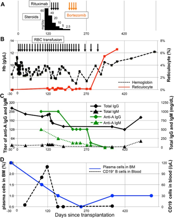
Fig 1.

Clinical course of the reported patient with immune hemolytic anemia. (A) Treatment course of steroids (black bars), rituximab (black arrows), and bortezomib (orange arrows). The height of the black bars shows the relative dose of steroids. (B) Graph of Hb (black dashed line) and reticulocyte percentage (red solid line) with timing of RBC transfusions (black arrows). (C) Graph of total IgG (black solid line with diamonds) and IgM (black dashed line with triangles) and anti-A IgG titers (green solid line with diamonds) and anti-A IgM titers (green dashed line with triangles). Semiquantitative titration of anti-A of both the IgM and the IgG subclasses were performed using standard methods. Briefly, 2% suspensions of A1 reagent RBCs were mixed with patient plasma in test tubes. For IgM detection, after 15 minutes of incubation at room temperature the samples were centrifuged and macroscopic agglutination was quantified; for IgG, samples were incubated for 60 minutes at 37°C, washed to remove unbound antibody, incubated with anti-human IgG, and then centrifuged to score agglutination.13 (D) Numbers of CD19+ cells in blood (black dashed line) were assessed by flow cytometry, and plasma cells percentage score for nucleated cells in bone marrow (blue solid line) were estimated blinded to case identical number and date.
