Abstract
Background
For laparoscopic Heller myotomy (LHM), the optimal myotomy length proximal to the esophagogastric junction (EGJ) is unknown. In this study, we used a functional lumen imaging probe (FLIP) to measure EGJ distensibility changes resulting from variable proximal myotomy lengths during LHM and peroral esophageal myotomy (POEM).
Methods
Distensibility index (DI) (defined as the minimum cross-sectional area at the EGJ divided by pressure) was measured with FLIP after each operative step. During LHM and POEM, each patient’s myotomy was performed in two stages: first, a myotomy ablating only the EGJ complex was created (EGJ-M), extending from 2cm proximal to the EGJ, to 3cm distal to it. Next, the myotomy was lengthened 4cm further cephalad to create an extended proximal myotomy (EP-M).
Results
Measurements were performed in 12 patients undergoing LHM and 19 undergoing POEM. LHM resulted in an overall increase in DI (1.6 ±1 vs. 6.3 ±3.4 mm2/mmHg, p<.001). Creation of an EGJ-M resulted in a small increase (1.6 to 2.3 mm2/mmHg, p<.01) and extension to an EP-M resulted in a larger increase (2.3 to 4.9 mm2/mmHg, p<.001). This effect was consistent, with 11 (92%) patients experiencing a larger increase after EP-M than after EGJ-M. Fundoplication resulted in a decrease in DI and deinsufflation an increase. POEM resulted in an increase in DI (1.3 ±1 vs. 9.2 ±3.9 mm2/mmHg, p<.001). Both creation of the submucosal tunnel and performing an EGJ-M increased DI, whereas lengthening of the myotomy to an EP-M had no additional effect. POEM resulted in a larger overall increase from baseline than LHM (7.9 ±3.5 vs. 4.7 ±3.3 mm2/mmHg, p<.05).
Conclusions
During LHM, an extended proximal myotomy was necessary to normalize distensibility, whereas during POEM, a myotomy confined to the EGJ complex was sufficient. In this cohort, POEM resulted in a larger overall increase in EGJ distensibility.
Keywords: achalasia, peroral endoscopic myotomy, laparoscopic Heller myotomy, functional lumen imaging probe, esophageal physiology
Introduction
In patients with achalasia, an immune-mediated loss of esophageal enteric neurons results in a failure of esophagogastric junction (EGJ) relaxation and aperistalsis of the esophageal body in response to swallowing. This esophageal dysmotility causes the characteristic symptoms of progressive dysphagia, regurgitation, and weight loss1. Procedural treatments for achalasia seek to disrupt the EGJ muscle complex, thus reducing EGJ pressure to allow for the passive transit of food boluses into the stomach. Current standard-of-care consists of either endoscopic pneumatic dilation or surgical laparoscopic Heller myotomy (LHM) with partial fundoplication. While a recent randomized trial suggested similar outcomes at two-years after these procedures2, considerable evidence exists that LHM results in more durable symptomatic relief, without the need for repeat interventions3,4. A recently introduced procedure, peroral esophageal myotomy (POEM), creates a surgical myotomy across the EGJ completely endoscopically, and has been shown in several series to result in excellent short-term symptomatic relief and reduction in EGJ pressure5–7.
The primary goal of any surgical myotomy (either LHM or POEM) is to divide the muscle bundles that make up the EGJ complex, in order to reduce esophageal outflow obstruction. However, there is little evidence regarding the optimal length of this myotomy for either procedure. A single retrospective study by Wright and colleagues compared LHM myotomy lengths distal to the EGJ and found that an extended distal length (at least 3 cm versus 1.5 cm) resulted in superior symptomatic outcomes8. Based on these results, such a distal myotomy extension is now considered standard-of-care9. The proximal extent of the myotomy during LHM is typically 6–8 cm cephalad to the EGJ2,10,11, but, to our knowledge, no study has compared outcomes between differential proximal myotomy lengths. This “standard” proximal length has been determined primarily by technical considerations, as it is typically the maximum length that can safely be achieved via a laparoscopic, transhiatal approach. However, this surgical convention has little physiologic basis, as the high-pressure zone of the EGJ complex is on average less than 4 cm in total length, with less than 2 cm lying cephalad to the squamocolumnar junction (SCJ)12,13.
If performing a shorter myotomy proximally that ablates just the EGJ complex could achieve the same normalization of EGJ physiology as a longer one, there could be several benefits to this modification. During LHM, less mediastinal dissection of the esophagus would be required, potentially decreasing the incidence of esophageal perforation, Vagus nerve injury, and pleural tears. During POEM, a shorter myotomy would allow for creation of a shorter submucosal tunnel, thus decreasing operative times and potentially lessening the incidence of mucosal perforations, capnothorax, and pneumoperitoneum. Additionally, there is emerging evidence that many patients regain a degree of esophageal peristalsis after both LHM and POEM14. Preservation of esophageal muscle fibers proximal to the EGJ complex might augment this effect, potentially reducing both dysphagia and iatrogenic gastroesophageal reflux (GER) postoperatively.
With this in mind, we sought to compare the physiologic effects of a myotomy ablating only the EGJ complex (extending from 2 cm cephalad to the EGJ, to 3 cm distal to it) with a myotomy with an additional proximal extension (from 6 cm cephalad to the EGJ, to 3 cm distal) for both LHM and POEM procedures. To allow for each patient to serve as their own control and to end each procedure with a conventional long proximal myotomy extension (our own standard practice prior to this study), during each operation we first performed an EGJ myotomy (EGJ-M) as described above, and then sequentially lengthened the myotomy 4 cm further cephalad to end with an extended proximal myotomy (EP-M). We used a functional lumen imaging probe (FLIP) to measure EGJ distensibility after each myotomy configuration, allowing for a comparison of their physiologic effects. Distensibility, as measured by FLIP, has been shown to better correlate with postoperative symptomatic results than manometric measurements15,16. Additionally, we have previously shown that FLIP can be used intraoperatively to measure distensibility changes after each of the key steps of both LHM and POEM17. We hypothesized that creation of an EGJ-M would be sufficient to normalize EGJ distensibility, thus providing initial evidence that an EP-M is not necessary during either LHM or POEM.
Methods
Patient selection
During the study period, patients presenting to the surgery clinic at a single institution for treatment of achalasia were counseled regarding the available options (pneumatic dilation, LHM, or POEM) and chose among them in consultation with their physicians. Patients undergoing LHM or POEM were approached regarding undergoing stepwise intraoperative FLIP measurements. Additional FLIP study eligibility criteria included age ≥18 years and a diagnosis of achalasia confirmed by esophageal manometry. All patients signed an informed consent for intraoperative FLIP measurements prior to their procedure and measurements were conducted according to a study protocol approved by the Northwestern Institutional Review Board.
Preoperative demographics and high-resolution manometry
Demographic and disease-specific information including age, sex, duration of symptoms, Eckardt symptom score18, prior endoscopic treatment for achalasia, and body mass index was collected prospectively. Patients underwent preoperative high-resolution manometry to confirm the diagnosis of achalasia. Manometry data were evaluated using esophageal pressure topography19 and interpreted according to the Chicago Classification of esophageal motility disorders20.
LHM operative technique
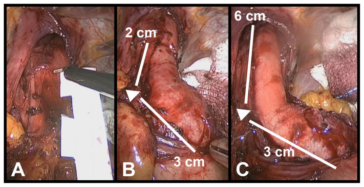
LHM operations were performed by one of two surgeons (E.H. and N.S.). Our operative technique for LHM has been previously described in detail21. Briefly, after creation of a pneumoperitoneum, trocar insertion, and liver retractor placement, the crura are opened widely and the esophagus is dissected high into the mediastinum. The short gastric vessels are divided to allow for subsequent partial fundoplication. For the purpose of this study, the myotomy was then performed in two stages: first, to create an EGJ-M and then, an EP-M. To perform the EGJ-M, a sterile ruler was used to measure from 2 cm proximal to the EGJ to 3 cm distal to the EGJ, and the anterior midline of the esophagus between these two points was marked with electrocautery. In the initial three LHM cases in this study, the EGJ was identified by visualizing the location of the SCJ on upper endoscopy and the angle of His laparoscopically. The axial location of these two markers matched in each of these cases, and this point additionally correlated with the minimum CSA measured by FLIP. From that point on in the series, the EGJ was defined laparoscopically by the angle of His, and only if uncertainty existed as to its location (e.g. due to a large or obscuring gastric fat pad), the position of the SCJ was confirmed endoscopically. A myotomy of both the longitudinal and circular muscle layers was then performed along the previously marked line. After a FLIP measurement was performed (described below), the ruler was used to measure 4 cm further cephalad and the myotomy was extended over this length to create the EP-M. Figure 1 illustrates this process of stepwise myotomy creation. A partial fundoplication was then performed. We prefer a posterior Toupet fundoplication, unless this configuration results in excessive anterior angulation of the EGJ, in which case an anterior Dor fundoplication was created.
Figure 1.

The stepwise myotomy creation during a laparoscopic Heller myotomy is shown here. First, a sterile ruler, with notches cut at 1 cm increments, is used to measure from 2 cm proximal to the esophagogastric junction (EGJ), to 3 cm distal to it (panel A). An EGJ-myotomy is then created between these two points (panel B). This myotomy is then lengthened 4 cm proximally to create the final extended proximal (EP) myotomy (panel C). The arrow points to the location of the EGJ in panels B and C.
POEM operative technique
POEM procedures were performed by one of the same two surgeons who performed LHM, or by both of these surgeons conjointly. Our operative technique closely follows that of Inoue and colleagues5, and has been previously described in detail7. A standard high-definition gastroscope with a transparent dissecting cap and CO2 insufflation was used. A saline solution was first injected into the anterior esophageal wall 12 cm proximal to the EGJ, in order to create a submucosal fluid bleb. A mucosotomy was then made overlying the fluid bleb and the scope maneuvered into the submucosal space. A submucosal tunnel was then dissected to at least 3 cm beyond the EGJ. A selective myotomy of the inner, circular muscle layer was then performed in two segments, in a fashion identical to LHM: first an EGJ-M (from 2 cm proximal to the EGJ, to 3 cm distal to it) and second an EP-M (extending the myotomy 4 cm further proximally). The endoscope shaft markings were used to measure myotomy distances in relation to the EGJ. After myotomy completion, the mucosotomy was closed with endoscopic clips.
FLIP
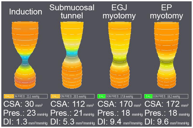
Intraoperative measurements were taken using a commercially available FLIP system (EndoFLIP; Crospon Ltd., Galway, Ireland) and probes (EF-325N; Crospon) that has previously been described in detail22,23. FLIP is a catheter-based probe that contains 17 ring-electrodes at 5 mm intervals, housed within a bag that can be variably inflated with saline solution. Impedance planimetry measurements are used to calculate bag cross-sectional areas (CSA) at the level of each electrode pair. Therefore, when the catheter is placed across the EGJ, these measurements represent the 16 CSAs at 5 mm intervals along an 8 cm segment of esophagus, EGJ, and stomach, giving a representation of luminal geometry (Figure 2). The probe also contains a solid-state pressure transducer that measures intra-bag pressure.
Figure 2.

FLIP measurements taken after each step of a POEM procedure: induction of anesthesia, creation of the submucosal tunnel, creation of an EGJ myotomy, and subsequent lengthening to an extended proximal (EP) myotomy. The primary outcome measure of distensibility index (DI) is calculated by dividing the minimum cross-sectional area (CSA) at the EGJ by intra-bag pressure.
The EGJ distensibility index (DI) is defined as the minimum CSA (i.e. narrowest portion of the EGJ) divided by intra-bag pressure. The median value of minimum CSA and intra-bag pressure measurements at each distension volume were calculated using MATLAB software (MathWorks; Natick, MA), and used for subsequent analysis when determining the DI.
Intraoperative FLIP protocol
Prior to probe insertion, an automated purge sequence was used to evacuate air from the FLIP probe and the pressure transducer was zeroed to atmospheric pressure. During both LHM and POEM, after induction of anesthesia, paralysis, and endotracheal intubation, an upper endoscopy was performed and the FLIP probe was advanced down the esophagus under direct endoscopic visualization. Probe position across the EGJ was confirmed endoscopically and by seeing an “hour glass” anatomic configuration on the FLIP monitor.
FLIP measurements were taken after each step of LHM and POEM, including EGJ-M and EP-M, using bag distension volumes of 30 ml and 40 ml. Between measurements the bag was deflated and the probe was advanced into the stomach. During LHM, FLIP measurements were performed at seven time points: 1) after induction of anesthesia, paralysis, and endotracheal intubation, 2) after creation of CO2 pneumoperitoneum to 15 mmHg, 3) after completion of hiatal dissection and esophageal mobilization, 4) after EGJ-M, 5) after EP-M, 6) after partial fundoplication, and 7) after final deinsufflation of pneumoperitoneum. During POEM, FLIP measurements were performed at four time points: 1) after induction of anesthesia, paralysis, and endotracheal intubation, 2) after submucosal tunnel creation, 3) after EGJ-M, and 4) after EP-M.
Statistical analysis
Data analysis was performed using SPSS software (version 22; IBM, Armonk, NY). For continuous variables, comparisons between time points within the same procedure were performed using a paired t-test. Comparisons between procedure types were performed using an independent t-test. Categorical variables were compared using a Fisher exact test. A two-tailed p-value of < .05 was used to determine statistical significance in all cases. Values throughout are presented as mean ± standard deviation.
Results
Stepwise myotomies with intraoperative FLIP measurements were performed in 12 patients undergoing LHM and 19 patients undergoing POEM. Table 1 shows the patients’ preoperative demographics, disease characteristics, and manometric measurements, which were all similar between the LHM and POEM groups. Baseline DI was also similar between the procedure groups. Of the four LHM patients with prior endoscopic treatment for achalasia, two had pneumatic dilations (one with a single dilation and one with two previous dilations) and two had botulinum toxin injections (one with a single injection and one with three prior injections). Of the four POEM patients with prior endoscopic treatment, two had a single pneumatic dilation and two had a single botulinum toxin injection. Patients with, and those without, prior endoscopic treatment had a similar baseline DI.
Table 1.
Preoperative demographic data, disease characteristics, and manometric measurements in the laparoscopic Heller myotomy (LHM) and POEM groups.
| Demographics | LHM | POEM | p-value |
|---|---|---|---|
| Number | 12 | 19 | |
| Female | 7 (58%) | 6 (32%) | .26 |
| Age | 54 ± 14 | 49 ± 16 | .41 |
| Body mass index (kg/m2) | 27 ± 5 | 25 ± 6 | .54 |
| Achalasia symptoms and prior treatment | |||
| Eckardt score (scale 0 – 12) | 6 ± 3 | 7 ± 2 | .27 |
| Duration of symptoms (years) | 6 ± 8 | 3 ± 4 | .41 |
| Prior endoscopic treatment | 4 (33%) | 4 (21%) | .68 |
| Manometric characteristics | |||
| 4-s integrated relaxation pressure (mmHg) | 24 ± 16 | 26 ± 13 | .75 |
| Achalasia subtype | .99 | ||
| I | 30% | 26% | |
| II | 60% | 63% | |
| III | 10% | 11% |
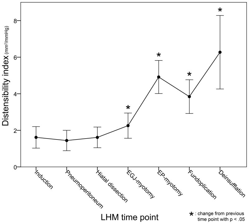
LHM resulted in an overall increase in DI at a distension volume of 40 ml (mean pre 1.6 ± 1 mm2/mmHg vs. mean post 6.3 ± 3.4 mm2/mmHg, p < .001). The stepwise changes in DI during LHM are shown in Table 2. Creation of pneumoperitoneum and hiatal dissection to mobilize the esophagus had no effect on DI. Creation of an EGJ-M resulted in a small but significant increase (mean 1.6 ± 1 mm2/mmHg to mean 2.3 ± 1.2 mm2/mmHg, p < .01) and extension of the myotomy to an EP-M resulted in a larger increase (mean 2.3 ± 1.2 mm2/mmHg to mean 4.9 ± 1.6 mm2/mmHg, p < .001), which is illustrated in Figure 3. This effect was consistent, with 11 of the 12 LHM patients experiencing a larger increase in DI after EP-M than after EGJ-M (mean increase after EGJ-M = 0.7 mm2/mmHg vs. mean increase after EP-M = 2.6 mm2/mmHg, p < .01). Partial fundoplication (5 Toupet, 7 Dor) then resulted in a decrease in DI and final deinsufflation of pneumoperitoneum an increase. Results at a distension volume of 30 ml were similar.
Table 2.
Distensibility index (DI) at each operative time point during laparoscopic Heller myotomy (LHM) at 30 ml and 40 ml distension volumes.
| 30 ml distension volume | 40 ml distension volume | |||
|---|---|---|---|---|
| LHM operative step | Mean DI (mm2/mmHg) | p-value versus previous time point | Mean DI (mm2/mmHg) | p-value versus previous time point |
| Induction | 2.2 ± 1.7 | - | 1.6 ± 1 | - |
| Insufflation | 1.4 ± 0.8 | .14 | 1.4 ± 1 | .58 |
| Hiatal dissection | 1.5 ± 0.8 | .53 | 1.6 ± 1 | .36 |
| EGJ myotomy (EGJ-M) | 1.9 ± 1 | .01* | 2.3 ± 1.2 | <.01* |
| Extended proximal myotomy (EP-M) | 4.7 ± 1.6 | <.001* | 4.9 ± 1.6 | <.001* |
| Partial fundoplication | 3.4 ± 1.9 | <.01* | 3.8 ± 1.6 | .001* |
| Deinsufflation | 6.9 ± 3.3 | <.001* | 6.3 ± 3.4 | <.01* |
| Overall change | 4.7 ± 2.8 | <.001* | 4.7 ± 3.3 | <.001* |
p < .05
Figure 3.
Stepwise changes in EGJ distensibility index (DI) are shown over the subsequent steps of laparoscopic Heller myotomy (LHM), measured at a 40 ml distension volume. Creation of pneumoperitoneum and hiatal dissection had no effect on DI. Creation of an EGJ myotomy resulted in a small but significant increase and extension of the myotomy to an extended proximal (EP) myotomy resulted in a larger increase. Partial fundoplication then resulted in a decrease in DI and final deinsufflation of pneumoperitoneum an increase.
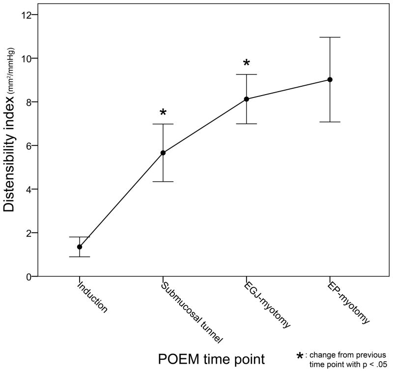
POEM also resulted in an overall increase in DI (mean pre 1.3 ± 1 mm2/mmHg vs. mean post 9.2 ± 3.9 mm2/mmHg, p < .001), and the stepwise changes during the procedure are shown in Table 3. Both creation of the submucosal tunnel and performing an EGJ-M resulted in increases in DI, whereas lengthening of the myotomy to an EP-M had no additional effect (Figure 4). Table 4 shows a comparison of overall changes in DI between procedures, with POEM resulting in a larger mean increase from baseline than LHM (7.9 ± 3.5 mm2/mmHg vs. 4.7 ± 3.3 mm2/mmHg, p < .05). Results were similar at a 30 ml distension volume.
Table 3.
Distensibility index (DI) at each operative time point during POEM at 30 ml and 40 ml distension volumes.
| 30 ml distension volume | 40 ml distension volume | |||
|---|---|---|---|---|
| POEM operative step | Mean DI (mm2/mmHg) | p-value versus previous time point | Mean DI (mm2/mmHg) | p-value versus previous time point |
| Induction | 1.8 ± 1.4 | - | 1.3 ± 1 | - |
| Submucosal tunnel | 6 ± 3.9 | <.001* | 5.7 ± 2.9 | <.001* |
| EGJ myotomy (EGJ-M) | 8.1 ± 2.8 | .01* | 8 ± 2.9 | .001* |
| Extended proximal myotomy (EP-M) | 9.3 ± 4.1 | .07 | 9.2 ± 3.9 | .12 |
| Overall change | 7.5 ± 3.4 | <.001* | 7.9 ± 3.5 | <.001* |
p < .05
Figure 4.
Stepwise changes in EGJ distensibility index (DI) are shown over the subsequent steps of POEM, measured at a 40 ml distension volume. Creation of the submucosal tunnel and performing an EGJ myotomy both increased DI, whereas lengthening to an extended proximal myotomy had no added effect.
Table 4.
Changes from baseline DI for LHM and POEM. POEM resulted in a larger increase than LHM at both 30 and 40 ml bag distension volumes.
| Mean change in DI from baseline (mm2/mmHg) | LHM | POEM | p-value |
|---|---|---|---|
| 30 ml distension volume | 4.7 ± 2.8 | 7.5 ± 3.4 | <.05 |
| 40 ml distension volume | 4.7 ± 3.3 | 7.9 ± 3.5 | <.05 |
Discussion
In this study we assessed the physiologic effects of two surgical myotomy configurations during operations for achalasia: one that ablates only the EGJ complex compared with a myotomy that extends an additional 4 cm proximally. Patients with achalasia experience symptoms due to a functional obstruction that occurs because of a non-relaxing EGJ. Therefore, we hypothesized that a myotomy confined to this area would be effective in normalizing EGJ distensibility, whereas the conventional surgical approach of extending this myotomy onto the lower esophagus would have no further physiologic benefit. For the EGJ-M, we chose a distal length of 3 cm based on prior work by Wright and colleagues suggesting the superiority of such an extension8, as it disrupts the sling fibers of the gastric cardia that augment EGJ pressurization. A proximal length of 2 cm was chosen because the high-pressure zone of the EGJ complex is less than 2 cm cephalad to the SCJ in the vast majority of patients12.
The most striking and surprising result of this study was that for LHM, our hypothesis appears to be incorrect. Although creating an EGJ-M resulted in a significant increase in DI (from 1.6 to 2.3 mm2/mmHg), this change was relatively small and not sufficient to normalize EGJ physiology. Prior studies have shown that healthy controls have an average DI between 5 and 8 mm2/mmHg15,16, with a lower limit of normal (10th percentile) of 2.9 mm2/mmHg16. More importantly, for patients with achalasia, post-intervention DI is strongly correlated with symptomatic outcomes15,16. Two studies established similar cutoff DI values of 2.8 and 2.9 mm2/mmHg for predicting treatment success, and patients with a DI of less than 2.9 mm2/mmHg were 12-fold more likely to have persistent symptoms (i.e. an Eckardt score > 3)16. In our study cohort, only 3 (25%) LHM patients had exceeded this DI cutoff after EGJ-M, whereas all patients reached it after cephalad lengthening to an EP-M. Based on these findings, it appears that it is necessary to perform an extended proximal myotomy during LHM in order to normalize EGJ distensibility.
Patients undergoing POEM exhibited a markedly different pattern of physiologic change during their operations. After performing only an EGJ-M, these patients had already achieved a mean DI of 8 mm2/mmHg and all 19 patients were above the aforementioned cutoff of 2.9 mm2/mmHg. In contrast to LHM, lengthening of the POEM myotomy to an EP-M configuration did not further augment DI. The etiology of this difference between POEM and LHM could lie in the step of submucosal dissection, which is unique to POEM. As we have previously shown17, creation of the submucosal tunnel (which starts 12 cm proximal to the EGJ), resulted in a profound increase in EGJ distensibility. Therefore in POEM patients, while the subsequent step of EGJ-M divided muscle to a point 2 cm proximal to the EGJ, the submucosal layer had already been disrupted further cephalad. This is in contrast to LHM, where at the completion of EGJ-M, no dissection (of either submucosa or muscle) had been performed further proximally. This difference provides evidence that the submucosal layer of the distal esophagus may play an important role in the architecture and structural integrity of the EGJ complex. Thus, the substantial increase in EGJ distensibility that we observed after EP-M during LHM may have been due to a disruption of the submucosa, rather than muscle, at this level. Following from this, because this submucosal segment had already been ablated during POEM, division of proximal muscle fibers during the EP-M step had no additional effect on EGJ physiology. During POEM, performing a myotomy above the EGJ complex may not be necessary, so long as the submucosal tunnel extends at least 6 cm cephalad. This modification would allow for creation of a shorter submucosal tunnel (starting 8 cm, rather than 12 cm above the SCJ), decreased operative times, and potentially fewer tunneling-related complications. However, the normalization of DI we observed after POEM EGJ-M may not be a durable effect, as postoperative scarring in the submucosal layer could cause re-adherence of the mucosa and muscle layers. Additionally, the increase in DI observed after tunneling may not be due to a disruption of the mechanical properties of the submucosa per se. Carbon dioxide has been shown to inhibit smooth muscle cell contraction in an in vitro model24, and thus exposure of the circular muscle layer of the esophagus to CO2 as a result of tunnel creation may have precipitated a transient increase in EGJ distensibility. For these reasons, potential future use of a shorter proximal myotomy during POEM should only be done in the context of a consented research study, ideally with patients randomized between short and traditional proximal myotomy lengths.
In addition to these differences in the pattern of change between the two procedures, POEM resulted in a significantly larger overall increase in DI than LHM. While only LHM includes the step of partial fundoplication, the decrease in DI it caused (of 1.1 mm2/mmHg) was not enough to entirely account for the difference in final DI between the two operations (LHM 6.3 mm2/mmHg vs. POEM 9.2 mm2/mmHg). In an earlier study, we found similar final DI measurements between the two operations17, and the higher DI achieved during POEM in the present cohort could be due to a learning curve effect. It is possible that with experience, the endoscopic visualization utilized during POEM allows for a more meticulous and complete muscle division or that the wide submucosal dissection performed during POEM is not fully achieved during LHM. However, it is not clear if the higher DI observed after POEM is of clinical benefit. While it has been shown that a low DI is associated with worse dysphagia post-intervention, too high a DI may predispose to iatrogenic GER. Patients with typical gastroesophageal reflux disease have been shown to have a higher DI than healthy controls25, but the relationship between EGJ distensibility and iatrogenic GER after interventions for achalasia has yet to be studied. Thus far, there has been only one non-randomized comparison of 24-h pH studies following LHM and POEM, which showed similar rates of pathologic esophageal acid exposure (LHM 32% vs. POEM 39%, p = ns)6, and further investigation is need to better understand the true incidence of, and risk factors for, GER after POEM.
This study has some limitations. Most importantly, we only compared intraoperative DI between the two myotomy constructions, and all patients ended their procedure with an EP-M, making further comparison of clinical outcomes impossible. While it has been shown that post-intervention DI correlates closely with symptomatic outcomes, it has yet to be demonstrated whether intraoperative DI measurements are predictive of postoperative symptomatic or physiologic treatment success. The differences observed between LHM and POEM in distensibility change after an EGJ-M in this study relied on intraoperative assessment of the location of the EGJ, and thus imprecision in identifying the EGJ accurately may have confounded these results. Lastly, patients in this study were not randomized to procedure type, and although their preoperative demographic and disease characteristics were similar, unmeasured differences between the two groups may have existed.
In conclusion, during LHM, an extended proximal myotomy to 6 cm cephalad to the EGJ was necessary to normalize EGJ distensibility. Conversely, during POEM, a myotomy confined to the EGJ complex was sufficient to normalize distensibility, so long as a proximal submucosal tunnel was created first. While both procedures resulted in significant increases in EGJ distensibility, the increase after POEM appeared to be larger. Further study is needed to correlate intraoperative FLIP measurements with postoperative symptomatic and physiologic outcomes.
Acknowledgments
The authors would like to acknowledge Rowena Martinez, RN, and Colleen Krantz, RN, for their help coordinating the clinical aspects of this study.
Grants
This work was supported by the National Institute of Diabetes and Digestive and Kidney Diseases Grant R01 DK-079902 (J.E. Pandolfino).
Footnotes
Disclosures
Nathaniel Soper is on the scientific advisory boards of TransEnterix and Miret Surgical, which are unrelated to this study. Ezra Teitelbaum, John Pandolfino, Peter Kahrilas, Lubomyr Boris, Frederic Nicodeme, Zhiyue Lin, and Eric Hungness have no conflicts of interest or financial ties to disclose.
To be presented at SAGES: Oral presentation number S016 - Foregut Session. April 5th, 2014.
References
- 1.Triadafilopoulos G, Boeckxstaens GE, Gullo R, Patti MG, Pandolfino JE, Kahrilas PJ, Duranceau A, Jamieson G, Zaninotto G. The Kagoshima consensus on esophageal achalasia. Dis Esophagus; 2012;25:337–48. doi: 10.1111/j.1442-2050.2011.01207.x. [DOI] [PubMed] [Google Scholar]
- 2.Boeckxstaens GE, Annese V, des Varannes SB, Chaussade S, Costantini M, Cuttitta A, Elizalde JI, Fumagalli U, Gaudric M, Rohof WO, Smout AJ, Tack J, Zwinderman AH, Zaninotto G, Busch OR European Achalasia Trial I . Pneumatic dilation versus laparoscopic Heller’s myotomy for idiopathic achalasia. N Engl J Med; 2011;364:1807–16. doi: 10.1056/NEJMoa1010502. [DOI] [PubMed] [Google Scholar]
- 3.Yaghoobi M, Mayrand S, Martel M, Roshan-Afshar I, Bijarchi R, Barkun A. Laparoscopic Heller’s myotomy versus pneumatic dilation in the treatment of idiopathic achalasia: a meta-analysis of randomized, controlled trials. Gastrointest Endosc; 2013;78:468–75. doi: 10.1016/j.gie.2013.03.1335. [DOI] [PubMed] [Google Scholar]
- 4.Campos GM, Vittinghoff E, Rabl C, Takata M, Gadenstatter M, Lin F, Ciovica R. Endoscopic and surgical treatments for achalasia: a systematic review and meta-analysis. Ann Surg; 2009;249:45–57. doi: 10.1097/SLA.0b013e31818e43ab. [DOI] [PubMed] [Google Scholar]
- 5.Inoue H, Minami H, Kobayashi Y, Sato Y, Kaga M, Suzuki M, Satodate H, Odaka N, Itoh H, Kudo S. Peroral endoscopic myotomy (POEM) for esophageal achalasia. Endoscopy; 2010;42:265–71. doi: 10.1055/s-0029-1244080. [DOI] [PubMed] [Google Scholar]
- 6.Bhayani NH, Kurian AA, Dunst CM, Sharata AM, Rieder E, Swanstrom LL. A Comparative Study on Comprehensive, Objective Outcomes of Laparoscopic Heller Myotomy With PerOral Endoscopic Myotomy (POEM) for Achalasia. Ann Surg. 2013 doi: 10.1097/SLA.0000000000000268. [DOI] [PubMed] [Google Scholar]
- 7.Hungness ES, Teitelbaum EN, Santos BF, Arafat FO, Pandolfino JE, Kahrilas PJ, Soper NJ. Comparison of perioperative outcomes between peroral esophageal myotomy (POEM) and laparoscopic Heller myotomy. J Gastrointest Surg; 2013;17:228–35. doi: 10.1007/s11605-012-2030-3. [DOI] [PubMed] [Google Scholar]
- 8.Wright AS, Williams CW, Pellegrini CA, Oelschlager BK. Long-term outcomes confirm the superior efficacy of extended Heller myotomy with Toupet fundoplication for achalasia. Surg Endosc; 2007;21:713–8. doi: 10.1007/s00464-006-9165-9. [DOI] [PubMed] [Google Scholar]
- 9.Stefanidis D, Richardson W, Farrell TM, Kohn GP, Augenstein V, Fanelli RD Society of American G, Endoscopic S . SAGES guidelines for the surgical treatment of esophageal achalasia. Surg Endosc; 2012;26:296–311. doi: 10.1007/s00464-011-2017-2. [DOI] [PubMed] [Google Scholar]
- 10.Zaninotto G, Annese V, Costantini M, Del Genio A, Costantino M, Epifani M, Gatto G, D’onofrio V, Benini L, Contini S, Molena D, Battaglia G, Tardio B, Andriulli A, Ancona E. Randomized controlled trial of botulinum toxin versus laparoscopic Heller myotomy for esophageal achalasia. Ann Surg; 2004;239:364–70. doi: 10.1097/01.sla.0000114217.52941.c5. [DOI] [PMC free article] [PubMed] [Google Scholar]
- 11.Rawlings A, Soper NJ, Oelschlager B, Swanstrom L, Matthews BD, Pellegrini C, Pierce RA, Pryor A, Martin V, Frisella MM, Cassera M, Brunt LM. Laparoscopic Dor versus Toupet fundoplication following Heller myotomy for achalasia: results of a multicenter, prospective, randomized-controlled trial. Surg Endosc; 2012;26:18–26. doi: 10.1007/s00464-011-1822-y. [DOI] [PubMed] [Google Scholar]
- 12.Pandolfino JE, Ghosh SK, Zhang Q, Jarosz A, Shah N, Kahrilas PJ. Quantifying EGJ morphology and relaxation with high-resolution manometry: a study of 75 asymptomatic volunteers. Am J Physiol Gastrointest Liver Physiol. 2006;290:G1033–40. doi: 10.1152/ajpgi.00444.2005. [DOI] [PubMed] [Google Scholar]
- 13.Mittal RK, Balaban DH. Mechanisms of disease: The esophagogastric junction. N Engl J Med; 1997;336:924–32. doi: 10.1056/NEJM199703273361306. [DOI] [PubMed] [Google Scholar]
- 14.Roman S, Kahrilas PJ, Mion F, Nealis TB, Soper NJ, Poncet G, Nicodeme F, Hungness E, Pandolfino JE. Partial Recovery of Peristalsis After Myotomy for Achalasia More the Rule Than the Exception. JAMA Surg; 2013;148:157–64. doi: 10.1001/2013.jamasurg.38. [DOI] [PMC free article] [PubMed] [Google Scholar]
- 15.Pandolfino JE, De Ruigh A, Nicodeme F, Xiao Y, Boris L, Kahrilas PJ. Distensibility of the esophagogastric junction assessed with the functional lumen imaging probe (FLIP) in achalasia patients. Neurogastroenterol Motil. 2013;25 doi: 10.1111/nmo.12097. [DOI] [PMC free article] [PubMed] [Google Scholar]
- 16.Rohof WO, Hirsch DP, Kessing BF, Boeckxstaens GE. Efficacy of Treatment for Patients With Achalasia Depends on the Distensibility of the Esophagogastric Junction. Gastroenterology; 2012;143:328–35. doi: 10.1053/j.gastro.2012.04.048. [DOI] [PubMed] [Google Scholar]
- 17.Teitelbaum EN, Boris L, Arafat FO, Nicodeme F, Lin ZY, Kahrilas PJ, Pandolfino JE, Soper NJ, Hungness ES. Comparison of esophagogastric junction distensibility changes during POEM and Heller myotomy using intraoperative FLIP. Surg Endosc; 2013;27:4547–55. doi: 10.1007/s00464-013-3121-2. [DOI] [PubMed] [Google Scholar]
- 18.Eckardt VF. Clinical presentations and complications of achalasia. Gastrointest Endosc Clin N Am. 2001;11:281–92. vi. [PubMed] [Google Scholar]
- 19.Clouse RE, Staiano A, Alrakawi A, Haroian L. Application of topographical methods to clinical esophageal manometry. Am J Gastroenterol; 2000;95:2720–30. doi: 10.1111/j.1572-0241.2000.03178.x. [DOI] [PubMed] [Google Scholar]
- 20.Bredenoord AJ, Fox M, Kahrilas PJ, Pandolfino JE, Schwizer W, Smout AJ International High Resolution Manometry Working G. Chicago classification criteria of esophageal motility disorders defined in high resolution esophageal pressure topography. Neurogastroenterol Motil. 2012;24(Suppl 1):57–65. doi: 10.1111/j.1365-2982.2011.01834.x. [DOI] [PMC free article] [PubMed] [Google Scholar]
- 21.Vaziri K, Soper NJ. Laparoscopic Heller myotomy: technical aspects and operative pitfalls. J Gastrointest Surg; 2008;12:1586–91. doi: 10.1007/s11605-008-0475-1. [DOI] [PubMed] [Google Scholar]
- 22.McMahon BP, Frokjaer JB, Kunwald P, Liao D, Funch-Jensen P, Drewes AM, Gregersen H. The functional lumen imaging probe (FLIP) for evaluation of the esophagogastric junction. Am J Physiol Gastrointest Liver Physiol. 2007;292:G377–84. doi: 10.1152/ajpgi.00311.2006. [DOI] [PubMed] [Google Scholar]
- 23.Perretta S, McAnena O, Botha A, Nathanson L, Swanstrom L, Soper NJ, Inoue H, Ponsky J, Jobe B, Marescaux J, Dallemagne B. Acta from the EndoFLIP(R) Symposium. Surg Innov. 2013 doi: 10.1177/1553350613513515. [DOI] [PubMed] [Google Scholar]
- 24.Fujimoto H, Shigemasa Y, Suzuki H. Carbon dioxide-induced inhibition of mechanical activity in gastrointestinal smooth muscle preparations isolated from the guinea-pig. J Smooth Muscle Res; 2011;47:167–82. doi: 10.1540/jsmr.47.167. [DOI] [PubMed] [Google Scholar]
- 25.Kwiatek MA, Pandolfino JE, Hirano I, Kahrilas PJ. Esophagogastric junction distensibility assessed with an endoscopic functional luminal imaging probe (EndoFLIP) Gastrointest Endosc; 2010;72:272–8. doi: 10.1016/j.gie.2010.01.069. [DOI] [PMC free article] [PubMed] [Google Scholar]


