Abstract
The genus Orthopyxis is widely known for its morphological variability, making species identification particularly difficult. A number of nominal species have been recorded in the southwestern Atlantic, although most of these records are doubtful. The goal of this study was to infer species boundaries in the genus Orthopyxis from the southwestern Atlantic using an integrative approach. Intergeneric limits were also tested using comparisons with specimens of the genus Campanularia. We performed DNA analyses using the mitochondrial genes 16S and COI and the nuclear ITS1 and ITS2 regions. Orthopyxis was monophyletic in maximum likelihood analyses using the combined dataset and in analyses with 16S alone. Four lineages of Orthopyxis were retrieved for all analyses, corresponding morphologically to the species Orthopyxis sargassicola (previously known in the area), Orthopyxis crenata (first recorded for the southwestern Atlantic), Orthopyxis caliculata (= Orthopyxis minuta Vannucci, 1949 and considered a synonym of O. integra by some authors), and Orthopyxis mianzani sp. nov. A re-evaluation of the traditional morphological diagnostic characters, guided by our molecular analyses, revealed that O. integra does not occur in the study area, and O. caliculata is the correct identification of one of the lineages occurring in this region, corroborating the validity of that species. Orthopyxis mianzani sp. nov. resembles O. caliculata with respect to gonothecae morphology and a smooth hydrothecae rim, although it shows significant differences for other characters, such as perisarc thickness, which has traditionally been thought to have wide intraspecific variation. The species O. sargassicola is morphologically similar to O. crenata, although they differ in gonothecae morphology, and these species can only be reliably identified when this structure is present.
Introduction
Hydroids of the family Campanulariidae Johnston, 1836 (Hydrozoa, Cnidaria) are ubiquitous in marine benthic communities, and in the southwestern Atlantic, they are frequently recorded in ecological and faunal studies [1,2,3,4,5,6,7,8,9,10,11,12,13]. Formal taxonomical studies of this family are relatively rare and mainly address the evolution of the medusa [14,15,16,17] and the delimitation of genera and species [7,18,19,20,21,22,23,24,25]. There has been a clear discordance regarding the diagnostic morphological characters used in the taxonomy of this group [19,26,27,28,29,30,31], mostly because the majority of these species have simple and similar morphologies that can be quite variable cf. [19]. In addition, the phylogenetic position of the family Campanulariidae among the Leptothecata cf. [32,33,34] is currently under dispute [17,35,36].
The genus Orthopyxis L. Agassiz, 1862 clearly illustrates the difficulties associated with taxa delimitation in the family. Many uncertainties exist concerning the validity of this genus e.g., [19,26,28,29,37,38], and it has been synonymized multiple times with the genus Campanularia Lamarck, 1816 based on their morphological similarities. In addition, species traditionally assigned to the genus Orthopyxis have very similar morphologies and few diagnostic characters, making delimitation difficult, particularly when only trophosomal characters are considered or available cf. [27,39]. Altogether, these practical issues—particularly the uncertain validity of the genus e.g., [19] (p.60) and many of its species e.g., [14,19]—demand different taxonomic approaches to reassess and establish species boundaries within Orthopyxis.
In the southwestern Atlantic, five species of the genus Orthopyxis have been recorded along the coast of Brazil by Vannucci-Mendes [40] and Vannucci [41,42], which were later re-identified as two species: Orthopyxis integra (Macgillivray, 1842) and Orthopyxis sargassicola (Nutting, 1915) [1,13,31] (Table 1). Vannucci-Mendes [40] and Vannucci [42] also recorded two species of Campanularia along the southeastern coast of Brazil, although both records are now considered dubious [8]. Unfortunately, a formal revision of these records is not possible, as most of the materials described by Vannucci have been lost [1]. Along the Argentinean coast, Blanco [43,44,45] recorded several species of Campanularia and Orthopyxis, some of which she subsequently re-identified as Campanularia subantarctica Millard, 1971 [46], which is currently considered to be a synonym of Campanularia lennoxensis Jäderholm, 1903 [47] (Table 1). Other records of Campanularia and Orthopyxis for the southwestern Atlantic are listed in Table 1. Most of them are considered dubious, requiring a revision of species records in this region.
Table 1. Records of species of Orthopyxis and Campanularia from the southwestern Atlantic, including their reidentifications, according to the literature.
| Record | Author of the record | Locality of the record | Reidentification | Author of the reindentification |
|---|---|---|---|---|
| Campanularia agas Cornelius, 1982 | [3,4,6,9,131, 132] | Uruguay and Argentina | - | - |
| Campanularia caliculata Hincks, 1853 | [133] | Strait of Magellan | Orthopyxis caliculata (Hincks, 1853) | [43] |
| Orthopyxis integra (Macgillivray, 1842) | [150] | |||
| ? Orthopyxis crenata (Hartlaub, 1901) | [47] | |||
| Campanularia clytioides (Lamouroux, 1824) | [133] | Strait of Magellan | - | - |
| Campanularia compressa Clark, 1876 | [134] | Tierra del Fuego and Falkland Islands | Campanularia integra Macgillivray, 1842 | [46,130] |
| Campanularia (Orthopyxis) everta Clark, 1876 | [45] | Tierra del Fuego, Argentina | Campanularia subantarctica Millard, 1971 | [46] |
| Orthopyxis mollis (Stechow, 1919) | [97,150] | |||
| Campanularia lennoxensis Jäderholm, 1903 | [47] | |||
| Orthopyxis hartlaubi El Beshbeeshy, 2011 | [138] | |||
| Campanularia hartlaubi (El Beshbeeshy, 2011) | [56] | |||
| [135] | Between Falkland Islands and Tierra del Fuego; Strait of Magellan | Campanularia subantarctica Millard, 1971 | [46] | |
| Orthopyxis mollis (Stechow, 1919) | [97] | |||
| Orthopyxis hartlaubi El Beshbeeshy, 2011 | [138] | |||
| Campanularia lennoxensis Jäderholm, 1903 | [56] | |||
| Campanularia everta Clark, 1876 | [130] | Argentina | - | - |
| Campanularia hesperia Torrey, 1904 | [8,40,89,136] | Santo Amaro Island, São Paulo, Brazil | ? Campanularia hesperia Torrey, 1904 | [1,8] |
| Campanularia hincksii Alder, 1856 | [10,12,53] | Rio de Janeiro and Bahia, Brazil | - | - |
| [3,6,9,57,58, 130, 137,138] | Argentina; Mar del Plata, Buenos Aires, Argentina | - | - | |
| Campanularia hincksii grandis Billard, 1906 | [139] | Quequén, Buenos Aires, Argentina | Campanularia hincksii Alder, 1856 | [46,57,138] |
| Campanularia hicksoni Totton, 1930 | [137] | Tierra del Fuego, Argentina | ? Campanularia hicksoni Totton, 1930 | [151] |
| [138,140] | Tierra del Fuego and Beagle Channel | - | - | |
| Campanularia integra Macgillivray, 1842 | [43,46,140] | Punta Peñas, Santa Cruz, Argentina and Beagle Channel | - | - |
| Campanularia (Campanularia) laevis Hartlaub, 1905 | [135] | Strait of Magellan, Argentina | Campanularia agas Cornelius, 1982 | [19,130] |
| Campanularia laevis Hartlaub, 1905 | [42] | Cabo Frio, Rio de Janeiro, Brazil | ? Campanularia agas Cornelius, 1982 | [1,8] |
| [137,138] | Buenos Aires, Argentina | Campanularia agas Cornelius, 1982 | [150] | |
| Campanularia lennoxensis Jäderholm, 1903 | [141,142] | Rio de Janeiro, Brazil | Orthopyxis crenata (Hartlaub, 1901) | [42] |
| ? Orthopyxis sargassicola (Nutting, 1915) | [1] | |||
| Campanularia longitheca Stechow, 1924 | [143] | Falkland Islands; Strait of Magellan | ? Campanularia (Orthopyxis) everta Clark, 1876 | [45] |
| Campanularia (Orthopyxis) norvegiae Broch, 1948 | [46,144] | South Georgia Islands | - | - |
| Campanularia sp. | [145] | Bahía San Sebastián, Tierra del Fuego, Argentina | - | - |
| Campanularia subantarctica Millard, 1971 | [6,46,57,58, 88,129,140] | Mar del Plata, Golfo San Matías, Golfo San Jorge, Tierra del Fuego, and Isla de los Estados, Argentina; Canal Beagle | - | - |
| Campanularia volubilis (Linnaeus, 1758) var. antarctica Ritchie, 1913 | [43,130] | Punta Peñas, San Julián, Argentina | ? Campanularia antarctica Ritchie, 1913 | [151] |
| Campanularia tincta Hincks, 1861 | [133] | Falkland Islands | ?Campanularia tincta Hincks, 1861 | [28] |
| Campanularia longitheca Stechow, 1924 | [143] | |||
| Campanularia subantarctica Millard, 1971 | [46] | |||
| Orthopyxis mollis (Stechow, 1919) | [97,150] | |||
| Orthopyxis hartlaubi El Beshbeeshy, 2011 | [138] | |||
| Campanularia hartlaubi (El Beshbeeshy, 2011) | [56] | |||
| [134] | Falkland Islands | ?Campanularia tincta Hincks, 1861 | [28] | |
| Campanularia longitheca Stechow, 1924 | [143] | |||
| Campanularia subantarctica Millard, 1971 | [46] | |||
| [146] | Falkland Islands | Campanularia longitheca Stechow, 1924 | [143] | |
| Campanularia subantarctica Millard, 1971 | [46] | |||
| [147] | Tierra del Fuego, Argentina | Campanularia longitheca Stechow, 1924 | [143] | |
| Campanularia subantarctica Millard, 1971 | [46] | |||
| Campanularia hartlaubi (El Beshbeeshy, 2011) | [56] | |||
| [43] | Punta Peñas, Santa Cruz, Argentina | Campanularia (Orthopyxis) everta Clark, 1876 | [45] | |
| Campanularia subantarctica Millard, 1971 | [46] | |||
| Campanularia tincta Hincks, 1861 var. eurycalyx Hartlaub, 1905 | [133] | Falkland Islands | Campanularia eurycalyx Stechow, 1924 | [130,143] |
| Campanularia subantarctica Millard, 1971 | [46] | |||
| Orthopyxis mollis (Stechow, 1919) | [150] | |||
| ? Campanularia lennoxensis Jäderholm, 1903 | [47] | |||
| Eucopella crenata Hartlaub, 1901 | [133] | Tierra del Fuego, Argentina | Orthopyxis lennoxensis (Jäderholm, 1903) | [40,130] |
| ? Campanularia (Orthopyxis) everta Clark, 1876 | [45,135] | |||
| Orthopyxis mollis (Stechow, 1919) | [150] | |||
| Campanularia lennoxensis Jäderholm, 1903 | [47] | |||
| Orthopyxis billardi Vannucci, 1954 | [42] | São João da Barra, Rio de Janeiro, Brazil | Orthopyxis sargassicola (Nutting, 1915) | [31](?), [1,8,13] |
| Orthopyxis caliculata (Hincks, 1853) | [43] | Puerto Madryn, Argentina | Campanularia integra Macgillivray, 1842 | [46,130,140] |
| Orthopyxis clytioides (Lamouroux, 1824) | [40,89] | Santos Bay, Santo Amaro Island and Itanhaém, São Paulo, Brazil | Orthopyxis sargassicola (Nutting, 1915) | [1](?), [8](?) |
| Orthopyxis integra (Macgillivray, 1842) | [13](?) | |||
| [90] | La Coronilla, Rocha, Uruguai | - | - | |
| Orthopyxis crenata (Hartlaub, 1901) | [42] | South of Cabo Frio, Brazil | Orthopyxis crenata (Hartlaub, 1901) | [97] |
| Orthopyxis sargassicola (Nutting, 1915) | [1,8,13,31] | |||
| Orthopyxis everta (Clark, 1976) | [44] | Puerto Madryn, Argentina | Campanularia (Orthopyxis) everta Clark, 1876 | [45] |
| Campanularia subantarctica Millard, 1971 | [46,130] | |||
| Orthopyxis mollis (Stechow, 1919) | [97] | |||
| Orthopyxis hartlaubi El Beshbeeshy, 2011 | [138] | |||
| Campanularia lennoxensis Jäderholm, 1903 | [56] | |||
| Orthopyxis hartlaubi El Beshbeeshy, 2011 | [137,138] | Santa Cruz and Tierra del Fuego, Argentina | Orthopyxis mollis (Stechow, 1919) | [97,150] |
| Campanularia hartlaubi (El Beshbeeshy, 2011) | [56] | |||
| Orthopyxis integra (Macgillivray, 1842) | [13,53,54,140, 149] | Rio de Janeiro, São Paulo, Paraná and Santa Catarina, Brazil; Beagle Channel | - | - |
| Orthopyxis lennoxensis (Jäderholm, 1903) | [40,89,148] | Santo Amaro and São Sebastião Islands, São Paulo, Brazil | Orthopyxis crenata (Hartlaub, 1901) | [42] |
| Orthopyxis sargassicola (Nutting, 1915) | [1,8,13,31] | |||
| Orthopyxis minuta Vannucci, 1949 | [41] | Brazil, Rio de Janeiro, Francês Island | Orthopyxis sargassicola (Nutting, 1915) | [1](?), [8,13] |
| Orthopyxis integra (Macgillivray, 1842) | [13](?) | |||
| Orthopyxis sargassicola (Nutting, 1915) | [1,10,13,48,51,54, 55] | Espírito Santo, Rio de Janeiro, São Paulo, Paraná and Santa Catarina, Brazil | - | - |
The symbol (?) indicate doubt in the identification, according to the original citations.
Currently, O. sargassicola and O. integra have been reported to occur in the southwestern Atlantic. In Brazil, O. sargassicola was recorded off the coast of Espírito Santo [10,48] and São Paulo states [1,49,50,51], and together with O. integra, it has been recorded along the coast of Rio de Janeiro [10,52,53], Paraná [54] and Santa Catarina states [13]. They are usually found in shallow waters, though have also been recorded in deeper areas of 35 and 70 meters [10,53], and frequently occur in epiphytic associations, often on macroalgae of the genus Sargassum C.Agardh, 1820 [1,13,50,51,54,55]. The species O. sargassicola, for instance, is among the most common and abundant species of hydroids in ephypytic environments in São Paulo and Paraná states [51,54]. In Argentina, O. caliculata (accepted as Campanularia integra, [46]) was recorded in Puerto Madryn, Chubut [43] and O. integra in Punta Peñas, Sán Julian ([46], as C. integra); a third species, O. everta (Clark, 1876), was recorded by Blanco [44,45] along the coast of Argentina, but it was later re-identified as C. subantarctica by Blanco [46] and is now thought to be two different species [47,56] (Table 1). Studies with Orthopyxis from Argentina are restricted to their original records, in which species are generally reported in epiphytic or epizoic associations, from shallow waters to depths of 157 meters [43,46]. Species of Campanularia, on the other hand, are frequently reported in epizoic associations in Argentina, often occurring on poriferans, bryozoans and abundantly on other hydroids, such as Amphisbetia operculata (Linnaeus, 1758) and Plumularia setacea (Linnaeus, 1758) [4,57,58, 59]. They are also found on molluscs, gorgonaceans and polychaete tubes, especially in areas where soft bottoms are predominant [6,9]. However, the distribution and substrate associations of Orthopyxis, and some species of Campanularia, from the southwestern Atlantic are not settled, since there are still many disagreements in the literature regarding the status of species records (Table 1). As well, the taxonomy of O. integra and O. sargassicola—two species traditionally found in the southwestern Atlantic—remains uncertain, casting doubts on the validity of their records.
Molecular data have been useful for analyzing interspecific boundaries in groups with difficult taxonomies e.g., [60,61,62,63]. For the Hydrozoa, the number of such molecular studies has increased over the last few years, particularly with respect to species delimitation e.g., [64,65,66,67,68,69,70,71,72,73,74] and misidentifications related to incomplete knowledge of morphology and life cycles e.g., [75]. Although there have been relatively few molecular studies involving representatives of the family Campanulariidae e.g., [14,23,24,25,76], these studies have provided important evidence for delimiting species boundaries within this family, suggesting the non-monophyly of Campanulariidae [14,73] and of some species of Clytia Lamouroux, 1812 and Orthopyxis [14,23,24,25].
The goal of this study was to reassess species boundaries within the genus Orthopyxis based on species models from the southwestern Atlantic. Furthermore, morphological characters associated with Orthopyxis are re-evaluated, one new species and one new record of Orthopyxis are described, and the intergeneric limits of Orthopyxis and Campanularia are reassessed.
Materials and Methods
Study Area and sampled taxa
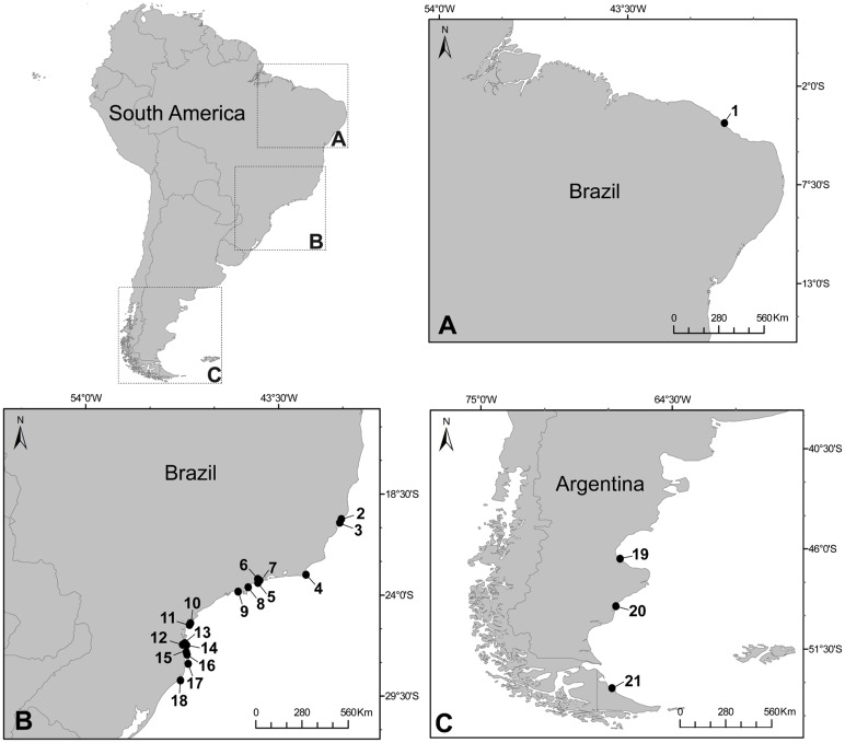
Specimens of the genus Orthopyxis and Campanularia were sampled in Brazil and Argentina (Fig. 1, Table 2). Samples were carried out in the northeastern (state of Ceará) and southeastern coast of Brazil (states of Espírito Santo, Rio de Janeiro, São Paulo, Paraná and Santa Catarina), and south of Argentina (provinces of Santa Cruz and Tierra del Fuego). All necessary permits were obtained for the field studies (sampling permits 16802–1 and 16802–2 SISBIO/ICMBio—Instituto Chico Mendes de Conservação da Biodiversidade), and no protected species were sampled. Colonies were collected during low tide on a variety of substrates, including rocks, algae (Sargassum sp. and Macrocystis pyrifera), mussel shells and other hydroid colonies (mainly species of Sertulariidae), and preserved in 95% ethanol. Species were identified based on taxonomic descriptions [19,31,47,77,78] and, whenever possible, by comparisons with type materials or other reference materials available in museums. Species vouchers were deposited in the Museu de Zoologia da Universidade de São Paulo (MZUSP), Brazil, and in the National Museum of Natural History, Smithsonian Institution (USNM), United States of America (Table 2). One specimen of the Campanulariinae genus Silicularia Meyen, 1834 from Argentina was included in several of the analyses because it is thought to be related to Orthopyxis cf. [14]. Two species of the genus Obelia Péron & Lesueur, 1810 (subfamily Obeliinae, sister group of Campanulariinae according to [14] and [73]) were used as outgroups in the phylogenetic analysis. All sequences were deposited in GenBank (accession numbers in Table 2). Additional data reported in this study (e.g. geographical coordinates, images) were deposited in the National Database Marine Biodiversity (available at https://marinebiodiversity.lncc.br/metacatui/).
Fig 1. Map of sampling areas in Brazil and Argentina.
Circles indicate specific sites were species were sampled. The numbers correspond to the records listed in Table 2.
Table 2. Codes, sampling sites, museum vouchers and GenBank acession numbers for the specimens included in the phylogenetic analyses.
| Species | Sampling site and specimen code in tree | Coordinates (number in Fig. 1) | Voucher | GenBank Acession Number | ||
|---|---|---|---|---|---|---|
| 16S | COI | ITS | ||||
| Obelia dichotoma | Sandwich Marina, Massachusetts, USA | 41°16′15″N 70°15′30″W | MZUSP 1776 | KM603472 | KM603473 | KM603474 |
| Obelia longissima | Gloucester State Pier, Massachusetts, USA | 42°36′51″N 70°39′06″W | MZUSP 1807 | KM603468 | KM603470 | KM603471 |
| Orthopyxis crenata | Caponga (CB), Cascavel, Ceará, Brazil | 04°02.348′S 38°11.572′W (1) | MZUSP 2633 | KM405590 | KM454926 | |
| Orthopyxis sargassicola | Praia Formosa (FB1), Aracruz, ES, Brazil | Specific coordinate unknown (2) | MZUSP 2629 | KM405610 | KM405542 | KM454946 |
| Orthopyxis sargassicola | Praia Formosa (FB2), Aracruz, ES, Brazil | Specific coordinate unknown (2) | MZUSP 2630 | KM405611 | KM405541 | |
| Orthopyxis sargassicola | Praia dos Padres (PB1), Aracruz, Espírito Santo (ES), Brazil | 19°55.941′S 40°07.327′W (3) | MZUSP 2617 | KM405622 | KM405531 | KM454957 |
| Orthopyxis sargassicola | Praia dos Padres (PB2), Aracruz, ES, Brazil | 19°55.941′S 40°07.327′W (3) | MZUSP 2618 | KM405623 | KM405530 | KM454958 |
| Orthopyxis sargassicola | Praia dos Padres (PB3), Aracruz, ES, Brazil | 19°55.941′S 40°07.327′W (3) | MZUSP 2619 | KM405624 | KM405529 | KM454959 |
| Orthopyxis sargassicola | Praia dos Padres (PB4), Aracruz, ES, Brazil | 19°55.941′S 40°07.327′W (3) | MZUSP 2620 | KM405625 | KM405528 | KM454960 |
| Orthopyxis sargassicola | Praia dos Padres (PB5), Aracruz, ES, Brazil | 19°55.941′S 40°07.327′W (3) | MZUSP 2627 | KM405626 | KM405527 | KM454961 |
| Orthopyxis sargassicola | Praia dos Padres (PB6), Aracruz, ES, Brazil | 19°55.941′S 40°07.327′W (3) | MZUSP 2628 | KM405627 | KM405526 | KM454962 |
| Orthopyxis sargassicola | Praia dos Padres (PB7), Aracruz, ES, Brazil | 19°55.941′S 40°07.327′W (3) | MZUSP 2632 | KM405525 | KM454963 | |
| Orthopyxis caliculata | Praia João Gonçalves (JGB1), Búzios, Rio de Janeiro (RJ), Brazil | Specific coordinate unknown (4) | MZUSP 2612 | KM405582 | KM454918 | |
| Orthopyxis caliculata | Praia João Gonçalves (JGB2), Búzios, RJ, Brazil | Specific coordinate unknown (4) | MZUSP 2613 | KM405583 | KM454919 | |
| Orthopyxis caliculata | Praia João Gonçalves (JGB3), Búzios, RJ, Brazil | Specific coordinate unknown (4) | MZUSP 2614 | KM405584 | KM405565 | KM454920 |
| Orthopyxis caliculata | Praia João Gonçalves (JGB4), Búzios, RJ, Brazil | Specific coordinate unknown (4) | MZUSP 2615 | KM405585 | KM454921 | |
| Orthopyxis sargassicola | Paraty (PTY1), RJ, Brazil | Specific coordinate unknown (5) | MZUSP 2605 | KM405628 | KM405524 | KM454964 |
| Orthopyxis sargassicola | Paraty (PTY2), RJ, Brazil | Specific coordinate unknown (5) | MZUSP 2606 | KM405629 | KM405523 | KM454965 |
| Orthopyxis sargassicola | Paraty (PTY3), RJ, Brazil | Specific coordinate unknown (5) | MZUSP 2607 | KM405630 | KM405522 | KM454966 |
| Orthopyxis sargassicola | Paraty (PTY4), RJ, Brazil | Specific coordinate unknown (5) | MZUSP 2608 | KM405631 | KM405521 | KM454967 |
| Orthopyxis sargassicola | Paraty (PTY5), RJ, Brazil | Specific coordinate unknown (5) | MZUSP 2609 | KM405632 | KM405520 | KM454968 |
| Orthopyxis sargassicola | Ilha dos Ratos (RI), Paraty, RJ, Brazil | 23°11.640′S 44°36.408′W (6) | MZUSP 2610 | KM405633 | KM405519 | KM454969 |
| Orthopyxis sargassicola | Ilha dos Meros (MI), Paraty, RJ, Brazil | 23°11.264′S 44°34.635′W (7) | MZUSP 2611 | KM405621 | KM405532 | KM454956 |
| Orthopyxis sargassicola | Praia do Lázaro (LB1), Ubatuba, SP, Brazil | 23°30′32.64″S 45°08′18.52″W (8) | MZUSP 2594 | KM405612 | KM405540 | KM454947 |
| Orthopyxis sargassicola | Praia do Lázaro (LB2), Ubatuba, SP, Brazil | 23°30′32.64″S 45°08′18.52″W (8) | MZUSP 2595 | KM405613 | KM405539 | KM454948 |
| Orthopyxis sargassicola | Praia do Lázaro (LB3), Ubatuba, SP, Brazil | 23°30′32.64″S 45°08′18.52″W (8) | MZUSP 2596 | KM405614 | KM405538 | KM454949 |
| Orthopyxis sargassicola | Praia do Lázaro (LB4), Ubatuba, SP, Brazil | 23°30′32.64″S 45°08′18.52″W (8) | MZUSP 2597 | KM405615 | KM405537 | KM454950 |
| Orthopyxis crenata | Praia do Lázaro (LB5), Ubatuba, SP, Brazil | 23°30′32.64″S 45°08′18.52″W (8) | MZUSP 2598 | KM405591 | KM454927 | |
| Orthopyxis sargassicola | Praia do Lázaro (LB6), Ubatuba, SP, Brazil | 23°30′32.64″S 45°08′18.52″W (8) | MZUSP 2599 | KM405616 | KM405536 | KM454951 |
| Orthopyxis sargassicola | Praia do Lázaro (LB7), Ubatuba, SP, Brazil | 23°30′32.64″S 45°08′18.52″W (8) | MZUSP 2600 | KM405617 | KM405535 | KM454952 |
| Orthopyxis crenata | Praia do Lázaro (LB8), Ubatuba, SP, Brazil | 23°30′32.64″S 45°08′18.52″W (8) | MZUSP 2601 | KM405592 | KM454928 | |
| Orthopyxis sargassicola | Praia do Lázaro (LB9), Ubatuba, SP, Brazil | 23°30′32.64″S 45°08′18.52″W (8) | MZUSP 2602 | KM405618 | KM405534 | KM454953 |
| Orthopyxis sargassicola | Praia do Lázaro (LB10), Ubatuba, SP, Brazil | 23°30′32.64″S 45°08′18.52″W (8) | MZUSP 2603 | KM405619 | KM454954 | |
| Orthopyxis sargassicola | Praia do Lázaro (LB11), Ubatuba, SP, Brazil | 23°30′32.64″S 45°08′18.52″W (8) | MZUSP 2604 | KM405620 | KM405533 | KM454955 |
| Orthopyxis sargassicola | Praia Preta, São Sebastião (SS), São Paulo (SP), Brazil | Specific coordinate unknown (9) | MZUSP 2593 | KM405634 | KM405518 | KM454970 |
| Orthopyxis mianzani | Praia do Miguel (MB1), Ilha do Mel, Paraná (PR), Brazil | 25°33′22.12"S 48°17′55.36"W (10) | MZUSP 2570 | KM405602 | KM405550 | KM454938 |
| Orthopyxis mianzani | Praia do Miguel (MB2), Ilha do Mel, PR, Brazil | 25°33′22.12"S 48°17′55.36"W (10) | MZUSP 2571 | KM405603 | KM405549 | KM454939 |
| Orthopyxis mianzani | Praia do Miguel (MB3), Ilha do Mel, PR, Brazil | 25°33′22.12"S 48°17′55.36"W (10) | MZUSP 2572 | KM405604 | KM405548 | KM454940 |
| Orthopyxis mianzani | Praia do Miguel (MB4), Ilha do Mel, PR, Brazil | 25°33′22.12"S 48°17′55.36"W (10) | MZUSP 2573 | KM405605 | KM405547 | KM454941 |
| Orthopyxis mianzani | Praia do Miguel (MB5), Ilha do Mel, PR, Brazil | 25°33′22.12"S 48°17′55.36"W (10) | MZUSP 2574 | KM405606 | KM405546 | KM454942 |
| Orthopyxis mianzani | Praia de Fora (FOB1), Ilha do Mel, PR, Brazil | 25°34′22.58"S 48°18′32.77"W (11) | MZUSP 2575 | KM405595 | KM405557 | KM454932 |
| Orthopyxis mianzani | Praia de Fora (FOB2), Ilha do Mel, PR, Brazil | 25°34′22.58"S 48°18′32.77"W (11) | MZUSP 2576 | KM405596 | KM405556 | KM454933 |
| Orthopyxis mianzani | Praia de Fora (FOB3), Ilha do Mel, PR, Brazil | 25°34′22.58"S 48°18′32.77"W (11) | USNM 1259970 | KM405597 | KM405555 | KM454934 |
| Orthopyxis mianzani | Praia de Fora (FOB4), Ilha do Mel, PR, Brazil | 25°34′22.58"S 48°18′32.77"W (11) | MZUSP 2577 | KM405598 | KM405554 | KM454935 |
| Orthopyxis mianzani | Praia de Fora (FOB5), Ilha do Mel, PR, Brazil | 25°34′22.58"S 48°18′32.77"W (11) | MZUSP 2578 | KM405599 | KM405553 | KM454936 |
| Orthopyxis mianzani | Praia de Fora (FOB6), Ilha do Mel, PR, Brazil | 25°34′22.58"S 48°18′32.77"W (11) | MZUSP 2579 | KM405600 | KM405552 | KM454937 |
| Orthopyxis mianzani | Praia de Fora (FOB7), Ilha do Mel, PR, Brazil | 25°34′22.58"S 48°18′32.77"W (11) | MZUSP 2580 | KM405601 | KM405551 | |
| Orthopyxis caliculata | Praia da Armação (AB), Penha, SC, Brazil | 26°47′S 48°37′W (12) | MZUSP 2565 | KM405578 | KM405567 | KM454914 |
| Orthopyxis caliculata | Praia da Paciência (PAB1), Penha, Santa Catarina (SC), Brazil | 26°46′38″S 48°36′10″W (13) | MZUSP 2550 | KM405586 | KM405564 | KM454922 |
| Orthopyxis crenata | Praia da Paciência (PAB2), Penha, SC, Brazil | 26°46′38″S 48°36′10″W (13) | MZUSP 2551 | KM405593 | KM405559 | KM454930 |
| Orthopyxis caliculata | Praia da Paciência (PAB3), Penha, SC, Brazil | 26°46′38″S 48°36′10″W (13) | MZUSP 2552 | KM405587 | KM405563 | KM454923 |
| Orthopyxis caliculata | Praia da Paciência (PAB4), Penha, SC, Brazil | 26°46′38″S 48°36′10″W (13) | MZUSP 2554 | KM405588 | KM405562 | KM454924 |
| Orthopyxis caliculata | Praia da Paciência (PAB5), Penha, SC, Brazil | 26°46′38″S 48°36′10″W (13) | MZUSP 2556 | KM405589 | KM405561 | KM454925 |
| Orthopyxis mianzani | Praia da Paciência (PAB6), Penha, SC, Brazil | 26°46′38″S 48°36′10″W (13) | MZUSP 2559 | KM405607 | KM405545 | KM454943 |
| Orthopyxis crenata | Praia da Paciência (PAB7), Penha, SC, Brazil | 26°46′38″S 48°36′10″W (13) | MZUSP 2560 | KM405594 | KM405558 | KM454931 |
| Orthopyxis caliculata | Praia Grande (GB), Penha, SC, Brazil | 26°46′S 48°35′W (14) | MZUSP 2563 | KM405581 | KM405566 | KM454917 |
| Orthopyxis caliculata | Praia de Bombas (BB), Bombinhas, SC, Brazil | 27o07′52.44″S 48°30′49.02″W (15) | MZUSP 4265 | KM405579 | KM454915 | |
| Orthopyxis caliculata | Praia da Conceição (COB), Bombinhas, SC, Brazil | 27°12′1.26″S 48°29′32.04″W (16) | MZUSP 4177 | KM405580 | KM454916 | |
| Orthopyxis sargassicola | Ilha Campeche (CI1), Florianópolis, SC, Brazil | 27°41′27″S 48°27′51″W (17) | MZUSP 4597 | KM405608 | KM405544 | KM454944 |
| Orthopyxis sargassicola | Ilha Campeche (CI2), Florianópolis, SC, Brazil | 27°41′27″S 48°27′51″W (17) | MZUSP 4599 | KM405609 | KM405543 | KM454945 |
| Orthopyxis crenata | Prainha, Laguna (LG), SC, Brazil | 28°36.097′S 48°48.957′W (18) | MZUSP 5055 | KM405560 | KM454929 | |
| Orthopyxis sp. indet. | Caleta Olivia, Argentina | 46°25.539′S 67°31.183′W (19) | MZUSP 2644 | KM405635 | KM454971 | |
| Campanulariidae sp. indet. | La Mina, Puerto San Julián (SJ1), Argentina | 49°09.413′S 67°37.987′W (20) | MZUSP 2638 | KM405576 | KM454912 | |
| Campanularia subantarctica | La Mina, Puerto San Julián (SJ2), Argentina | 49°09.413′S 67°37.987′W (20) | MZUSP 2639 | KM405574 | KM405569 | KM454910 |
| Campanulariidae sp. indet. | La Mina, Puerto San Julián (SJ3), Argentina | 49°09.413′S 67°37.987′W (20) | MZUSP 2640 | KM405577 | KM454913 | |
| Campanularia sp. | La Mina, Puerto San Julián (SJ4), Argentina | 49°09.413′S 67°37.987′W (20) | MZUSP 2641 | KM405572 | KM405571 | KM454908 |
| Campanularia sp. | La Mina, Puerto San Julián (SJ5), Argentina | 49°09.413′S 67°37.987′W (20) | MZUSP 2642 | KM405573 | KM405570 | KM454909 |
| Campanularia subantarctica | La Mina, Puerto San Julián (SJ6), Argentina | 49°09.413′S 67°37.987′W (20) | MZUSP 2643 | KM405575 | KM405568 | KM454911 |
| Silicularia rosea | Río Grande, Cabo Santo Domingo, Argentina | 53°41.330′S 67°50.673′W (21) | MZUSP 2645 | KM405636 | KM454972 | |
Molecular data
Nuclear DNA and mitochondrial DNA were extracted using Instagene (Bio-Rad Laboratories, Hercules, California, USA), according to the manufacturer’s protocol. Portions of the mitochondrial 16S ribosomal RNA gene and the cytochrome oxidase subunit I (COI) gene as well as the entire nuclear Internal Transcribed Spacer (ITS) region (ITS1, 5.8S ribosomal RNA gene and ITS2) were amplified by PCR and verified on 1.5% agarose gels (PCR conditions and primers are described in Table 3). PCR products were purified using the AMPure purification kit (Agencourt Bioscience Corporation, Beckman Coulter, Beverly, Massachusetts, USA), and purified products were prepared for sequencing using the Big Dye Terminator v3.1 Cycle Sequencing kit (Applied Biosystems, Foster City, California, USA) and the same PCR primers. The sequencing reactions were carried out using an ABI PRISM 3100 Genetic Analyzer (Applied Biosystems, Foster City, California, USA).
Table 3. Primers and PCR conditions for DNA amplification.
| Genes | Primers | Reference | Primers Sequence (5′-3′) | PCR conditions | Fragment Size (approx.) |
|---|---|---|---|---|---|
| 16S | C&B1 (F) 1 | [152] | TCGACTGTTTACCAAAAACATAGC | Init. Denat.: 94°C, 3min; 5 cycles: 94°C, 30sec; 45°C, 50sec; 72°C, 1min; 30 cycles: 95°C, 30sec; 50°C, 45sec; 72°C, 1min; Fin. Ext.: 72°C, 5min; 10°C | 610 bp |
| C&B2 (R) | [152] | ACGGAATGAACTCAAATCATGTAAG | |||
| 2Hydrom (R) | Ale E, LEM 2 | CTGTTATCCCTAAGGTAGC | 475 bp | ||
| COI | LCO1490 (F) 1 | [153] | -GGTCAACAAATCATAAAGATATTGG- | Init. Denat.: 94°C, 2min; 10 cycles: 94°C, 30sec; 48°C, 1min; 72°C, 1min20sec; 25 cycles: 94°C, 30sec; 50°C, 40sec; 72°C, 1min20sec; Fin. Ext.: 72°C, 7min; 10°C | 660 bp |
| HCO2198 (R) | [153] | -TAAACTTCAGGGTGACCAAAAAATCA- | |||
| HCOcato (R) | [117] | -CCTCCAGCAGGATCAAAGAAAG | 630 bp | ||
| ITS1–5.8S-ITS2 | CAS18sF1 (F) | [154] | TACACACCGCCCGTCGCTACTA | Init. Denat.: 94°C, 3min; 35 cycles: 95°C, 30sec; 50°C, 45sec; 72°C, 1min; Fin. Ext.: 72°C, 7min; 4°C | 765 bp |
| F5′ (F) | [118] | TAACAAGGTTTCCGTAGG | 630 bp | ||
| ITS1A (F) | [155] | -GTAACAAGGTTTCCGTAGGTG | 630 bp | ||
| CAS28sB1d (R) 1 | [154] | TTCTTTTCCTCCSCTTAYTRATATGCTTAA | |||
| jfITS1–5F (F) | [116] | -GGTTTCCGTAGGTGAACCTGCGGAAGGATC | Init. Denat.: 94°C, 2min; 35 cycles: 94°C, 30sec; 55°C, 45sec; 72°C, 1min; Fin. Ext.: 72°C, 7min; 4°C | 680 bp | |
| ITS-R-28S-15 (R) | Maronna MM, LEM 2 | ACTCGCCGTTACTAGGGGAATCCTTGTTAG |
(F) Forward (R) Reverse.
1Used in conjunction with different forward or reverse primers.
2Primers designed by members of the Laboratory of Marine Evolution (LEM), University of Sao Paulo, Brazil.
Sequences were assembled and edited using Geneious (version 7.1 created by Biomatters, Auckland, New Zealand), and aligned using MAFFT [79]. The obtained sequences were compared with those deposited in GenBank using the Basic Local Alignment Search Tool (BLAST, [80]) to confirm genes and species of interest. Additionally, the ITS1 and ITS2 regions were extracted from the complete ITS sequences using the sequence from Hydra circumcincta [81] in GenBank (GU722663) as a guide to delimit the ITS1 sequences and the ITS2 Database [82] to delimit the ITS2 sequences. The coding sequences of COI were translated and compared with the complete mitochondrial genome of Laomedea flexuosa [83] (GenBank NC_016463) to ensure pseudogenes were not amplified. Since not all sequences of the same marker had the same length (see Table 3), some portions of the longer sequences were excluded from the alignments to adjust all sequences to the same length.
Phylogenetic analysis
Phylogenetic analyses were performed on (a) individual markers, (b) combined mitochondrial markers (16S+COI), (c) combined nuclear markers (ITS1+ITS2), and (d) the entire combined dataset (16S+COI+ITS1+ITS2), using maximum likelihood (ML) and parsimony (P) criteria. The datasets were built using unique haplotypes, and the combined datasets included only those specimens with sequences available for all markers (details of the analyses in Table 4).
Table 4. Details of the datasets used in the phylogenetic analyses.
| Total | 16S+COI | ITS1+ITS2 | 16S | COI | ITS1 | ITS2 | |
|---|---|---|---|---|---|---|---|
| Number of characters | 1553 | 1046 | 509 | 476 | 575 | 263 | 242 |
| Number of informative sites (P) | 665 | 261 | 390 | 113 | 153 | 214 | 163 |
| Number of most parsimonious trees (P) | 74 | 116 | 4115 | 3 | 11 | 5 | 2130 |
| Minimum length (P) | 1276 | 511 | 1056 | 284 | 304 | 623 | 365 |
| Model of nucleotide evolution (ML) | GTR+G | GTR+I+G | GTR+G | GTR+I | GTR+G | GTR+G | SYM+G |
(P) Parsimony, (ML) Maximum Likelihood.
Sequences of nuclear DNA with ambiguous sites (17 ITS1 and 22 ITS2 sequences) were treated using IUPAC ambiguity codes. The maximum number of ambiguous sites recorded for one sequence was five (the ITS2 sequence of a specimen from Penha, Santa Catarina), and 46% of the sequences had only one ambiguous site. Sequences with identical IUPAC codes at identical positions were considered as the same haplotype in the analyses.
Phylogenetic analyses using parsimony (P) criteria were performed using the PAUP 4.0b10 [84] and TNT [85] programs. Analyses consisted of 1000 unweighted heuristic searches using a random algorithm and branch-swapping using the TBR (tree bisection-reconnection) algorithm. Gaps were considered as a fifth state. Branch support was estimated in TNT with bootstrapping on 1000 replicates. Phylogenetic analyses using Maximum Likelihood (ML) criteria were performed using PALM (Phylogenetic Reconstruction by Automatic Likelihood Model Selector, [86]) with the most appropriate model of nucleotide evolution for each dataset based on Akaike Information Criterion (AIC, Table 4). Branch support was estimated with bootstrapping on 1000 replicates. Phylogenetic p-distances (uncorrected) were calculated using the PAUP 4.0b10 program.
Morphological analysis
We performed Principal Component Analysis (PCA, [87]) on a correlation matrix based on 37 different measures of the trophosome (S1 Table) of the voucher specimens of O. caliculata and O. mianzani sp. nov. (the same specimens used in the phylogenetic analyses). For both species, we did not include any characters from the gonothecae in the PCA, as not all colonies presented this reproductive structure. This analysis was performed to better delimitate the species by assessing the degree of variation for their morphological characters and by identifying their most relevant diagnostic characters.
Nomenclatural acts
The electronic edition of this article conforms to the requirements of the amended International Code of Zoological Nomenclature, and hence the new names contained herein are available under that Code from the electronic edition of this article. This published work and the nomenclatural acts it contains have been registered in ZooBank, the online registration system for the ICZN. The ZooBank LSIDs (Life Science Identifiers) can be resolved and the associated information viewed through any standard web browser by appending the LSID to the prefix “http://zoobank.org/”. The LSID for this publication is: urn:lsid:zoobank.org:pub:280AC2D0–9DCE-4BCE-AF85–2586B3951522. The electronic edition of this work was published in a journal with an ISSN, and has been archived and is available from the following digital repositories: PubMed Central and LOCKSS.
Results
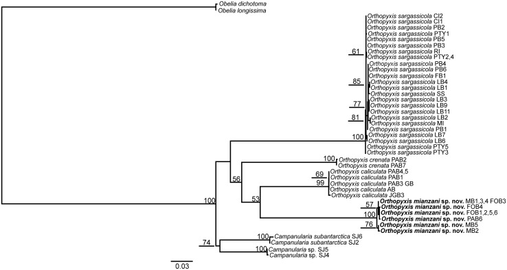
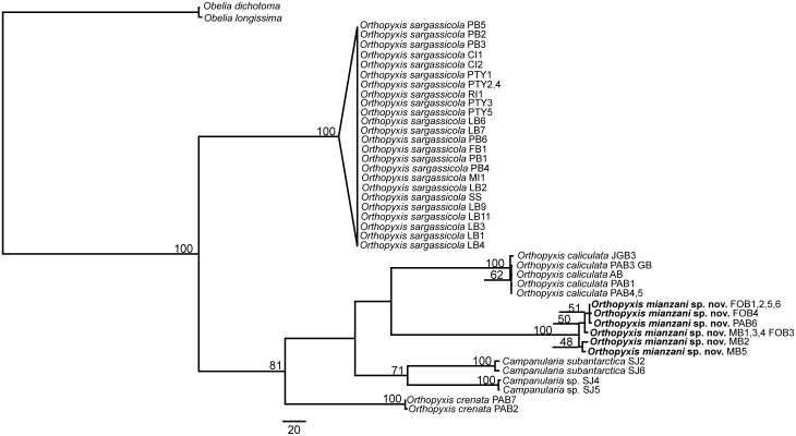
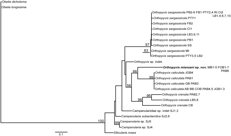
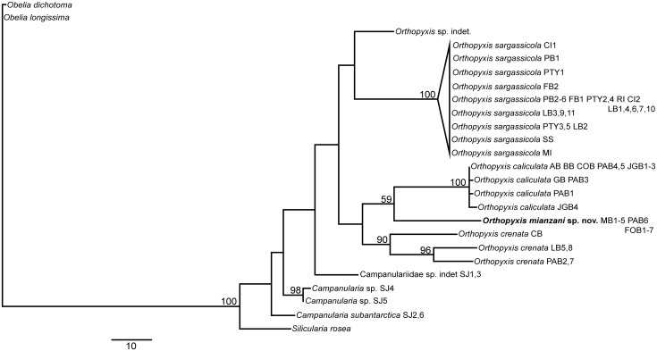
Nearly all the topologies obtained using the different datasets identified six well-defined clades with high branch support values. However, these topologies did present some incongruencies with respect to the phylogenetic relationships among these clades. The individual and combined nuclear datasets showed low resolution and low values for branch support, whereas the combined mitochondrial datasets showed higher resolution but also had low branch support (S1–S10 Figs.). The combined dataset involving all four markers revealed the best definition of the relationships among the lineages, with a higher frequency of well supported nodes (all six less inclusive clades with bootstrap = 99–100, Figs. 2–3). In addition, the 16S topologies showed the most congruent results (Figs. 4–5). Therefore the topologies involving the combined and the 16S datasets represented the most robust hypothesis for our data and are used as our working hypothesis for discussions.
Fig 2. Maximum Likelihood tree based on 16S, COI, ITS1 and ITS2 data.
Bootstrap values are shown for each node. Nodes without numbers indicate support below 50.
Fig 3. One of the 74 most parsimonious trees based on 16S, COI, ITS1 and ITS2 data.
These trees are only different in the position of the haplotypes within O. sargassicola clade, which is collapsed. Bootstrap values are shown for each node. Nodes without numbers indicate support below 50.
Fig 4. Maximum Likelihood tree based on 16S data.
Bootstrap values are shown for each node. Nodes without numbers indicate support below 50.
Fig 5. One of the three most parsimonious trees based on 16S data.
These trees are only different in the position of the haplotypes within O. sargassicola clade, which is collapsed. Bootstrap values are shown for each node. Nodes without numbers indicate support below 50.
The genera Orthopyxis and Campanularia
The genus Orthopyxis was monophyletic according to the 16S topologies and the ML topology with the combined dataset, although with low support value (bootstrap<50, Figs. 2, 4–5). Orthopyxis was not monophyletic in the P topology with the combined dataset, in which species assigned to Campanularia fell within Orthopyxis as a sister group to Orthopyxis caliculata (Hincks, 1853)+Orthopyxis mianzani sp. nov. (Fig. 3). Although not conclusive, Orthopyxis was monophyletic in the majority of our topologies, a hypothesis we follow in this study. However, this hypothesis requires further testing with the addition of more representatives from the genus Campanularia.
Campanularia was monophyletic only in topologies derived from the combined dataset. One of the lineages of Campanularia corresponds morphologically to Campanularia subantarctica Millard, 1971, and it is characterized by the deep hydrothecae with bluntly rounded marginal teeth, subhydrothecal spherule present; gonothecae oval-elongated arising from hydrorhiza, with distal aperture on top of a low collar [77,88], ([47], as C. lennoxensis). The second lineage of Campanularia is also morphologically similar to C. subantarctica, but we were unable to identify this lineage to the species level due to the lack of gonothecae. Additionally, these two lineages showed genetic distances of up to 7.83% for mitochondrial markers and 26.38% for nuclear markers (Table 5), indicating that they likely represent two distinct species.
Table 5. Minimum and maximum p-distances (uncorrected) (%) from the mithocondrial dataset (low left corner) and nuclear dataset (up right corner).
| Species | Orthopyxis sargassicola | Orthopyxis caliculata | Orthopyxis mianzani | Orthopyxis crenata | Campanularia subantarctica | Campanularia sp. | Campanulariidae sp. indet. | Orthopyxis sp. indet. |
|---|---|---|---|---|---|---|---|---|
| Orthopyxis sargassicola | 0.35/0.51 | 17.51–44.05 | 19.05–43.19 | 9.66–41.07 | 13.69–45.42 | 18.39–44.10 | 22.02–40.72 | 12.30–36.72 |
| Orthopyxis caliculata | 8.68–12.87 | 0.30/0.00 | 18.62–27.86 | 13.22–31.76 | 9.84–29.57 | 14.74–25.30 | 15.81–28.67 | 15.38–23.61 |
| Orthopyxis mianzani | 9.33–16.17 | 7.81–15.65 | 0.17/1.28 | 14.90–35.54 | 17.87–28.60 | 24.10–24.73 | 19.98–33.19 | 17.70–29.36 |
| Orthopyxis crenata | 9.33–13.39 | 8.68–13.74 | 9.33–15.48 | 4.43/3.31 | 7.81–10.96 | 7.81–10.61 | 17.13–34.58 | 2.52–32.52 |
| Campanularia subantarctica | 7.38–9.74 | 6.72–11.65 | 8.24–14.78 | 11.19–38.43 | 0.69/0.51 | 12.80–26.38 | 16.26–38.79 | 13.13–33.48 |
| Campanularia sp. | 7.38–9.74 | 6.72–10.09 | 7.81–14.78 | 13.06–40.55 | 2.60–7.83 | 0.10/0.00 | 19.25–33.31 | 14.66–27.52 |
| Campanulariidae sp. indet. | 7.38–8.03 | 7.16–7.38 | 8.24 | 7.38–8.03 | 4.77 | 4.77 | 0.00 | 17.57–30.13 |
| Orthopyxis sp. indet. | 5.86–6.51 | 6.07–6.29 | 8.24 | 7.38–8.46 | 4.56 | 5.21 | 3.69 | 0.00 |
Values in the diagonal indicate mean intraspecific distances (mithocondrial/nuclear markers).
Species of the genus Orthopyxis
We delimited four lineages of the genus Orthopyxis in the southwestern Atlantic, three of which correspond morphologically to Orthopyxis sargassicola (Nutting, 1915), Orthopyxis crenata (Hartlaub, 1901), and Orthopyxis caliculata (Hincks, 1853) (considered a synonym of Orthopyxis integra (Macgillivray, 1842) by some authors; see discussion below), and one of which is new to science (Figs. 2–5). These species showed genetic distances ranging from 7.81–16.17% and 9.66–44.05% for mitochondrial and nuclear makers, respectively (Table 5).
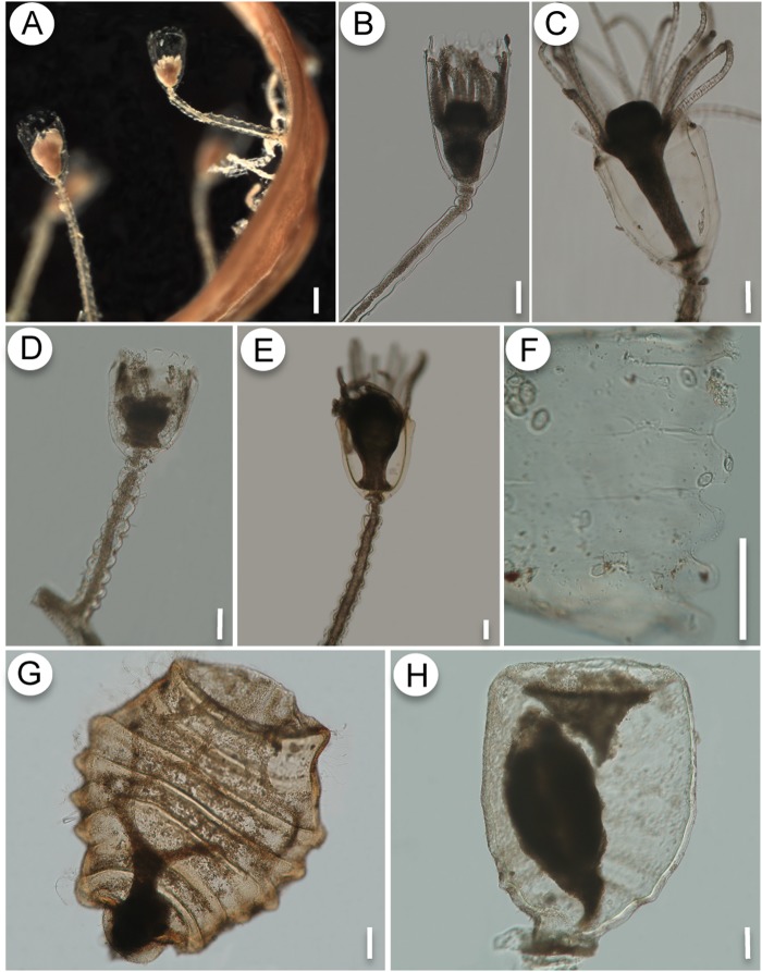
The specimens of O. sargassicola and O. crenata recorded here have the general features of Orthopyxis, such as a thick perisarc (variable to some extent), campanulate hydrothecae, sinuous pedicels, and subhydrothecal spherule (Fig. 6). The presence of rounded hydrothecal cusps and a laterally compressed, completely ribbed gonotheca, are distinctive characters of O. sargassicola [1,13,31] (Fig. 6A-C, G), whereas O. crenata is characterized by low, rounded hydrothecal cusps and laterally compressed, smooth gonothecae [29,47] (Fig. 6D-F, H). Although these species can be readily distinguished by comparing their gonothecae, morphological variation in the size and shape of the hydrothecal cusps may cause these diagnostic characters to overlap when the gonotheca is absent, hampering identification. This is the first record of Orthopyxis crenata in the southwestern Atlantic, although previous authors may have overlooked this species due to its morphological similarity with O. sargassicola.
Fig 6. A-C, G: Orthopyxis sargassicola.
A: general view of the colony on Sargassum sp.; B-C: detail of the trophosome, showing variation in perisarc thickness of hydrotheca; G: gonotheca. D-F, H: Orthopyxis crenata. D-E: detail of the trophosome; F- detail of the hydrothecal cusps; H- gonotheca. Scales: A—200 μm; B-H—100 μm.
The species O. caliculata and O. mianzani sp. nov., although highly genetically divergent (Table 5), have similar morphologies that could be traditionally associated with Orthopyxis integra (Macgillivray, 1842). Both species have stolonal colonies, sinuous pedicels, subhydrothecal spherule, campanulate hydrotheca with rim even, and gonotheca roughly cylindrical, with wide aperture, truncated on top [19,78]. However, they are morphologically distinct with respect to characters usually assumed to show wide intraspecific variation, such as perisarc thickness and length of the hydrothecae and pedicels.
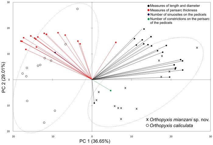
PCA performed using the morphometric data for O. caliculata and O. mianzani sp. nov. (Fig. 7) showed that the two lineages are clearly separated by perisarc thickness and polyp general dimensions. Specimens of O. caliculata have a thicker perisarc and smaller general dimensions (length and diameter of the hydrothecae, pedicels, and subhydrothecal spherule) of the polyp (Fig. 7). These results show that, although variable to some extent, perisarc thickness and polyp dimensions can be used to delimitate these species. Therefore, we believe the name Orthopyxis caliculata (Hincks, 1853) is the correct identification of one of these lineages, and we corroborate the validity of that species.
Fig 7. Correlation biplot of the first and second principal components of the PCA based on morphometric variables of Orthopyxis caliculata and Orthopyxis mianzani sp. nov. from the southwestern Atlantic.
The percentage of variation explained by each principal component is shown in parentheses.
Systematic Account
Orthopyxis caliculata (Hincks, 1853)
(Fig. 8)
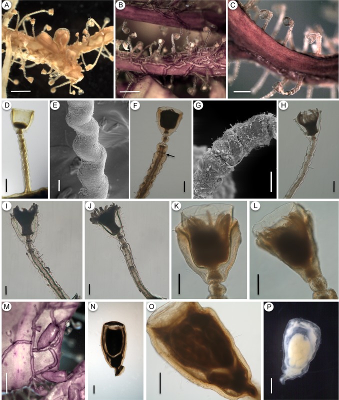
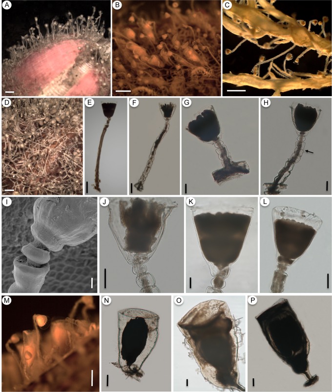
Fig 8. Orthopyxis caliculata.
A-C: general view of the colony (A-MZUSP 4177; B,C- MZUSP 1563); D-H: detail of the trophosome with the sinuosities of the pedicel (E) and constrictions in the perisarc (arrow in F, G) (D-MZUSP 2550; E-MZUSP 2565; F-MZUSP 2554; G-MZUSP 4177; H-MZUSP 2552); I-J: positions of maximum (I) and minimum (J) perisarc thickness of the trophosome (MZUSP 2615); K-L: detail of the hydrotheca, showing two different forms due to compression (MZUSP 2554); M: general view of gonothecae on algae (MZUSP 2563); N: detail of male gonotheca (MZUSP 2554); O-P: detail of female gonothecae (O-MZUSP 2563; P-MZUSP 2613). Scales: A,B,M—1 mm; C—500 μm; D,F,H,O—200 μm; E—20 μm; G—50 μm; I,J,K,L—100 μm; N,P—300 μm.
Orthopyxis clytioides—Vannucci-Mendes, 1946 [40]: 546, Est.1, Figs. 6,7.—Vannucci, 1951 [89]: 111 [not Orthopyxis clytioides (Lamouroux, 1824)].
Orthopyxis minuta Vannucci, 1949 [41]: 234, t.1, Figs.15–17, t.2, Fig.18.—Vannucci, 1951 [89]: 108. (syn. nov.)
Orthopyxis caliculata—Blanco, 1964 [43]: 157, L.1, Figs. 4,9.
Fig 9. Orthopyxis mianzani sp. nov.
A-C: general view of the colony (A-MZUSP 2575; B-MZUSP 2580; C-MZUSP 2559); D: detail of the hydrorhiza (USNM 1259970); E-H: details of the trophosome, showing variation in pedicels from smooth (E) to sinuous (F), variation in the length of the pedicels (compare E, F and G) and constrictions of the perisarc (H, arrow) (E-MZUSP 2576; F-MZUSP 2572; G-MZUSP 2570; H-MZUSP 2574); I: detail of subhydrothecal spherule (USNM 1259970); J-L: detail of hydrothecae (J-MZUSP 2572; K-MZUSP 2576; L-MZUSP 2579); M: general view of the gonotheca on natural substrate (MZUSP 2580); N-P: detail of female gonothecae (N-MZUSP 2572; O-USNM 1259970; P-MZUSP 2580). Scales: A-D—1 mm; E—300 μm; F, N, P—200 μm; G, H, J-L, O—100 μm; I—20 μm; M—500 μm.
Orthopixis clytioides—Milstein, 1976 [90]: 77, Figs. 8,9,11 [not Orthopyxis clytioides (Lamouroux, 1824)].
Campanularia integra—Blanco, 1994 [46]: 192 [not Campanularia integra Macgillivray, 1842].
Orthopyxis integra—Miranda et al., 2011 [13]: 347, Fig. 25a-d [not Orthopyxis integra (Macgillivray, 1842)].
Material examined. Brazil, Santa Catarina (SC), Penha, Praia Grande, 26°46’S 48°35’W, 0–1 m, 08.vii.2009, with female gonothecae, on algae, coll. E.C. Bornancin, MZUSP 2563; SC, Penha, Praia da Paciência, 26°46’38”S 48°36’10”W, 3 m, 02.vii.2009, without gonothecae, on algae, coll. A.F. Cunha, MZUSP 2550; SC, Penha, Praia da Paciência, 26°46’38”S 48°36’10”W, 0–1 m, 02.vii.2009, without gonothecae, on algae, with some colonies of Obelia sp., coll. A.F. Cunha, MZUSP 2552; SC, Penha, Praia da Paciência, 26°46’38”S 48°36’10”W, 3 m, 01.vii.2009, with male gonothecae, on algae, coll. A.F. Cunha, MZUSP 2554; SC, Penha, Praia da Paciência, 26°46’38”S 48°36’10”W, 3 m, 01.vii.2009, without gonothecae, on algae, coll. A.F. Cunha, MZUSP 2556; SC, Penha, Praia da Armação, 0–1 m, 07.vii.2009, without gonothecae, on algae, coll. E.C. Bornancin, MZUSP 2565; SC, Bombinhas, Praia de Bombas, 27°07’52.44”S 48°30’49.02”W, 0–2 m, 03.xii.2006, with female gonothecae, on algae, coll. A.C. Marques & T.P. Miranda, MZUSP 4265; SC, Bombinhas, Praia da Conceição, 27°12’1.26”S 48°29’32.04”W, 0–2 m, 02.xii.2006, with male and female gonothecae (two colonies), on algae, coll. A.C. Marques, E. Ale, M.A. Imazu & T.P. Miranda, MZUSP 4177; Rio de Janeiro, Búzios, Praia de João Gonçalves, coordinate unknown, 20.viii.2009, with few female gonothecae, on algae, coll. L.S. Miranda, A.C. Morandini & S.N. Stampar, MZUSP 2612, MZUSP 2613, MZUSP 2614 and MZUSP 2615.
Additional material examined. Argentina, Chubut, Puerto Madryn, Orthopyxis caliculata (Hincks, 1853), O.M. Blanco det., Museo de La Plata, MLP 47 to MLP 54; Santa Cruz, San Julián, Punta Peñas, Orthopyxis caliculata (Hincks, 1853), O.M. Blanco det., MLP 55; Santa Cruz, Punta Peñas, Campanularia integra Macgillivray, 1842, O.M. Blanco, det., MLP 8536. Uruguay, Rocha, La Coronilla, Orthopixis clytioides (Lamouroux, 1824) [incorrect subsequent spelling], det. A. Milstein. United States, Alaska, Aleutian Islands, Orthopyxis integra (Macgillivray, 1842), A. Govindarajan det., National Museum of Natural History, USNM 1106184. Kara Sea, Campanularia integra Macgillivray, 1842, USNM 17834.
Description. Colonies stolonal, up to 1.6 mm high. Hydrothecae and pedicels laterally compressed, amount of compression varying according to perisarc thickness. Pedicels arise from creeping, flattened hydrorhiza at irregular intervals. Hydrorhiza with very thick perisarc (31–47.5 μm). Pedicels sinuous, with 5–13 sinuosities (crenations) throughout their length, forming a “zig-zag” on pedicels, not spiral, as commonly assumed (Fig. 8E). Occasionally 1–4 constrictions, usually on upper portion of pedicels (most likely regions of growth) (Fig. 8F-G). Pedicels 588–1260 μm in length, usually with thick perisarc (23.54 μm on average) but also colonies with thinner perisarc occur (11.5–30 μm, Fig. 8H). Subhydrothecal spherule present immediately below hydrotheca, slightly smaller than pedicel in diameter, with thick perisarc (14–32.5 μm). Hydrotheca campanulate, 230–374 μm in length, rim smooth, sometimes slightly everted (Fig. 8H) and occasionally growing beyond the thick hydrothecal walls (Fig. 8K-L). Hydrotheca laterally compressed, more conspicuous when perisarc is very thick. Hydrotheca may show two different forms in relation to the compression: (1) when viewed from its broader aspect (i.e., position of maximum perisarc thickness), hydrotheca with thick, straight and parallel walls, gradually projecting inwards towards base, where the perisarc reaches maximum thickness and forms an interior chamber, in which the hydranth rests (Fig. 8K); (2) when viewed from its narrower aspect (i.e., position of minimum perisarc thickness), the much thinner walls are oblique, tapering towards the base (Fig. 8L). Hydranth with 22–26 tentacles. Male and female gonothecae with similar morphology, up to 1.2 mm high, arising from hydrorhiza on short, smooth pedicels, usually growing parallel to substrate. Young gonothecae short and conical, truncated on top, with wide aperture; mature gonothecae with walls oblique at base but gradually elongating and straightening to become parallel, upper portion also truncated, with wide aperture. Gonothecae laterally compressed, perisarc thick (25–46 μm), with somewhat wavy outline, sometimes more pronounced (Fig. 8M-P). Gonophore with two medusa buds, inferior one smaller, superior one larger, and developing gonads in longitudinal rows.
Remarks. Orthopyxis caliculata (Hincks, 1853) has been considered a synonym of O. integra (Macgillivray, 1842) by many authors. Levinsen [91] was likely the first to assign Hincks’ species to O. integra (as Campanularia integra), arguing that he possessed colonies of O. integra that presented intermediate characters from both species, referring in particular to the thickness of the perisarc of the hydrothecae and the presence of annulations on the gonothecae. Many subsequent authors followed this proposal [19,29,78,92,93,94,95,96,97], also arguing that the characters used to distinguish these species are actually intraspecific variations of the same character.
Hincks [98] noted the shape of the hydrothecae and the presence of a “double cup” and “double” pedicel as the main characters that distinguish O. integra and O. caliculata. He subsequently amended his description by arguing that the appearance of a “double” hydrotheca and pedicel is a result of the considerable perisarc thickening in this species [26]. The widely accepted notion that these characters represent variations within the same species has prevented many authors from accepting them as informative (as stated above), although some authors who agree with Hincks [26,98] in regarding O. integra and O. caliculata as separate species point out characters such as the size and shape of the hydrothecae and gonothecae, as important differences between these species e.g., [27,28,99,100]. Indeed, the name O. caliculata is currently used as a valid name in some studies [101, 102], based on similar opinions.
Neither species was originally described with gonothecae [98,103], although subsequent descriptions of these species represented the gonothecae of O. integra as clearly different from those of O. caliculata. The gonothecae of O. integra is described as cylindrical, completely spirally grooved throughout, and truncated on top, whereas the gonothecae of O. caliculata is described as smooth, oval-elongated, laterally compressed, also truncated on top, and with a wide aperture [26,28,99]. Authors who advocate the synonymy of O. integra and O. caliculata consider both types of gonothecae as variations within O. integra (see [78]). Despite this, Millard [29] notes that she never recorded polyps of O. integra in South Africa with spirally grooved gonothecae, and many other records of O. integra include only specimens with oval-elongated, smooth gonothecae e.g., [13,97,104,105,106]. Indeed, cylindrical, spirally grooved gonothecae appear to be restricted to northern records of O. integra e.g., [38,107,108,109,110], as noted by Bale [99].
We studied non-type material of O. integra that presented spirally grooved gonotheca (USNM 17834 from Kara Sea, and 1106184 from Alaska, Aleutian Islands)—in contrast with the oval-elongated, smooth gonotheca of our material—and we have concluded that these two types of gonotheca indicate two different species. These non-type materials of O. integra also differ from our specimens of O. caliculata in the thickness of the perisarc of the hydrothecae and pedicels, as well as in the length of the hydrothecae, which is larger in O. integra (see comparisons on Table 6). Many of these differences have already been noted and discussed by Bale [99], and more recently by Calder et al. [102]. Our molecular analysis revealed two different lineages presenting the traditional morphological characters associated to O. integra. A re-evaluation of the morphological characters of these two lineages demonstrates that their most consistent differences rely on characters previously considered to be intraspecific variations by many authors. Therefore, we conclude that the two completely different gonothecae morphologies should not be considered as variations within O. integra.
Table 6. Comparative measurements of Orthopyxis caliculata, Orthopyxis mianzani (mean±standard error [range]) and specimens of Orthopyxis integra from the National Museum of Natural History, Smithsonian Institution.
| Measurements (μm) | Orthopyxis caliculata (Np = 12; Ng = 5) | Orthopyxis mianzani (Np = 13; Ng = 4) | O. integra** (Np = 3; Ng = 4) | O. integra*** (Np = 4; Ng = 4) |
|---|---|---|---|---|
| Total length of the trophosome | 1213.83±81.58 [840–1658] | 1566.77±156.01 [600–2380] | 2082.98±197.57 [1695.38–2343.28] | 3949.79±718.48 [2437.92–5605.39] |
| Hydrorhiza | ||||
| Diameter | 84.17±3.37 [65–100] | 88.77±3.22 [75–114] | 135.29±5.77 [126.08–145.92] | 139.80±8.66 [116.24–157.08] |
| Perisarc thickness | 39.92±2.24 [31–47.5] | 24.46±1.17 [12.5–30.5] | * | * |
| Pedicel | ||||
| Length | 825.08±70.62 [588–1260] | 943.15±127.76 [190–1870] | 1405.38±223.40 [959.41–1652.07] | 3337.79±677.82 [1938.89–4958.59] |
| Diameter | 95.71±4.53 [68.5–118] | 108±4.15 [89–145] | 99.14±7.32 [85.40–110.39] | 90.80±5.36 [82.96–106.55] |
| Perisarc thickness | 23.54±1.75 [11.5–30] | 11±0.60 [7.5–12.5] | 10.32±0.61 [9.01–10.86] | 8.31±0.64 [6.97–9.65] |
| Maximum number of sinuosities | 7.97±0.80 [5–13] | 4.29±0.76 [0–12] | * | 0 (all pedicels smooth troughout) |
| Subhydrothecal spherule | ||||
| Length | 63.30±3.43 [48–78] | 70.69±5.41 [50–120] | 74.13±4.94 [65.64–82.75] | 52.09±7.81 [33.74–68.08] |
| Diameter | 84.55±2.45 [72–93] | 101±3.74 [85–130] | 100.80±5.15 [91.28–108.96] | 78.63±8.40 [55.14–93.26] |
| Perisarc thickness | 22.35±1.53 [14–32.5] | 14.69±1.09 [7.5–22.5] | 12.49±2.29 [7.95–15.32] | 6.17±0.68 [5.03–8.13] |
| Hydrotheca | ||||
| Length | 318.33±11.85 [230–374] | 418.69±17.74 [328–520] | 667.51±22.46 [622.58–690.37] | 604.02±56.53 [448.80–717.15] |
| Diameter at rim | 283.17±5.63 [263–312] | 369.54±14.97 [304–490] | 420.87±4.93 [414.50–430.58] | 500.03±25.76 [452.88–569.57] |
| Diameter at base | 157.83±5.90 [120–175] | 173.19±2.85 [160–200] | 180.33±12.37 [168.41–205.06] | 237.48±31.33 [174.94–322.64] |
| Length:Diameter ratio | 1.26±0.04 [0.96–1.60] | 1.44±0.04 [1.22–1.71] | 1.78±0.05 [1.72–1.88] | 1.44±0.16 [0.98–1.65] |
| Perisarc thickness | 29.46±2.22 [15.25–36.5] | 7.75±0.80 [2.5–12.5] | 9.27±1.32 [7.12–11.66] | 4.87±0.79 [2.95–6.23] |
| Hydranth | ||||
| Number of tentacles | 24±0.58 [22–26] (N = 10) | 32.46±5.31 [23–43] | * | * |
| Gonotheca | ||||
| Length | 1166.42±30.75 [1096–1262.5] | 1210±64.16 [1090–1390] | 1422.12±96.79 [1202.46–1651.57] | 2086.71±87.53 [1933.13–2278.53] |
| Maximum Diameter | 650.33±31.48 [552–772] | 722.50±16.52 [690–760] | 522.97±19.92 [474.07–571.46] | 620.34±15.58 [590.20–659.85] |
| Length:Diameter ratio | 1.82±0.07 [1.57–2.09] | 1.79±0.07 [1.66–1.99] | 2.74±0.26 [2.10–3.17] | 3.37±0.20 [2.95–3.86] |
| Perisarc thickness | 39.21±3.24 [25–46] | 21.25±1.25 [20–25] | * | * |
| Nematocysts | ||||
| Microbasic mastigophores A type (LenghtxDiam.) | 5.14±0.06 [4–6] x 1.80±0.04 [1.5–3] (N = 60) | 5.13±0.06 [5–6] x 1.79±0.05 [1.5–2] (N = 40) | * | * |
| Microbasic mastigophores B type (LengthxDiam.) | 10.21±0.11 [9–12] x 2.89±0.05 [2–4] (N = 60) | 10.24±0.08 [9–11] x 2.81±0.04 [2.5–3] (N = 40) | * | * |
Np = number of polyps measured; Ng = number of gonothecae measured (N = when different number). The measures of diameter and perisarc thickness were obtained from the position of maximum perisarc thickness (broad view).
*Information not obtained.
**USNM17834.
***USNM1106184.
Considering this, we believe that none of the species described in this study, nor the records included in our synonym, should be assigned to O. integra; instead, they should be assigned to O. caliculata. We understand that it is not simple to delimit these two species morphologically; therefore, we did not include in the synonymy materials we could not access. The one exception is Vannucci’s material [40,41], which is most likely lost (see [1]), for which we tentatively attribute the specimens she described with gonothecae to O. caliculata. The specimen Campanularia integra recorded by Blanco [43] consists of only one microslide with one polyp without gonothecae, and the hydrotheca of this specimen differs from the typical hydrotheca of O. caliculata, being more elongated and cylindrical, similar to the hydrothecae of many species of the genus Campanularia. It is unclear whether this morphology is a preparation artifact or an actual morphological difference, so we therefore decided not to include this record in the synonym of O. caliculata, pending more detailed study. However, the specimens of C. integra recorded by Blanco [46] correspond to the description of O. caliculata. Milstein [90] described specimens with gonothecae that also correspond to O. caliculata. The records of O. integra by Miranda et al. [13] came from localities very close to our records of O. caliculata, and examination of their material leaves no doubt that it should be assigned to O. caliculata.
Type locality. Pegwell Bay, England [98].
Records from the southwestern Atlantic. Brazil, São Paulo, Santos Bay, Santo Amaro Island, Itanhaém [40,89]; Rio de Janeiro, Francês Island [41,89], and Búzios (this study); Santa Catarina, Penha (this study) and Bombinhas [13] (and this study). Uruguay, Rocha, La Coronilla [90]. Argentina, Chubut, Puerto Madryn [43], Santa Cruz, San Julián and Punta Peñas [46].
Orthopyxis mianzani Cunha, Genzano & Marques sp. nov. urn:lsid:zoobank.org:act:A6F4A8FB-FDCC-4BE9–8368–6BFE29CAECC4
(Fig. 9)
?Orthopyxis integra—Grohmann et al., 2011 [53]: 195, Fig. 3F, 1–4 [not Orthopyxis integra (Macgillivray, 1842)].
Material examined. Holotype: Brazil, Paraná (PR), Ilha do Mel, Praia de Fora, 25°34’22.58”S 48°18’32.77”W, 0–1 m, 27.vii.2010, with female gonothecae, on mussel shell and cirriped, coll. E.C. Bornancin & A.F. Cunha, MZUSP 2580; Paratypes: PR, Ilha do Mel, Praia do Miguel, 25°33’22.12”S 48°17’55.36”W, 0–1 m, 26.vii.2010, without gonothecae, on mussel shell, coll. E.C. Bornancin & A.F. Cunha, MZUSP 2571, MZUSP 2573; with female gonothecae, MZUSP 2572, MZUSP 2574; without gonothecae, on mussel shell and cirriped, MZUSP 2570; PR, Ilha do Mel, Praia de Fora, 25°34’22.58”S 48°18’32.77”W, 0–1 m, 27.vii.2010, without gonothecae, on mussel shell and cirriped, coll. E.C. Bornancin & A.F. Cunha, MZUSP 2575, MZUSP 2579; with female gonothecae, USNM 1259970; without gonothecae, on mussel shell, MZUSP 2576; without gonothecae, on Phragmatopoma sp., MZUSP 2577; without gonothecae, on cirriped, MZUSP 2578; Santa Catarina, Penha, Praia da Paciência, 26°46’38”S 48°36’10”W, 0–1 m, 05.vii.2009, without gonothecae, on algae, coll. A.F. Cunha, MZUSP 2559.
Etymology. This species is named after Dr. Hermes W. Mianzan (CONICET and Instituto Nacional de Investigación y Desarollo Pesquero—INIDEP, Mar del Plata, Argentina) for his dedication and commitment to the study of South American cnidarians, and his leadership towards the integration of Latin American marine scientists. Unfortunately, our great “amigo” Hermes passed away during the writing of this manuscript.
Diagnosis. Hydrothecae, pedicels and gonothecae with thin perisarc. Lateral compression only on gonothecae, nearly no compression detectable on hydrothecae or pedicels, both usually longer when compared with other species of Orthopyxis. Reduced amount of sinuosities on pedicels, sometimes almost completely smooth. Gonothecae smooth and different from other Orthopyxis species with ribbed gonothecae.
Description. Colonies stolonal, up to 2.3 mm high. Gonothecae laterally compressed but rarely hydrothecae (compression better observed in hydrothecae with thicker perisarc). Pedicels arise from creeping, flattened hydrorhiza at irregular intervals. Hydrorhiza with moderately thick perisarc (12.5–30.5 μm) and large (diameter 75–114 μm, Fig. 9D). Pedicels usually with slight sinuosities at base and smooth throughout their length, sometimes either sinuous throughout (up to 12 tenuous sinuosities) (Fig. 9F) or with 1–4 marked perisarc constrictions at upper portion (Fig. 9H). Pedicels usually long, rarely small, 190–1870 μm in length, with moderately thick perisarc (7.5–12.5 μm). Subhydrothecal spherule present right below hydrotheca, slightly smaller than pedicel in diameter, with moderately thick perisarc (7.5–22.5 μm). Hydrotheca campanulate, 328–520 μm in length, rim smooth. Perisarc thickness is poorly correlated with hydrothecal form, although hydrotheca may be slightly compressed when perisarc is thicker. Hydrothecal walls slightly oblique with moderately thick perisarc, tapering towards base where perisarc reaches its maximum thickness, forming an interior chamber in which the hydranth rests (Fig. 9J-L). Hydranth with 23–43 tentacles. Female gonothecae up to 1.39 mm high, arising from hydrorhiza on short, smooth pedicels. Young gonotheca short, conical, truncated on top, with wide aperture; mature gonotheca with rounded walls at base, gradually elongating and straightening until parallel, truncated on top, with a wide aperture (Fig. 9N-P). Gonothecae laterally compressed, with moderately thick perisarc (20–25 μm) and a somewhat wavy outline. Gonophore with two medusa buds, inferior one smaller, superior one larger and developing gonads in longitudinal rows.
Remarks. Although this species resembles several nominal species of Orthopyxis, it presents important morphological differences. With respect to the trophosome, it resembles that of the widely known Orthopyxis integra (Macgillivray, 1842), but they differ significantly in gonothecae shape (see remarks of O. caliculata; also see [26,28,111]). The gonothecae of O. mianzani sp. nov. is also very similar to that of O. caliculata (Hincks, 1853), but the length of the pedicels and hydrothecae in O. mianzani sp. nov. is 100 μm greater (on average) compared with O. caliculata (Table 6; Fig. 7), and its perisarc is, on average, two to three times thinner than that of O. caliculata (Table 6; Figs. 8 and 9).
Indeed, a thin perisarc is a good diagnostic character for this species, as it does not appear to be as variable as in other species of Orthopyxis. Although there is some variation in perisarc thickness (2.5–12 μm on hydrothecae, 7.5–22.5 μm on subhydrothecal spherule and 7.5–12.5 μm on pedicels), it is never as thick as in O. caliculata or as described and illustrated for many other species of Orthopyxis, such as Orthopyxis pacifica Stechow, 1919, Orthopyxis angulata Bale, 1914 (see also [101]) and Orthopyxis compressima (Kubota & Yamada, 1992). Even among species currently considered to be synonyms of O. integra [19], the perisarc is frequently described as very thick or variable in thickness (e.g., Orthopyxis compressa Clark, 1877; Orthopyxis asymmetrica Stechow, 1919); in cases where the species is represented with a thin perisarc, other characters appear to differ from those of O. mianzani sp. nov., such as the gonothecae (e.g., Campanularia integriformis Marktanner-Turneretscher, 1890, Orthopyxis wilsoni Bale, 1914).
The slightly sinuous pedicels of O. mianzani sp. nov. may also prove to be a good diagnostic character, particularly for distinguishing this species from O. caliculata, as these sinuosities are never so marked as in the latter species. This character also differentiates O. mianzani sp. nov. from Orthopyxis clytioides (Lamouroux, 1824). The pedicels of O. clytioides, represented by Lamouroux [112] as real annulations, are quite different from the sinuosities found in O. mianzani sp. nov. and other species of Orthopyxis, such as O. integra and O. caliculata [26,28,78]. Orthopyxis clytioides, however, still has a doubtful taxonomic status and some authors suggest it may be related to the genus Obelia [19,99].
The specimens belonging to O. integra recorded by Grohmann et al. [53] in Rio de Janeiro, Brazil, closely resemble this new species, particularly with respect to the thin perisarc and shape of hydrothecae and gonothecae. They are tentatively assigned here to O. mianzani sp. nov., pending future study of the material of Grohmann et al. [53].
Type locality. Ilha do Mel, Paraná, Brazil.
Other records from the southwestern Atlantic. Brazil, Santa Catarina, Penha (this study), Rio de Janeiro [53].
Silicularia, Orthopyxis sp. indet., and Campanulariidae sp. indet.
Silicularia rosea Meyen, 1834 and unidentified specimens were only included in the 16S phylogenies, as we were unable to amplify COI fragments from these specimens. In the 16S phylogenies, Silicularia rosea has a basal position relative to the other genera. The highly supported clade Silicularia+Campanularia+Orthopyxis corroborates the close relationships between these genera, although this may have been affected by using a relatively distant root species (Obelia dichotoma, O. longissima).
The specimens from San Julián, Argentina (Campanulariidae sp. indet.) are morphologically similar to Orthopyxis mianzani sp. nov., but their ambiguous position among the different phylogenies (Figs. 4–5; S3–S4, S7–S10 Figs.) makes it difficult to determine their true identity. Considering only the 16S phylogenies, they occupied a basal position among Orthopyxis. The specimen from Caleta Olivia, Argentina (Orthopyxis sp. indet.) is morphologically similar to Orthopyxis crenata, but it lacks gonothecae, which would have allowed for better comparisons, and it also had an ambiguous position in the phylogenies, hampering its identification. This specimen, however, was consistently positioned among the species of the genus Orthopyxis. As reliable information for the identification of these specimens was lacking, they were left unidentified until more information is available to determine their taxonomic status.
Discussion
Our results reinforce the importance of using mitochondrial markers, particularly the 16S rRNA gene, for phylogenetic inferences at many taxonomic levels. The use of 16S to define genera and species is common in studies with the Hydrozoa [24,25,64,66,67,69,76,113], and its potential for barcoding has been demonstrated [71,114]. The resolution levels provided by this gene are also adequate for phylogenetic inferences among putative superfamilies, orders and even subclasses e.g., [65], including the Hydroidolina [16]. In this study, the phylogenetic signal from 16S proved crucial for defining the relationships among the species and genera in these analyses, corroborating the monophyly of the genus Orthopyxis and delimiting the four species that occur in the southwestern Atlantic.
By contrast, the nuclear ITS markers are not often used for phylogenetic inferences in studies of the Hydrozoa e.g., [81,115], being more common in studies of the Scyphozoa [116,117,118,119]. Species of the genus Aurelia [116,119] and many other invertebrates [120] (Insecta), [121] (Decapoda), [122] (Anthozoa) show great variability in the ITS region, and as a consequence, the ITS markers are generally considered inadequate for supraspecific phylogenetic inferences e.g., [121]. Our ITS analyses corroborate the results obtained with the mitochondrial markers by identifying the same six clades in nearly all analyses. However, the high genetic distance values of the ITS region (Table 5) provide important evidence that phylogenetic information based on ITS on more inclusive levels of the trees is inadequate.
Many molecular studies have characterized cryptic lineages, such as in the genera Aurelia (7–9 lineages with genetic distances of 13–24% for COI and 7.8–14.5% for 16S [116,118]) and Tamoya (2 lineages with genetic distances of 4.4–4.5% for COI and 2.1–2.5% for 16S [123]). Similar results were obtained for species of the genera Coryne, Turritopsis and Cordylophora, in which interspecific distances ranged from 12.35–15.3% for COI and 3.7–9.2% for 16S [67,69,72]. The genetic distances among the species O. sargassicola, O. crenata, O. caliculata and O. mianzani sp. nov. agree with those studies, ranging from 12.35–16% for COI and 7.81–10.2% for 16S. It is important to note, however, that specimens with the diagnostic features of the species O. integra, which are commonly reported in the study region, represented two different lineages, neither of which was diagnosed as O. integra after a reexamination of their morphological characters. Additionally, the commonly recorded species C. subantarctica appears to include two different lineages, although we could not assess the taxonomic status of these lineages due to the low number of specimens. The discovery of different lineages, sometimes in presumably cosmopolitan species, has been recurrent in the family Campanulariidae [14,23,76] and even in genera with extensive revisions aiming to establish interspecific limits (e.g., Obelia [18,20]).
Although it is possible to assess species boundaries in the genus Orthopyxis using molecular methods, this task is not straightforward using morphological characters, primarily due to wide intraspecific variation. Molecular studies involving morphologically variable groups reveal that morphological characters used to delimit species are frequently misinterpreted, and some traditional diagnostic characters are proving to be inadequate e.g., [124,125]. Despite this, many misleading assumptions regarding the variability of morphological characters in the genus Orthopyxis still remain, and conclusions are frequently based on partial or non-formal analyses, derived either from the study of relatively few specimens or from repetition of the opinions of different authors, which are sometimes not based on actual voucher specimens. Indeed, this appears to be the case for the species O. integra in the southwestern Atlantic. The intraspecific variation of O. integra has been widely documented [19,29,78,91,92,109], and this species is traditionally assumed to be cosmopolitan [19,28,97], but it is clear that the amplitude of intraspecific variation of certain O. integra morphological characters has been overestimated. Perisarc thickness, for instance, is an important diagnostic character for the species of O. caliculata and O. mianzani sp. nov. delimited in this study, although this character is frequently considered too variable to be relevant for diagnostic purposes [19,29,39,47]. Furthermore, we believe that other characters, such as the presence of annulations on the gonothecae, may also be useful diagnostic characters for different lineages within O. integra and that they should be investigated more closely. A worldwide revision of O. integra is particularly timely, as it appears many of its synonyms may in fact represent true species.
Orthopyxis sargassicola, a species widely known in the western Atlantic [1,13,28,31], also appeared as one of the lineages of Orthopyxis delimited here. We recorded this species along the southeastern coast of Brazil, and it is known to occur in different regions along the Brazilian coast e.g.,[1,8,13]. There are no records of O. sargassicola in Argentina. Other records are from the Gulf Stream (type locality, [28]), east of cape Hatteras [108], and in Aruba, Bonaire and Curaçao [126]. Orthopyxis crenata, another lineage delimited in this study, is first recorded for the southwestern Atlantic. Previous records attributed to this species (Table 1) are misidentifications or still have a doubtful taxonomic status. Specimens of O. crenata were recorded for Brazil in the states of Ceará (Fortaleza), São Paulo (Ubatuba) and Santa Catarina (Penha and Laguna); other global records include Chile [47,127], New Zealand [39,97,128], South Africa [29] (as Campanularia crenata) and Japan [100] (as C. crenata). There have been many discussions of the variability of the hydrothecal cusps of O. crenata, which vary from slight crenations on the margin of the hydrotheca to well-developed cusps [19,29,39,47,97,100], commonly overlapping with the morphology of the cusps of the species O. sargassicola. Calder [31] highlighted the morphological similarities between these two species, which are distinguished by the presence of annulations on the gonothecae of O. sargassicola, and by their absence in O. crenata. Migotto [1] also noted that some of the specimens he identified as O. sargassicola from São Sebastião (SP), Brazil, had morphological similarities to O. crenata, particularly with respect to the hydrothecal cusps and medusoids. Neither species can be identified with any certainty in the absence of gonothecae, and therefore, the records of O. sargassicola without gonothecae in the southwestern Atlantic should be considered with caution.
Specimens assigned to the genus Campanularia here are morphologically similar to the species of Orthopyxis, from which they can be distinguished by gonothecae morphology. With respect to the trophosome, the specimens of Campanularia do not possess a thickened perisarc on the hydrotheca and pedicels, as is observed in many species of Orthopyxis. Galea et al. [47] considered Campanularia subantarctica Millard, 1971 to be a synonym of the species Campanularia lennoxensis Jäderholm, 1903 based on the argument that their specimens presented gonothecae features found in both species and that perisarc thickness is a variable feature in the Campanulariidae. As already discussed, Campanulariidae is well known for its morphological variability e.g., [19], but we show that perisarc thickness may be a relevant character for delimiting certain species, at least when included in a detailed analysis with a wide range of specimens. Additionally, descriptions of C. subantarctica for the study area resemble the specimens described by Millard [77] (e.g., with a thinner perisarc [88,129]). Considering this, we believe the proposed synonymy is premature without more complete evidence, and we regard C. subantarctica Millard, 1971 as a valid species, pending more detailed study.
The difficulties in identifying species of Orthopyxis and Campanularia in the study area are noteworthy, particularly considering the high number of nominal species described and the uncertain synonymies e.g., [1,6,13,31,40,41,42,43,44,45,46,130]. Our analysis corroborates the monophyly of Orthopyxis and delimits four species in the southwestern Atlantic, consistent with an assessment of their morphological characters. These findings are crucial to our understanding of the intergeneric limits and species boundaries in the family Campanulariidae. We believe that this integrative approach clarifies many taxonomic difficulties associated with the species of Orthopyxis, and we hope that it may serve as a model for the delimitation of other species within the Campanulariidae.
Supporting Information
Bootstrap values are shown for each node. Nodes without numbers indicate support below 50.
(TIF)
Bootstrap values are shown for each node. Nodes without numbers indicate support below 50.
(TIF)
Bootstrap values are shown for each node. Nodes without numbers indicate support below 50.
(TIF)
Bootstrap values are shown for each node. Nodes without numbers indicate support below 50.
(TIF)
Bootstrap values are shown for each node. Nodes without numbers indicate support below 50.
(TIF)
Bootstrap values are shown for each node. Nodes without numbers indicate support below 50.
(TIF)
Bootstrap values are shown for each node. Nodes without numbers indicate support below 50.
(TIF)
Bootstrap values are shown for each node. Nodes without numbers indicate support below 50.
(TIF)
Bootstrap values are shown for each node. Nodes without numbers indicate support below 50.
(TIF)
Bootstrap values are shown for each node. Nodes without numbers indicate support below 50.
(TIF)
(DOCX)
Acknowledgments
The authors would like to thank all the colleagues from LEM (Laboratory of Marine Evolution) and LEMol (Laboratory of Molecular Evolution) of the University of São Paulo, Brazil, for their valuable help and support during the development of this study, and particularly MM Maronna for the outgroup sequences of Obelia, and MA Mendoza-Becerril for helping with the map of the sampling sites. Also, many thanks to Tito MC Lotufo, Helena Matthews-Cascon and their students for assistance during fieldwork in Fortaleza, Ceará. We would also like to thank M Cristina Damborenea, Museo de La Plata, Argentina; Fabrizio Scarabino, Museo Nacional de Historia Natural, Montevideo, Uruguay; and Allen G Collins and W Geoff Keel, National Museum of Natural History, Smithsonian Institution, United States, for providing museum specimens for study. Finally, we would like to thank Dale R Calder and an anonymous reviewer for their helpful comments on a previous version of this manuscript.
Data Availability
All relevant data are within the paper and its Supporting Information files. DNA sequences are available from GenBank (accession numbers KM405518-KM405636 and KM454908-KM454972). Additional data reported in this study (e.g. geographical coordinates, images) are also available from the National Database Marine Biodiversity (https://marinebiodiversity.lncc.br/metacatui/).
Funding Statement
This study was supported by Coordenação de Aperfeiçoamento de Pessoal de Nível Superior (CAPES) (AFC), CAPES Procad (ACM), Conselho Nacional de Desenvolvimento Científico e Tecnológico (CNPq) (grant no. 562143/2010-6; 563106/2010-7; 564945/2010-2; 477156/2011-8; 458555/2013-4; 305805/2013-4) (ACM), São Paulo Research Foudation (FAPESP) (grant no. 2004/09961-4; 2010/52324-6; 2011/50242-5; 2013/50484-4 – ACM, 2011/22260-9 – AFC), and Consejo Nacional de Investigaciones Científicas y Técnicas (CONICET) (grant no. PIP 0152 CONICET) (GNG). The funders had no role in study design, data collection and analysis, decision to publish, or preparation of the manuscript.
References
- 1. Migotto AE (1996) Benthic shallow-water hydroids (Cnidaria, Hydrozoa) of the coast of São Sebastião, Brazil, including a checklist of Brazilian hydroids. Zool Verhand 306: 1–125. [Google Scholar]
- 2. Genzano GN, Rodriguez GM (1998) Association between hydroid species and their substrates from the intertidal zone of Mar del Plata (Argentine). Misc Zool 21(1): 21–29. [Google Scholar]
- 3. Zamponi MO, Genzano GN, Acuña FG, Excoffon AC (1998) Studies of benthic cnidarians taxocenes along a transect off Mar del Plata (Argentina). Russ J Mar Biol 24(1): 7–13. [Google Scholar]
- 4. Genzano GN, Zamponi MO, Excoffon AC, Acuña FH (2002) Hydroid populations from sublittoral outcrops off Mar del Plata, Argentina: abundance, seasonality and reproductive periodicity. Ophelia 56(3): 161–170. [Google Scholar]
- 5. Genzano GN, Mianzan H, Diaz-Briz L, Rodriguez C (2008) On the occurrence of Obelia medusa blooms and empirical evidence of unusual massive accumulations of Obelia and Amphisbetia hydroids on the Argentina shoreline. Lat Am J Aquat Res 36(2): 301–307. [Google Scholar]
- 6. Genzano GN, Giberto D, Schejter L, Bremec C, Meretta P (2009) Hydroid assemblages from the Southwestern Atlantic Ocean (34–42°S). Mar Ecol 30: 33–46. [Google Scholar]
- 7. Lindner A, Migotto AE (2002) The life cycle of Clytia linearis and Clytia noliformis: metagenic campanulariids (Cnidaria: Hydrozoa) with contrasting polyp and medusa stages. J Mar Biol Assoc UK 82: 541–553. [Google Scholar]
- 8. Migotto AE, Marques AC, Morandini AC, Silveira FL (2002) Checklist of the Cnidaria Medusozoa of Brazil. Biota Neotrop 2(1):1–31. [Google Scholar]
- 9. Genzano GN, Zamponi MO (2003). Hydroid assemblages from Mar del Plata, Argentina, at depths between 0 and 500m. Distribution and biological substrata. Oceanol Acta 25: 303–313. [Google Scholar]
- 10. Grohmann PA, Nogueira CC, Silva VMA (2003) Hydroids (Cnidaria, Hydrozoa) collected on the continental shelf of Brazil during the Geomar X Oceanographic Operation. Zootaxa 299: 1–19. [Google Scholar]
- 11. Marques AC, Morandini AC, Migotto AE (2003) Synopsis of knowledge on Cnidaria Medusozoa from Brazil. Biota Neotrop 3(2): 1–18. [Google Scholar]
- 12. Kelmo F, Attrill MJ (2003) Shallow-water Campanulariidae (Hydrozoa, Leptothecata) from Northern Bahía, Brazil. Rev Biol Trop 51(1):123–146. [PubMed] [Google Scholar]
- 13. Miranda TP, Haddad MA, Shimabukuro V, Dubiaski-Silva J, Marques AC (2011) Fauna de hidroides (Cnidaria, Hydrozoa) da região de Bombinhas, Santa Catarina, Brasil. Biota Neotrop 11(3): 331–353. [Google Scholar]
- 14. Govindarajan AF, Boero F, Halanych KM (2006) Phylogenetic analysis with multiple markers indicates repeated loss of the adult medusa stage in Campanulariidae (Hydrozoa, Cnidaria). Mol Phylogenet Evol 38: 820–834. [DOI] [PubMed] [Google Scholar]
- 15. Boero F, Bucci C, Colucci AMR, Gravili C, Stabili L (2007) Obelia (Cnidaria, Hydrozoa, Campanulariidae): a microphagous, filter-feeding medusa. Mar Ecol 28(Suppl. 1): 178–183. [Google Scholar]
- 16. Cartwright P, Evans NM, Dunn CW, Marques AC, Miglietta MP, et al. (2008) Phylogenetics of Hydroidolina (Hydrozoa: Cnidaria). J Mar Biol Assoc UK 88: 1663–1672. [Google Scholar]
- 17. Leclère L, Schuchert P, Cruaud C, Couloux A, Manuel M (2009) Molecular phylogenetics of Thecata (Hydrozoa, Cnidaria) reveals long-term maintenance of life history traits despite high frequency of recent character changes. Syst Biol 58(5): 509–526. 10.1093/sysbio/syp044 [DOI] [PubMed] [Google Scholar]
- 18. Cornelius PFS (1975) The hydroid species of Obelia (Coelenterata, Hydrozoa: Campanulariidae), with notes on the medusa stage. Bull Br Mus Nat Hist Zool 28(6): 249–293. [Google Scholar]
- 19. Cornelius PFS (1982) Hydroids and medusae of the family Campanulariidae recorded from the eastern North Atlantic, with a world synopsis of genera. Bull Br Mus Nat Hist Zool 42(2): 37–148. [Google Scholar]
- 20. Cornelius PFS (1990) European Obelia (Cnidaria, Hydroida): systematics and identification. J Nat Hist 24: 535–578. [Google Scholar]
- 21. Östman C (1982) Nematocysts and taxonomy in Laomedea, Gonothyraea and Obelia (Hydrozoa, Campanulariidae). Zool Scr 11(4): 227–241. [Google Scholar]
- 22. Östman C (1999) Nematocysts and their value as taxonomic parameters within the Campanulariidae (Hydrozoa). A review based on light and scanning electron microscopy. Zoosyst Rossica 1: 17–28. [Google Scholar]
- 23. Lindner A, Govindarajan AF, Migotto AE (2011) Cryptic species, life cycles, and the phylogeny of Clytia (Cnidaria: Hydrozoa: Campanulariidae). Zootaxa 2980: 23–36. [Google Scholar]
- 24. Zhou K, Zheng L, He J, Lin Y, Cao W, et al. (2013) Detection of a new Clytia species (Cnidaria: Hydrozoa: Campanulariidae) with DNA barcoding and life cycle analyses. J Mar Biol Assoc UK 93(8): 2075–2088. [Google Scholar]
- 25. He J, Zheng L, Zhang W, Lin Y, Cao W (2014) Morphology and molecular analyses of a new Clytia species (Cnidaria: Hydrozoa: Campanulariidae) from the East China Sea. J Mar Biol Assoc UK. pp. 1–12. 10.1017/S0025315414000836. [DOI] [Google Scholar]
- 26. Hincks T (1868) A history of the British hydroid zoophytes. Volume I—Text. London: John Van Voorst, Paternoster Row; 338p. [Google Scholar]
- 27. Bale WM (1914) Further notes on Australian hydroids. Part I. Proc R Soc Vic 27: 72–93. [Google Scholar]
- 28. Nutting CC (1915) American Hydroids. Part III The Campanularidae and the Bonneviellidae. Washington: Government Printing Office; 126p. [Google Scholar]
- 29. Millard NAH (1975) Monograph on the Hydroida of Southern Africa. Ann South Afr Mus 68: 1–513. [Google Scholar]
- 30. Bouillon J (1985) Essai de classification des Hydropolypes-Hydroméduses (Hydrozoa-Cnidaria). Indo-Malayan Zool 2: 29–243. [Google Scholar]
- 31. Calder DR (1991) Shallow-water hydroids of Bermuda: the Thecatae, exclusive of Plumularioidea. R Ont Mus Life Sci Contrib 154: 140p. [Google Scholar]
- 32. Marques AC, Collins AG (2004) Cladistic analysis of Medusozoa and cnidarians evolution. Invertebr Biol 123(1): 23–42. [Google Scholar]
- 33. Collins AG, Schuchert P, Marques AC, Jankowski T, Medina M, et al. (2006) Medusozoan phylogeny and character evolution clarified by new large and small subunit rDNA data and an assessment of the utility of phylogenetic mixture models. Systematic Biol 55(1): 97–115. [DOI] [PubMed] [Google Scholar]
- 34. van Iten H, Leme JM, Simões MG, Marques AC, Collins AG (2006) Reassessment of the phylogenetic position of Conulariids (?Ediacaran-Triassic) within the subphylum Medusozoa (Phylum Cnidaria). J Syst Palaeontol 4(2): 109–118. [Google Scholar]
- 35. Marques AC (1996) A critical analysis of a cladistic study of the genus Eudendrium (Cnidaria: Hydrozoa), with some comments on the family Eudendriidae. J Comp Biol 1(3–4): 153–162. [Google Scholar]
- 36. Collins AG (2000) Towards understanding the phylogenetic history of Hydrozoa: hypothesis testing with 18S gene sequence data. Sci Mar 64(Supl. 1): 5–22. [Google Scholar]
- 37. Arai MN, Brinckmann-Voss A (1980) Hydromedusae of British Columbia and Puget Sound. Can Bull Fish Aquat Sci 204: 192p. [Google Scholar]
- 38. Schuchert P (2001) Hydroids of Greenland and Iceland (Cnidaria, Hydrozoa). Medd Grønl Biosci 53: 1–184. 5837205 [Google Scholar]
- 39. Ralph PM (1957) New Zealand thecate hydroids. Part I. Campanulariidae and Campanulinidae. T Roy Soc New Zeal 84(4): 811–854. [Google Scholar]
- 40. Vannucci-Mendes M (1946) Hydroida Thecaphora do Brasil. Arq Zool Est São Paulo 4(14): 535–598. [Google Scholar]
- 41. Vannucci M (1949) Hydrozoa do Brasil. Bol Fac Filos Cienc Letr Univ São Paulo 99(14): 219–266. [Google Scholar]
- 42. Vannucci M (1954) Hydrozoa e Scyphozoa existentes no Instituto Oceanográfico. Bolm Inst Oceanogr 5: 95–149. [Google Scholar]
- 43. Blanco OM (1964) Algunos Campanularidos argentinos. Rev Mus La Plata 7: 149–171. [Google Scholar]
- 44. Blanco OM (1967) Contribución al conocimiento de los hidrozoarios argentinos. Rev Mus La Plata 9: 243–297. [Google Scholar]
- 45. Blanco OM (1976) Hidrozoos de la expedición “Walther Herwig". Rev Mus La Plata 12: 27–74. [Google Scholar]
- 46. Blanco OM (1994) Enumeración sistemática y distribución geográfica preliminar de los hidroides de la República Argentina, Suborden Athecata (Gymnoblastea, Anthomedusae), Thecata (Calyptoblastea, Leptomedusae) y Limnomedusae. Rev Mus La Plata 14: 181–216. [Google Scholar]
- 47. Galea HR, Häussermann V, Försterra G (2009) New additions to the hydroids (Cnidaria: Hydrozoa) from the fjords region of southern Chile. Zootaxa 2019: 1–28. [Google Scholar]
- 48. Grohmann PA, Souza MM, Nogueira CC (1997) Hydroids from the vicinity of a large industrial area in Vitória, Espírito Santo, Brazil. Proc 6th Intl Conf Coel Biol 1995: 227–232. [Google Scholar]
- 49. Rosso S, Marques AC (1997) Patterns of intertidal hydrozoan distribution along the coast of São Paulo State, southeastern Brazil. Proc 6th Intl Conf Coel Biol 1995: 145–422. [Google Scholar]
- 50. Oliveira OMP, Marques AC (2007) Epiphytic hydroids (Hydrozoa: Anthoathecata and Leptothecata) of the world. Check List 3(1): 21–38. [Google Scholar]
- 51. Cunha AF, Jacobucci GB (2010) Seasonal variation of epiphytic hydroids (Cnidaria: Hydrozoa) associated to a subtropical Sargassum cymosum (Phaeophyta: Fucales) bed. Zoologia 27(6): 945–955. [Google Scholar]
- 52. Nogueira CC, Grohmann PA, Silva VMAP (1997) Hydroids from the vicinity of a nuclear power plant site (CNAAA-Unidade I) at Angra-dos-Reis, Rio de Janeiro, southeastern Brazil. Proc 6th Intl Conf Coel Biol 1995: 365–369. [Google Scholar]
- 53. Grohmann PA, Nogueira CC, Silva VMAP (2011) Hydroids (Cnidaria, Hydrozoa) collected on the inner continental shelf of the state of Rio de Janeiro, Brazil, during Oceanographic Operations GEOCOSTA RIO I and II. Biota Neotrop 11(2): 193–201. [Google Scholar]
- 54. Haddad MA, Chiaverini AP (2000) Repartição de espaço entre hidroides (Cnidaria, Hydrozoa) epifíticos em Sargassum stenophylum (Phaeophyta, Fucales) de Guaratuba, Paraná. An V Simp de Ecossist Bras: Conserv 2: 101–109. [Google Scholar]
- 55. Oliveira OMP, Marques AC (2011) Global and local patterns in the use of macrophytes as substrata by hydroids (Hydrozoa: Anthoathecata and Leptothecata). Mar Biol Res 7:786–795. [Google Scholar]
- 56. Galea HR, Schories D (2012) Some hydrozoans (Cnidaria) from Central Chile and the Strait of Magellan. Zootaxa 3296: 19–67. [Google Scholar]
- 57. Genzano GN (1990) Hidropólipos (Cnidaria) de Mar del Plata, Argentina. Nerítica 5: 35–54. [Google Scholar]
- 58. Genzano GN (1994) Organismos epizoicos de Amphisbetia operculata (L.) (Cnidaria, Hydrozoa). Iheringia Sér Zool 76: 3–8. [Google Scholar]
- 59. Meretta PE, Genzano GN (2014) Epibiont community variation on two morphologically different colonies: Amphisbetia operculata and Plumularia setacea (Cnidaria, Hydrozoa). Mar Biol Res, 10.1080/17451000.2014.923101 [DOI] [Google Scholar]
- 60. Aguilar C, Sánchez JA (2007) Molecular morphometrics: contribution of ITS2 sequences and predicted RNA secondary structures to octocoral systematics. B Mar Sci 81(3): 335–349. [Google Scholar]
- 61. Benzoni F, Stefani F, Pichon M, Galli P (2010) The name game: morpho-molecular species boundaries in the genus Psammocora (Cnidaria, Scleractinia). Zool J Linn Soc 160: 421–456. [Google Scholar]
- 62. Budd AF, Romano SL, Smith ND, Barbeitos MS (2010) Rethinking the phylogeny of scleractinian corals: a review of morphological and molecular data. Integr Comp Biol 50(3): 411–427. 10.1093/icb/icq062 [DOI] [PubMed] [Google Scholar]
- 63. Schmidt-Roach S, Lundgren P, Miller KJ, Gerlach G, Noreen AME, et al. (2013) Assessing hidden species diversity in the coral Pocillopora damicornis from Eastern Australia. Coral Reefs 32: 161–172. [Google Scholar]
- 64. Schierwater B, Ender A (2000) Sarsia marii n. sp. (Hydrozoa, Anthomedusae) and the use of 16S rDNA sequences for unpuzzling systematic relationships in Hydrozoa. Sci Mar 64(Supl. 1): 117–122. [Google Scholar]
- 65. Collins AG, Winkelmann S, Hadrys H, Schierwater B (2005) Phylogeny of Capitata (Cnidaria, Hydrozoa) and Corynidae (Capitata) in light of mitochondrial 16S rDNA data. Zool Scr 34: 91–99. [Google Scholar]
- 66. Govindarajan AF, Piraino S, Gravili C, Kubota S (2005) Species identification of bivalve-inhabiting marine hydrozoans of the genus Eugymnanthea . Invertebr Biol 124(1): 1–10. [Google Scholar]
- 67. Schuchert P (2005) Species boundaries in the hydrozoan genus Coryne . Mol Phylogenet Evol 36: 194–199. [DOI] [PubMed] [Google Scholar]
- 68. Galea HR, Leclére L (2007) On some morphologically aberrant, auto-epizootic forms of Plumularia setacea (Linnaeus, 1758) (Cnidaria: Hydrozoa) from southern Chile. Zootaxa 1484: 39–49. [Google Scholar]
- 69. Miglietta MP, Piraino S, Kubota S, Schuchert P (2007) Species in the genus Turritopsis (Cnidaria, Hydrozoa): a molecular evaluation. J Zool Syst Evol Res 45(1): 11–19. [Google Scholar]
- 70. Miglietta MP, Schuchert P, Cunningham CW (2009) Reconciling genealogical and morphological species in a worldwide study of the family Hydractiniidae (Cnidaria, Hydrozoa). Zool Scr 38: 403–430. [Google Scholar]
- 71. Moura CJ, Harris DJ, Cunha MR, Rogers AD (2008) DNA barcoding reveals cryptic diversity in marine hydroids (Cnidaria, Hydrozoa) from coastal and deep-sea environments. Zool Scr 37: 93–108. [Google Scholar]
- 72. Folino-Rorem NC, Darling JA, D’Ausilio CA (2009) Genetic analysis reveals multiple cryptic invasive species of the hydrozoan genus Cordylophora . Biol Invasions 11: 1869–1882. [Google Scholar]
- 73. Peña Cantero AL, Sentandreu V, Latorre A (2010) Phylogenetic relationships of the endemic Antarctic benthic hydroids (Cnidaria, Hydrozoa): what does the mitochondrial 16S rRNA tell us about it? Polar Biology 33: 41–57. 20626218 [Google Scholar]
- 74. Nawrocki AM, Schuchert P, Cartwright P (2010) Phylogenetics and evolution of Capitata (Cnidaria: Hydrozoa), and the systematics of Corynidae. Zool Scr 39: 290–304. [Google Scholar]
- 75. Miranda LS, Collins AG, Marques AC (2010) Molecules clarify a cnidarian life cycle—The “Hydrozoan” Microhydrula limopsicola is an early stage of the Staurozoan Haliclystus antarcticus . PLoS ONE 5(4): e10182 10.1371/journal.pone.0010182 [DOI] [PMC free article] [PubMed] [Google Scholar]
- 76. Govindarajan AF, Halanych KM, Cunningham CW (2005) Mitochondrial evolution and phylogeography in the hydrozoan Obelia geniculata (Cnidaria). Mar Biol 146: 213–222. [Google Scholar]
- 77. Millard NAH (1971) Hydrozoa In: Bakker EMZ, Winterbotton JM, Dyer RA, editors. Marion and Prince Edward Islands. Cape Town: AA Balkema; pp. 396–408. [Google Scholar]
- 78. Cornelius PFS (1995) North-West European Thecate hydroids and their medusae. Part 2. Sertulariidae to Campanulariidae. Synopses of the British Fauna (New Series) 50 (2): 1–386. [Google Scholar]
- 79. Katoh K, Misawa K, Kuma K-i, Miyata T (2002) MAFFT: a novel method for rapid multiple sequence alignment based on fast Fourier transform. Nucleic Acids Res 30(14): 3059–3066. [DOI] [PMC free article] [PubMed] [Google Scholar]
- 80. Altschul SF, Gish W, Miller W, Myers EW, Lipman DJ (1990) Basic local alignment search tool. J Mol Biol 215(3): 403–410. [DOI] [PubMed] [Google Scholar]
- 81. Martínez DE, Iñiguez AR, Percell KM, Willner JB, Signorovitch J, et al. (2010) Phylogeny and biogeography of Hydra (Cnidaria: Hydridae) using mitochondrial and nuclear DNA sequences. Mol Phylogenet Evol 57: 403–410. 10.1016/j.ympev.2010.06.016 [DOI] [PubMed] [Google Scholar]
- 82. Schultz J, Müller T, Achtziger M, Seibel PN, Dandekar T, et al. (2006) The internal transcribed spacer 2 database—a web server for (not only) low level phylogenetic analyses. Nucleic Acids Res 34: 704–707. [DOI] [PMC free article] [PubMed] [Google Scholar]
- 83. Kayal E, Bentlage B, Collins AG, Kayal M, Pirro S, et al. (2012) Evolution of linear mitochondrial genomes in Medusozoan Cnidarians. Genome Biol Evol 4(1): 1–12. 10.1093/gbe/evr123 [DOI] [PMC free article] [PubMed] [Google Scholar]
- 84. Swofford DL (2002) PAUP* (Phylogenetic Analysis Using Parsimony and Other Methods) Version 4.0b10 Massachusetts: Sinauer Associates, Sunderland. [Google Scholar]
- 85. Goloboff PA, Farris JS, Nixon KC (2008) TNT, a free program for phylogenetic analysis. Cladistics 24: 774–786. [Google Scholar]
- 86. Chen S-H, Su S-Y, Lo C-Z, Chen K-H, Huang T-J, et al. (2009) PALM: A paralleled and integrated framework inference with automatic likelihood model selectors. PLoS ONE 4(12): e8116 10.1371/journal.pone.0008116 [DOI] [PMC free article] [PubMed] [Google Scholar]
- 87. Jolliffe IT (2002) Principal Components Analysis. Second Edition New York: Springer Series in Statistics; 457p. [Google Scholar]
- 88. Blanco OM (1984) Symplectoscyphus marionensis Millard, 1971 (Hydroida Thecata) y sus epizoicos. Rev Mus La Plata 13: 261–267. [Google Scholar]
- 89. Vannucci M (1951) Distribuição dos Hydrozoa até agora conhecidos nas costas do Brasil. Bolm Inst Oceanogr, 2, 105–124. [Google Scholar]
- 90. Milstein A (1976) Hydroidea de las costas uruguayas. Dusenia 9(3): 77–93. [Google Scholar]
- 91. Levinsen GMR (1893) Meduser, Ctenophorer og Hydroider fra Grønlands Vestkyst, tilligemed Bemaerkninger om Hydroidernes Systematik. Vid Medd Naturh For Kjöbenhavn 1892: 143–212, 215–220. [Google Scholar]
- 92. Broch H (1909) Die Hydroiden der Arktischen Meere. Fauna Arctica 5: 129–247. [Google Scholar]
- 93. Broch H (1918) Hydroida (Part II). Danish Ingolf-Exped 5(7): 205p. [Google Scholar]
- 94. Kramp PL (1935) Polypdyr (Coelenterata). I. Ferskvandspolypper og Goplepolypper. Danmarks Fauna 41:1–207. [Google Scholar]
- 95. Vervoort W (1946) Hydrozoa (C1) A. Hydropolypen. Fauna van Nederland 14: 1–36. [Google Scholar]
- 96. Vervoort W (1949) Notes on a small collection of hydroids from Jersey (Channel Islands). Zool Meded 30(11): 133–162. [Google Scholar]
- 97. Vervoort W, Watson JE (2003) The Marine Fauna of New Zealand: Leptothecata (Cnidaria: Hydrozoa) (Thecate Hydroids). Wellington: NIWA Biodiversity Memoir 119 538p. [Google Scholar]
- 98. Hincks T (1853) Further notes on British Zoophytes, with descriptions of new species. Ann Mag Nat Hist 11(63): 178–185. [Google Scholar]
- 99. Bale WM (1934) Note on Campanularia integra and Orthopyxis caliculata . Proc Linn Soc N S W 59: 273–276. [Google Scholar]
- 100. Hirohito ES (1995) The hydroids of Sagami Bay II. Thecata. Tokyo: Publications of the Biological Laboratory Imperial Household; 355p. [Google Scholar]
- 101. Watson JE (2005) Hydroids of the Archipelago of the Recherche and Esperance, Western Australia: annotated list, redescription of species and description of new species In: Well FE, Walker DI, Kendrick GA, editors. The Marine Flora and Fauna of Esperance, Western Australia. Perth: Western Australian Museum; pp. 495–612. [Google Scholar]
- 102. Calder DR, Choong HHC, Carlton JT, Chapman JW, Miller JA, et al. (2014) Hydroids (Cnidaria: Hydrozoa) from Japanese tsunami marine debris washing ashore in the northwestern United States. Aquat Invasions 9(4): in press. [Google Scholar]
- 103. Macgillivray J (1842) Catalogue of the marine zoophytes of the neighbourhood of Aberdeen. Ann Mag Nat Hist 1(9): 462–469. [Google Scholar]
- 104. Gili JM, Vervoort W, Pagès F (1989) Hydroids from the West African coast: Guinea Bissau, Namibia and South Africa. Scient Mar 53(1): 67–112. [Google Scholar]
- 105. Vervoort W (1993) Report on hydroids (Hydrozoa, Cnidaria) in the collection of the Zoological Museum, University of Tel-Aviv, Israel. Zool Med Leiden 67(40): 537–565. [Google Scholar]
- 106. Peña Cantero AL, García Carrascosa AM (2002) The benthic fauna of the Chafarina Islands (Alborán Sea, western Mediterranean). Zool Verh Leiden 337: 1–180. [Google Scholar]
- 107. Clark SF (1877) Report on the hydroids collected on the coast of Alaska and the Aleutian Islands, by W.H. Dall, U.S. Coast Survey, and party, from 1871 to 1874 inclusive. Proc Acad Nat Sci Philadelphia 1876: 209–238. [Google Scholar]
- 108. Fraser CM (1944) Hydroids of the Atlantic Coast of North America. Toronto: The University of Toronto Press; 634p. [Google Scholar]
- 109. Naumov DV (1969) Hydroids and Hydromedusae of the USSR. Jerusalem: Israel: Program for Scientific Translations; 660p. [Google Scholar]
- 110. Calder DR (1970) Thecate hydroids from the shelf waters of northern Canada. J Fish Res Bd Canada 27: 1501–1547. [Google Scholar]
- 111. Johnston G (1847) A History of the British Zoophytes. Vol. I London: John Van Voorst, Paternoster Row; 488p. [Google Scholar]
- 112. Lamouroux JVF (1824) Description des polypiers flexibles In: Quoy JRC, Gaimard JP, editors. Voyage autour du monde, entrepris par ordre du Roi, exécuté sur les corvettes de S.M. l’Uranie et la Physicienne, pendant les années 1817, 1818, 1819 et 1820. Paris: Pillet Aîné; pp. 603–643. [Google Scholar]
- 113. Moura CM, Cunha MR, Porteiro FM, Yesson C, Rogers AD (2011) Evolution of Nemertesia hydroids (Cnidaria: Hydrozoa: Plumulariidae) from the shallow and deep Waters of the NE Atlantic and western Mediterranean. Zool Scr 41(1): 79–96. [Google Scholar]
- 114. Moura CJ, Cunha MR, Porteiro FM, Rogers AD (2011) The use of the DNA barcode gene 16S mRNA for the clarification of taxonomic problems within the Family Sertulariidae (Cnidaria, Hydrozoa). Zool Scr 40(5): 520–537. [Google Scholar]
- 115. Fritz GB, Pfannkuchen M, Reuner A, Schill RO, Brümmer F (2009) Craspedacusta sowerbii, Lankester 1880—population dispersal analysis using COI and ITS sequences. J Limnol 68(1): 46–52. [Google Scholar]
- 116. Dawson MN, Jacobs DK (2001) Molecular evidence for cryptic species of Aurelia aurita (Cnidaria, Scyphozoa). Biol Bull 200: 92–96. [DOI] [PubMed] [Google Scholar]
- 117. Dawson MN (2005) Incipient speciation of Catostylus mosaicus (Scyphozoa, Rhizostomeae, Catostylidae), comparative phylogeography and biogeography in south-east Australia. J Biogeogr 32: 515–533. [Google Scholar]
- 118. Schroth W, Jarms G, Streit B, Schierwater B (2002) Speciation and phylogeography in the cosmopolitan marine moon jelly, Aurelia sp. BMC Evol Biol 2:1 [DOI] [PMC free article] [PubMed] [Google Scholar]
- 119. Ki J-S, Hwang D-S, Shin K, Yoon WD, Lim D, et al. (2008) Recent moon jelly (Aurelia sp. 1) blooms in Korean coastal waters suggest global expansion: examples inferred from mitochondrial COI and nuclear ITS-5.8S rDNA sequences. ICES J Mar Sci 65: 443–452. [Google Scholar]
- 120. Vogler AP, DeSalle R (1994) Evolution and phylogenetic information content of the ITS-1 region in the tiger beetle Cicindela dorsalis . Mol Biol Evol 11(3): 393–405. [DOI] [PubMed] [Google Scholar]
- 121. Harris DJ, Crandall KA (2000) Intragenomic variation within ITS1 and ITS2 of freshwater crayfishes (Decapoda: Cambaridae): implications for phylogenetic and microsatellite studies. Mol Biol Evol 17(2): 284–291. [DOI] [PubMed] [Google Scholar]
- 122. Nu-Wei VW, Wallace CC, Dai C-F, Pillay KRM, Chen CA (2006) Analyses of the ribosomal internal transcribed spacers (ITS) and the 5.8S gene indicate that extremely high rDNA heterogeneity is a unique feature in the scleractinian coral genus Acropora (Scleractinia; Acroporidae). Zool Stud 45(3): 404–418. [Google Scholar]
- 123. Collins AG, Bentlage B, Gillan WB, Lynn TH, Morandini AC, et al. (2011) Naming the Bonaire banded box jelly, Tamoya ohboya, n.sp. (Cnidaria: Cubozoa: Carybdeida: Tamoyidae). Zootaxa 2753: 53–68. [Google Scholar]
- 124. Bo M, Bavestrello G, Barucca M, Makapedua DM, Poliseno A, et al. (2012) Morphological and molecular characterization of the problematic whip black coral genus Stichopathes (Hexacorallia: Antipatharia) from Indonesia (North Sulawesi, Celebes Sea). Zool J Linn Soc 166: 1–13. [Google Scholar]
- 125. Alurralde G, Torre L, Schwindt E, Castilla JC, Tatián M (2013) A re-evalution of morphological characters of the invasive ascidian Corella eumyota reveals two different species at the tip of South America and in the South Shetland Islands, Antarctica. Polar Biol 36: 957–968. [Google Scholar]
- 126. Leloup E (1935) Hydraires Calyptoblastiques des Indes Occidentales. Mém Mus r his nat Belg 10(2): 1–73. [Google Scholar]
- 127. Leloup E (1974) Hydropolypes calyptoblastiques du Chili. Report n 48 of the Lund University of Chile Expedition 1948–1949. Sarsia 55: 1–61. [Google Scholar]
- 128. Hartlaub C (1901) Hydroiden aus dern Stillen ocean. Ergebnisse einer Reise nach dem Pacific. Zool Jahrb 14: 349–379. [Google Scholar]
- 129. Genzano GN, Cuartas E, Excoffon A (1991) Porifera y Cnidaria de la Campaña Oca Balda 05/88. Thalassas 9: 63–78. [Google Scholar]
- 130. Genzano GN, Zamponi MO (1997) Frecuencia de estudio y diversidad de los hidrozoos bentónicos de la plataforma continental argentina. Cienc Mar 23(3): 285–302. [Google Scholar]
- 131. Genzano GN (1995) New records of hydropolyps (Cnidaria, Hydrozoa) from south-western Atlantic Ocean. Misc Zool 18:1–8. [Google Scholar]
- 132. Genzano GN, Giberto D, Bremec C (2011) Benthic survey of natural and artificial reefs off Mar del Plata, Argentina, southwestern Atlantic. Lat Am J Aquat Res 39: 553–566. [Google Scholar]
- 133. Hartlaub C (1905) Die Hydroiden der magalhaensischen Region und chilenischen Küste. Zool Jahrb 6(3): 497–714. [Google Scholar]
- 134. Jäderholm E (1905) Hydroiden aus antarktischen und subantarktischen Meeren gesarnmelt von der schewedischen Südpolarexpedition. Wiss Ergebn Schwed Südpol Exped 1901–1903 5: 1–41. [Google Scholar]
- 135. Vervoort W (1972) Hydroids from the Theta, Vema and Yelcho cruises of the Lamont- Doherty Geological Observatory. Zool Verh 120: 1–247. [Google Scholar]
- 136. Silveira FL, Morandini AC (2011) Checklist dos Cnidaria do Estado de São Paulo, Brasil. Biota Neotrop 11: 1–10. [Google Scholar]
- 137.El Beshbeeshy M (1991) Systematische, morphologische und zoogeographische unter—suchungen an den Thekaten Hydroiden des Patagonoschen schelfs. PhD. Thesis, Hamburg University.
- 138. El Beshbeeshy M (2011) Thecate hydroids from the Patagonian shelf (Coelenterata, Hydrozoa, Thecata). Edited by Jarms G.. Verh Naturwiss Ver Hamburg 46: 19–233. [Google Scholar]
- 139. Blanco OM (1968) Nueva contribución al conocimiento de la fauna marina hidroide. Rev Mus La Plata 10: 195–224. [Google Scholar]
- 140.Seo M (2003) Diversidad, ecología y distribución de los Hydroida bentónicos (Cnidaria, Hydrozoa) en Canal Beagle, Tierra del Fuego. Undergraduate Thesis, Universidad de Buenos Aires.
- 141. Stechow E (1914) Zur Kenntnis neuer oder seltener Hydroidpolypen, meist Campanulariden, aus Amerika und Norwegen. Zool Anz 45: 120–136. [Google Scholar]
- 142. Stechow E (1919) Zur Kenntnis der Hydroidenfauna des Mittelmeeres, Amerikas und anderer Gebiete, nebst Angaben über einige Kirchenpauer’sche Typen von Plumulariden. Zool Jahrb 42: 1–172. [Google Scholar]
- 143. Stechow E (1924) Diagnosen neuer Hydroiden aus Australien. Zool Anz 59: 57–69. [Google Scholar]
- 144. Broch H (1948) Antarctic hydroids. Sci Res Norw Antarct Exped 1927–1928 28: 1–23. [Google Scholar]
- 145. López-Gappa J, Sueiro MC (2007) The subtidal macrobenthic assemblages of Bahía San Sebastián (Tierra del Fuego, Argentina). Polar Biol 30: 679–687. [Google Scholar]
- 146. Ritchie J (1907) The hydroids of the Scottish National Antarctic Expedition. T Roy Soc Edin 45: 519–545. [Google Scholar]
- 147. Jäderholm E (1917) Hydroids from the South Seas. Redog Norrköp H Allm Läroverk Läsaret 1916–1917: 1–25. 11308317 [Google Scholar]
- 148. Vannucci M (1951) Hydrozoa e Scyphozoa existentes no Instituto Paulista de Oceanografia. Bolm Inst Oceanogr 2: 69–99. [Google Scholar]
- 149.Oliveira OMP (2003) Diversidade e sazonalidade de hidróides (Cnidaria, Hydrozoa) epifíticos do Canal de São Sebastião, SP. M.Sc. Thesis, Universidade de São Paulo.
- 150. Galea H (2007) Hydroids and hydromedusae (Cnidaria, Hydrozoa) from the fjords region of southern Chile. Zootaxa 1597: 1–116. [Google Scholar]
- 151. Peña Cantero AL, Svoboda A, Vervoort W (2004) Antarctic hydroids (Cnidaria, Hydrozoa) of the families Campanulinidae, Lafoeidae and Campanulariidae from recent Antarctic expeditions with R.V. Polarstern, with the description of a new species. J Nat Hist 38: 2269–2303. [Google Scholar]
- 152. Cunningham CW, Buss LW (1993) Molecular evidence for multiple episodes of paedomorphosis in the family Hydractiniidae. Biochem Syst Ecol 21(1): 57–69. [Google Scholar]
- 153. Folmer O, Black M, Hoeh W, Lutz R, Vrijenhoek R (1994) DNA primers for amplification of mitochondrial cytochrome c oxidase subunit I from diverse metazoan invertebrates. Mol Mar Biol Biotech 3(5): 294–299. [PubMed] [Google Scholar]
- 154. Ji Y-J, Zhang D-X, He L-J (2003) Evolutionary conservation and versatility of a new set of primers for amplifying the ribosomal internal transcribed spacer regions in insects and other invertebrates. Mol Ecol Notes 3: 581–585. [Google Scholar]
- 155. Matejusová I, Gelnar M, McBeath AJA, Collins CM, Cunningham CO (2001) Molecular markers for gyrodactylids (Gyrodactylidae: Monogenea) from five fish families (Teleostei). Int J Parasitol 31: 738–745. [DOI] [PubMed] [Google Scholar]
Associated Data
This section collects any data citations, data availability statements, or supplementary materials included in this article.
Supplementary Materials
Bootstrap values are shown for each node. Nodes without numbers indicate support below 50.
(TIF)
Bootstrap values are shown for each node. Nodes without numbers indicate support below 50.
(TIF)
Bootstrap values are shown for each node. Nodes without numbers indicate support below 50.
(TIF)
Bootstrap values are shown for each node. Nodes without numbers indicate support below 50.
(TIF)
Bootstrap values are shown for each node. Nodes without numbers indicate support below 50.
(TIF)
Bootstrap values are shown for each node. Nodes without numbers indicate support below 50.
(TIF)
Bootstrap values are shown for each node. Nodes without numbers indicate support below 50.
(TIF)
Bootstrap values are shown for each node. Nodes without numbers indicate support below 50.
(TIF)
Bootstrap values are shown for each node. Nodes without numbers indicate support below 50.
(TIF)
Bootstrap values are shown for each node. Nodes without numbers indicate support below 50.
(TIF)
(DOCX)
Data Availability Statement
All relevant data are within the paper and its Supporting Information files. DNA sequences are available from GenBank (accession numbers KM405518-KM405636 and KM454908-KM454972). Additional data reported in this study (e.g. geographical coordinates, images) are also available from the National Database Marine Biodiversity (https://marinebiodiversity.lncc.br/metacatui/).


