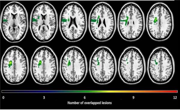
Figure 1.
Lesion overlap of the 12 participants identified with ipsilesional neglect. Each lesion was plotted onto a normal template brain using MRIcroN® software (Rorden, Karnath,&Bonilha, 2007). Colors denoting increasing numbers of participants having a lesion in a specific region, from “black” (n=1) to “red” (n=12).

