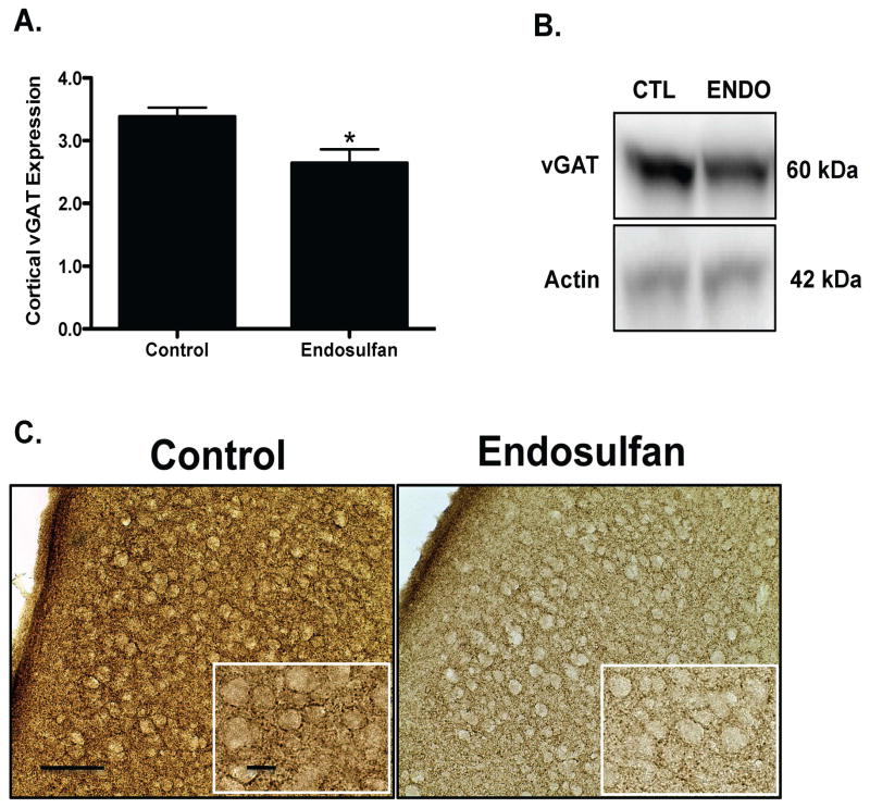
Figure 3.
Developmental exposure of male offspring to endosulfan significantly reduces the expression of vGAT in the frontal cortex. Female mice were administered either 0 (control) or 1 mg/kg endosulfan throughout gestation and lactation. Expression of vGAT in the frontal cortex was determined by immunoblot (A) in male offspring. Representative immunoblot (B) and immunohistochemical (C) images for vGAT are included. Data represent mean ± SEM (6–8 animals each from a different litter per treatment group). *Values for animals that are significantly different from controls (p < 0.05). Scale Bars: 100 μm and 50 μM, respectively.

