Abstract
Anaplasma marginale is the causative agent of anaplasmosis in cattle. Transposon mutagenesis of this pathogen using the Himar1 system resulted in the isolation of an omp10 operon insertional mutant referred to as the omp10::himar1 mutant. The work presented here evaluated if this mutant had morphological and/or growth rate defects compared to wild-type A. marginale. Results showed that the morphology, developmental cycle, and growth in tick and mammalian cell cultures are similar for the mutant and the wild type. Tick transmission experiments established that tick infection levels with the mutant were similar to those with wild-type A. marginale and that infected ticks successfully infected cattle. However, this mutant exhibited reduced infectivity and growth in cattle. The possibility of transforming A. marginale by transposon mutagenesis coupled with in vitro and in vivo assessment of altered phenotypes can aid in the identification of genes associated with virulence. The isolation of deliberately attenuated organisms that can be evaluated in their natural biological system is an important advance for the rational design of vaccines against this species.
INTRODUCTION
Anaplasma marginale is a tick-associated bacterium and the etiologic agent of bovine anaplasmosis, a disease that causes considerable losses to both dairy and beef industries worldwide (1, 2). Although organisms of this species are principally pathogenic to cattle, they are also found in other ruminants, such as water buffalo and deer (3).
The transmission cycle of A. marginale has been well documented and indicates that the success of this pathogen depends on its ability to adapt to its invertebrate and vertebrate hosts. In the tick, during its transit from the midgut to the salivary glands, A. marginale has to overcome different tissue barriers and defense mechanisms in order to ensure its transmission to the vertebrate host (4–7). In cattle, A. marginale replicates within mature erythrocytes, producing an acute disease characterized by hemolytic anemia. However, one of the most important features of the biology of these bacteria is the lifelong persistent infection of its ruminant host, achieved by evasion of the immune system using a mechanism of antigenic variation in which different variants of outer membrane proteins Msp2 and Msp3 are expressed. These persistently infected cattle remain a reservoir of A. marginale organisms for continued tick transmission (8–11).
The ability of A. marginale to thrive in such diverse environments is mediated by differential gene transcription (12). Hence, the identification and characterization of these genes using recombinant DNA technologies is not only central to understanding the biology and pathogenesis of these organisms but also for the development of drug therapies and vaccines for the control of anaplasmosis. Recently, the use of transposon mutagenesis in the A. marginale Virginia strain to create insertional mutations was demonstrated (13). Delivery of a plasmid containing the Himar1 transposon and the A7 transposase into host cell-free A. marginale resulted in the isolation of mCherry fluorescent and spectinomycin- and streptomycin-resistant bacteria. Molecular characterization of these isolated mutant organisms established that the Himar1 transposon sequences were integrated within the omp10 gene and that its insertion altered not only the expression of this gene but also the expression of the omp9, omp8, and omp7 downstream genes. These recombinant A. marginale organisms, referred to as omp10::himar1 mutants, are capable of infecting tick cell cultures, suggesting that these genes are not essential for the survival of A. marginale in this environment (13).
The omp7 to omp10 genes are members of the msp2 superfamily and are incorporated into pfam01617, a family of bacterial surface antigens (14, 15). RNA sequencing demonstrated that in A. marginale-infected erythrocytes, omp10 is transcribed as part of an operon with AM1225 at the 5′ end and with omp9, omp8, omp7, and omp6 in tandem at the 3′ end (16). Similarly, during infection of tick cells, reverse transcription (RT)-PCR experiments showed that A. marginale expresses these genes as a polycistronic message and that these genes are downregulated during tick cell culture relative to transcription levels during blood stage infection (12, 13). Omp6 is a truncated version of Omp10 and is thought not to be expressed as a functional protein (15). Omp7 to Omp9 appear as tandem repeats with 70 to 75% amino acid identity between paralogs, while Omp10 is more distantly related, with ∼30% amino acid identity to Omp7 to Omp9. AM1225 encodes a protein of unknown function (14). omp7 to omp10 are each ∼1,200 bp, encoding proteins of ∼400 amino acids. The protein expression of Omp10 appears to be lower than that of Omp7 to Omp9 (15). Omp7 to Omp9 are part of the protective outer membrane protein complexes that are capable of inducing complete protection (17). They have been identified as leading vaccine candidates, as they induce CD4+ T cell responses (17–19). Given the potential role of omp10, omp9, omp8, and omp7 in the pathogenesis of A. marginale, we hypothesized that the reduction of the expression of these outer membrane protein genes caused by insertion of the Himar1 transposon sequences into omp10 could result in an altered phenotype. Therefore, we wanted to determine if the A. marginale omp10::himar1 mutant has morphological and/or growth rate defects compared to wild-type A. marginale, to determine whether the transposon insert is stable without antibiotic selection pressure, and finally to evaluate if these mutant bacteria could infect and colonize mammalian cells.
MATERIALS AND METHODS
Cell lines and Anaplasma marginale cultivation.
For this work, two cell lines were used. ISE6 tick cells derived from embryonated eggs of the blacklegged tick, Ixodes scapularis, were kept at 34°C in nonvented 25-cm2 cell culture flasks (Nunc) and maintained in tick L-15B300 medium supplemented with 10% heat-inactivated fetal bovine serum (FBS) (Fisher Scientific) and 5% tryptose phosphate broth (BD Diagnostics) (20). The RF/6A (ATCC CRL-1780) mammalian cells from the retina choroid endothelium of a rhesus monkey (Macaca mulatta) were kept at 37°C in vented 25-cm2 cell culture flasks (Corning) and maintained in RPMI 1640 medium (HyClone) supplemented with 10% heat-inactivated FBS, 2 mM l-glutamine (Life Technologies), 0.25% NaHCO3 (Sigma-Aldrich), and 25 mM HEPES (Sigma-Aldrich) (21).
The A. marginale Virginia wild-type parental strain and the omp10::himar1 mutant transformed with a transposon bearing the mcherry and aadA genes, for mCherry fluorescent protein expression and spectinomycin/streptomycin resistance (13), were maintained in tick ISE6 cells at 34°C in tick cell medium supplemented with 0.1% Lipogro (Rocky Mountain Biologicals), 25 mM HEPES (Sigma-Aldrich), and 0.25% NaHCO3 (Sigma-Aldrich) (20). Infected RF/6A endothelial cells with wild-type and omp10::himar1 A. marginale were maintained as described above.
Immunoblots.
Binding pattern and specificity of the rabbit R883 antibody (monospecific antibody to affinity-purified Msp2) (22) for the A. marginale wild type and omp10::himar1 mutant were assessed by sodium dodecyl sulfate-polyacrylamide gel electrophoresis (SDS-PAGE) and immunoblotting using equal amounts (108 cells) of host-free bacteria. Polyvinylidene difluoride (PVDF) membranes (Thermo Scientific) were incubated with polyclonal R883 anti-Msp2 antibody and normal rabbit serum (NRS). This last antibody served as a negative control. Final dilutions of each antibody were 1:10,000. Antibody binding was detected with horseradish peroxidase-conjugated protein G (Life Technologies) diluted to 1:75,000 and SuperSignal West Femto chemiluminescent substrate (Thermo Scientific) as described in the manufacturer's instructions.
Immunoelectron microscopy of the A. marginale omp10::himar1 mutant in tick cell culture.
Cultures of ISE6 tick cells 50% infected with wild-type or omp10::himar1 mutant A. marginale as estimated by microscopy analysis of slides stained with Diff-Quik were fixed with electron microscopy grade 4% paraformaldehyde and 1% glutaraldehyde in 1× phosphate-buffered saline (PBS), pH 7.2. Fixed cells were processed with the aid of a Pelco BioWave Prolaboratory microwave (Ted Pella, Inc.). Samples were washed in 1× PBS (pH 7.2), washed in water, and dehydrated in a graded ethanol series (25%, 50%, 75%, 95%, 100%, 100%), infiltrated in HM-20 acrylic resin (Electron Microscopy Sciences), and UV cured at −10°C for 48 h. Cured resin blocks were trimmed, ultrathin sectioned, and collected on Ni-coated Formvar 400-mesh grids (Electron Microscopy Sciences). Ultrathin sections were immunolabeled at room temperature as follows: grids were treated with 200 mM NH4Cl in 1× high-salt Tween (HST; 0.5 M NaCl, 0.02 M, 0.1% Tween 20 [pH 7.2]) for 20 min, rinsed in HST, incubated for 1 h with blocking solution (1.5% bovine serum albumin [BSA], 0.5% cold-water fish skin gelatin, 0.01% Tween 20 in HST [pH 7.2]), and incubated with R883/anti-Msp2 in a 1:10,000 dilution or NRS in a 1:10,000 dilution overnight at 4°C. The following day, the grids were washed in PBS 3 times for 10 min each and incubated for 1 h at 21°C in 18-nm-colloidal-gold affinity-purified goat anti-rabbit IgG (Jackson Immuno Research) diluted 1:30 in PBS solution. Subsequent washes were in PBS and distilled water, and then samples were poststained with 2% aqueous uranyl acetate and Reynolds' lead citrate. Sections were examined with a Hitachi H-7000 transmission electron microscope.
Infection of endothelial RF/6A cells with omp10::himar1 mutant and wild-type A. marginale.
For infection of RF/6A endothelial cells, the A. marginale omp10::himar1 mutant and the wild type in ISE6 cells were used as the primary inoculum. ISE6 cells heavily infected (>90%) with wild-type or omp10::himar1 A. marginale were transferred into a sterile centrifuge tube and briefly vortexed for 30 s to release bacteria. One milliliter of inoculum from each strain was added to three wells (triplicate) of a six-well plate (Corning), and cultures were incubated at 37°C in a 5% CO2 environment. Two days postinfection, the inoculum was removed and the medium was replaced with fresh supplemented RPMI 1640 medium. Medium was replaced twice a week, and infection was monitored daily by phase-contrast and fluorescence microscopy and once a week by Diff-Quik staining.
Infection of cattle and tick transmission.
Successful infection of cattle by intravenous inoculation of A. marginale has been reported with doses ranging from 104 up to 108 organisms in A. marginale strain St. Maries and the attenuated A. marginale subsp. centrale (23, 24). A spleen-intact Holstein calf (number 42362) was inoculated intravenously with approximately 105 A. marginale omp10::himar1 cells, as determined by qPCR using the single-copy msp5 gene (25). Cells were scraped from a heavily infected 25-cm2 cell culture flask and passed 20 times through a 20-gauge needle prior to injection. Calf number 35371 was infected with wild-type A. marginale using 51 infected Dermacentor andersoni ticks and a 7-day-transmission feed. The calves were monitored for signs of infection by Giemsa-stained blood smears to detect the percent parasitized erythrocytes (PPE), by PCR and Southern analysis to detect Msp5 (25), and by competitive enzyme-linked immunosorbent assays (cELISA) (26). The splenectomized calf (number 43817) was inoculated with 1010 mutant organisms and monitored as described above for 75 days. Adult male D. andersoni ticks from the Reynolds Creek stock were applied to calf number 43817 on day 51 and fed for 12 days, while the animal was experiencing parasitemias ranging from 0.28 to 2.5 PPE. The ticks were held for 5 days to allow for digestion of the bloodmeal, and then 100 ticks were applied to spleen-intact calf number C1417 and allowed to feed for 7 days. Ticks were dissected for collection of midguts and salivary glands, and the infection levels were quantitated using both msp5 and mcherry, as described previously (25). Calf number C1417 was monitored as described above for 72 days, when the parasitemia had returned to undetectable levels.
Immunofluorescence microscopy.
Salivary glands from three adult male Dermacentor andersoni ticks acquisition-fed on a calf infected with the mCherry fluorescent protein-transformed A. marginale and three control midguts and salivary glands from ticks fed on a calf infected with wild-type A. marginale were fixed in freshly prepared 4% paraformaldehyde (Sigma-Aldrich) for 10 min at room temperature. The tissues were incubated in 1% bovine serum albumin and 0.05% Triton X-100 in PBS for 30 min. This was followed by incubation with 2 μg/ml anti-MSP2 primary antibody overnight at 4°C, followed by 10 μg/ml of Alexa Fluor 488 goat anti-rabbit IgG-H&L secondary antibody for 2 h at room temperature. Tissues were washed five times in 1% PBS after each incubation step. Images were acquired using a Nikon Eclipse Ti inverted microscope system using a 20× objective.
Dual immunofluorescence of RF/6A cells 10 days postinfection with the omp10::himar1 mutant and wild-type A. marginale strains was performed using the ANAF16C1 (anti-Msp5; 2 μg/ml) (27) monoclonal antibody and the rabbit anti-human von Willebrand factor (Dako; 1:2 dilution) (28, 29) or a mixture of equal parts of both antibodies. Cells were fixed by adding 300 μl of acetone and incubated at room temperature for 10 min. Samples were blocked by adding 100 μl of 5% BSA diluted in PBS and incubated in a humid atmosphere at room temperature for 1 h and subsequently washed once with 0.05% Tween 20 (Sigma-Aldrich) in PBS.
Samples were then reacted with primary and secondary antibodies as described previously (28–30). Negative-control monoclonal Tryp1E1 antibody that exhibits specificity for a variable surface glycoprotein of Trypanosoma brucei was used at a concentration of 2 μg/ml (31), and NRS was used at a 1:2 dilution. Samples were then washed with 500 μl of 0.05% Tween 20 (Sigma-Aldrich) in PBS three times for 5 min at room temperature and then reacted with secondary antibodies: Alexa Fluor 488 goat anti-mouse IgG antibody (Life Technologies) to detect a reaction with the primary ANAF16C1 and Alexa Fluor 568 goat anti-rabbit IgG antibody (Life Technologies) to detect a reaction with von Willebrand factor. Both secondary antibodies were used at a 1:400 dilution. Afterward, slides were mounted using ProLong Gold antifade reagent with DAPI (4′,6-diamidino-2-phenylindole) dihydrochloride (Life Technologies).
Anaplasma marginale wild-type and omp10::himar1 mutant growth curves in tick cell culture.
The growth kinetics of the A. marginale wild type and the omp10::himar1 mutant were determined by TaqMan quantitative real-time PCR (qPCR) of total extracted DNA to determine genome equivalents (GE). Two groups of 12 25-cm2 cell culture flasks, each with confluent monolayers of uninfected ISE6 cells, were inoculated with 1 ml of ISE6 cells that were >90% infected with the A. marginale wild type or the omp10::himar1 mutant. Twenty-four hours after inoculation, the inoculum was removed and replaced with 5 ml of fresh supplemented tick cell medium, and this time was considered 0 h postinfection (p.i.). Duplicate cultures of infected cells with each A. marginale strain were harvested at different time points (0, 3, 5, 9, 11, and 13 days) p.i. Infected cells were scraped from the growth surface with a cell scraper into the supernatant and centrifuged at 100 × g for 7 min at room temperature and washed twice in 1× PBS. The final pellet was used for DNA extraction (final volume of 50 μl DNA per flask), qPCR assay of 200 ng extracted DNA, and determination of A. marginale GE per flask. Statistical differences in growth rates were evaluated using Student's t test with SigmaPlot (Systat Software).
DNA extraction and qPCR.
DNA isolation from tick ISE6 cells infected with wild-type and omp10::himar1 mutant A. marginale strains was performed using the QIAamp DNA minikit (Qiagen) per the manufacturer's instruction. DNA concentration of each sample was determined using the Qubit double-stranded DNA (dsDNA) HS assay kit (Life Technologies) on a Qubit fluorometer (Life Technologies). DNA isolation from bovine blood or tick midguts and salivary glands employed the DNeasy blood and tissue kit (Qiagen).
Quantitation of the A. marginale wild type and the omp10::himar1 mutant in ISE6 tick cell cultures was performed by qPCR using two sets of primers and probes: the forward and reverse primers AB1242 and AB1243 and the probe AB1250, which target the single-copy gene opag2 (32), and the forward and reverse primers AB1345 and AB1346 and the probe AB1347, which target the aadA or spectinomycin resistance gene found in the omp10::himar1 mutant (Table 1).
TABLE 1.
TaqMan qPCR oligonucleotides used in this study
| Oligonucleotide | Sequence (5′ to 3′) | Target | Size (bp) | Reference |
|---|---|---|---|---|
| AB1242 | AAAACAGGCTTACCGCTCCAA | opag2 | 151 | 32 |
| AB1243 | GGCGTGTAGCTAGGCTCAAAGT | |||
| AB1250a | CTCTCCTCTGCTCAGGGCTCTGCG | |||
| AB1345 | GGTGACCGTAAGGCTTGATG | aadA | 279 | This study |
| AB1346 | ACCAAGGCAACGCTATGTTC | |||
| AB1347b | ACCATTGTTGTGCACGACGACA | |||
| nMSP5F | AAGTTGTAAGTGAGGGCATAGCCTCC | msp5 | 188 | 25 |
| nMSP5R | AACTTATCGGCATGGTCGCCTAGT | |||
| RFPF | TACGGTGACGCAGGATTCATCACTG | mcherry | 227 | This study |
| RFPR | ATGTCGTCTTGACCTCGGCATCAT |
Oligonucleotide labeled with 6-carboxyfluorescein (6-FAM) at the 5′ end and tetramethylrhodamine (TAMRA) at the 3′ end.
Oligonucleotide labeled with tetrachlorofluorescein (TET) at the 3′ end and black hole quencher 1 (BHQI) at the 3′ end.
Tenfold serial dilutions of the opag2/pCR-TOPO or the pHimarcisA7mCherry-SS plasmids were used for standard curve preparation, and the opag2 and aadA gene copy numbers were calculated based on the standard curve.
Triplicate reactions from cultures were analyzed. Samples were normalized by loading the same amount of total DNA (200 ng). Reaction mixtures of 25 μl containing 5 μl of DNA from wild-type or omp10::himar1 mutant A. marginale, 1× QuantiTect probe PCR mix (Qiagen), 0.4 μM forward and reverse primers, and 0.2 μM probe were used for amplification in a Bio-Rad DNA Engine Opticon thermal cycler with the following conditions: 95°C for 15 min and 40 cycles of 94°C for 1 min, 54°C for 15 s, and 60°C for 1 min.
Quantitation of the A. marginale wild type and the omp10::himar1 mutant to determine the dose of the A. marginale omp10::himar1 mutant and wild-type inoculum from infected ISE6 tick cells, in blood and tick tissues, was performed by qPCR using the forward and reverse primers nMSP5F, nMSP5R, RFPF, and RFPR, targeting the msp5 and mcherry genes (Table 1). Triplicate reaction mixtures from each sample were used. Reaction mixtures of 25 μl containing 1 μl of DNA from blood or tick tissue preparations, 1× Master mix for Express SYBR GreenER (Life Technologies), and 200 nM forward and reverse primers were used for amplification in a Bio-Rad C1000 Touch thermal cycler with the following conditions: 95°C for 5 min and 35 cycles of 95°C for 30s, 60°C for 30s, and 72°C for 1 min.
Tenfold serial dilutions of cloned control templates were used for standard curve preparation, and the A. marginale copy number was calculated based on the standard curve. The no-template control, uninfected cells, and DNA from Anaplasma phagocytophilum were used as negative controls.
Transposon insertion stability of the A. marginale omp10::himar1 mutant in tick cell culture.
The stability of the transposon insertion without antibiotic (spectinomycin/streptomycin)-selective pressure in A. marginale omp10::himar1 mutants was compared by measuring the copy number of aadA (spectinomycin/streptomycin resistance gene-containing insert) and opag2 using qPCR during 7 serial passages.
RESULTS
Electron microscopy of the Anaplasma marginale omp10::himar1 mutant in tick cell culture.
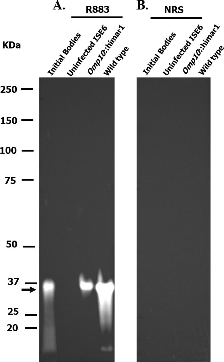
The polyclonal anti-Msp2 R883 antibody binding pattern and specificity was evaluated by immunoblotting. A. marginale omp10::himar1 mutant and wild-type copy numbers per sample were quantified by qPCR using the opag2 single-copy gene. For this, 108 organisms of the A. marginale wild type and the omp10::himar1 mutant were loaded per lane. A. marginale strain Virginia initial bodies and uninfected ISE6 cells were used as positive and negative controls, respectively. R883 specifically reacted with the major surface protein Msp2 (36 kDa) in A. marginale omp10::himar1 mutant, wild-type, and initial bodies (Fig. 1A). A similar reaction was not detected in the uninfected ISE6 cells or by using negative-control NRS (Fig. 1B).
FIG 1.
Immunoblotting of host cell-free omp10::himar1 mutant and wild-type A. marginale using the specific antibody R883. Proteins from equal amounts of host cell-free wild-type and omp10::himar1 A. marginale from infected ISE6 tick cells were separated by SDS-PAGE. Immunoblotted PVDF membranes of transferred proteins reacted with specific antibodies, and reactions were visualized by chemiluminescence. (A) Polyclonal antibody R883 (1:10,000) with specificity to the Msp2 protein (36 kDa) (black arrow); (B) negative control, normal rabbit serum (1:10,000). A. marginale strain Virginia initial bodies and uninfected ISE6 cells were used as positive and negative controls, respectively.
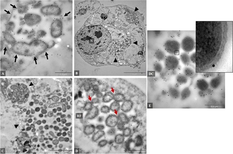
Transmission electron microscopy (TEM) of omp10::himar1 mutant A. marginale immunogold labeled with polyclonal anti-Msp2 (R883) antibody was used for the localization, visualization, and analysis of these organisms to determine if their morphology was characteristic of what has been described for wild-type A. marginale (33, 34). Based on this analysis, morphological defects in the omp10::himar1 mutant that could alter its development in infected ISE6 cells were not found. Instead, several of the characteristics common to the prototypical wild-type A. marginale were visualized; for example, the A. marginale omp10::himar1 mutant grew within membrane-bound inclusions or intravacuolar microcolonies (morulae) (20, 33, 35) (Fig. 2A). Also, the ISE6 cells were infected with several morulae, an indication of multiple omp10::himar1 mutant A. marginale invasion events (Fig. 2B and C) typical of A. marginale infection (33, 35). The two distinct morphological forms of A. marginale were also visualized in ISE6 cells infected with the omp10::himar1 mutant: enlarged oval and electron-translucent or reticulate forms (RF) (Fig. 2D) and round or coccoid electron-dense or dense-core (DC) forms (Fig. 2E). Several RF of omp10::himar1 mutant A. marginale were undergoing cell division by binary fission consistent with previous TEM descriptions of A. marginale in ISE6 cells (Fig. 2D). RF and DC forms of omp10::himar1 mutant A. marginale have an outer and inner membrane separated by a periplasmic space (Fig. 2E). These observations suggest that the morphology and development of the A. marginale omp10::himar1 mutant in tick cell culture are similar to what has been observed for the wild type (20, 35).
FIG 2.
Transmission electron microscopy of the A. marginale omp10::himar1 mutant, immunogold labeled with a polyclonal anti-Msp2 antibody (R883). (A) Anaplasma organisms within membrane-bound vacuole (arrows); (B and C) cell infected with multiples colonies (arrowheads); (D) colony formed by RF, reticulated forms with some organisms dividing by binary fission (red arrows); (E) colony formed by DC, dense-core organisms; inset showing double-layered membrane. N, nucleus. A. marginale was grown in ISE6 tick cells.
Growth curves of omp10::himar1 mutant Anaplasma marginale versus the wild type in tick cell culture.
The A. marginale omp10::himar1 mutant was further characterized by comparing the in vitro growth rate to that of wild-type A. marginale. Duplicate cultures of ISE6 cells infected with the A. marginale omp10::himar1 mutant and the wild type maintained without antibiotic selection were evaluated to determine the increase of GE by qPCR over a period of 13 days. At day 0 p.i., the mean copy numbers of GE for omp10::himar1 mutant and wild-type A. marginale were 8.87 × 10⁶ copies/flask (95% confidence interval [CI], 6.83 × 10⁶ to 1.09 × 10⁷) and 1.22 × 10⁷ copies/flask (95% CI, 8.64 × 10⁶ to 1.58 × 10⁷), respectively. At day 13 p.i., the omp10::himar1 and wild-type GE copy numbers were 4.70 × 10⁸ copies/flask (95% CI, 4.551 × 10⁸ to 4.85 × 10⁸) and 5.01 × 10⁸ copies/flask (95% CI, 4.26 × 10⁸ to 5.76 × 10⁸), respectively. The A. marginale omp10::himar1 mutant and the wild type had log increases of 1.72 and 1.67 GE, respectively (Fig. 3). The growth rates of the omp10::himar1 mutant and wild-type A. marginale in ISE6 culture were not significantly different (P = 0.527), and the doubling times were 54.7 h (2.3 days) for omp10::himar1 and 58 h (2.4 days) for wild-type A. marginale. Pearson correlation coefficients for wild-type and omp10::himar1 mutant A. marginale strains were 0.949 and 0.977, respectively.
FIG 3.
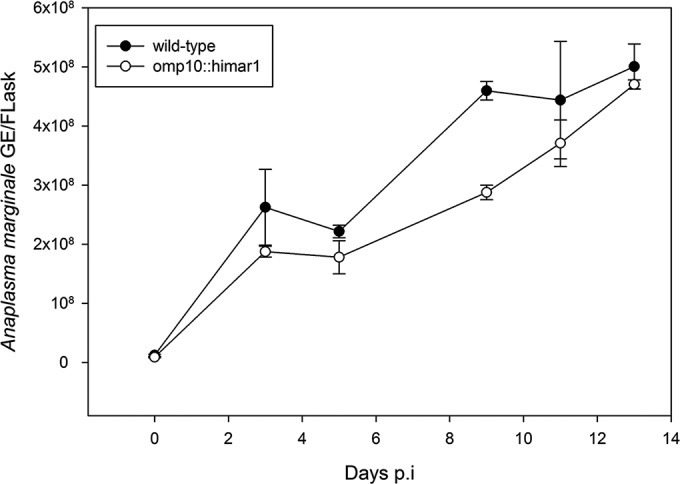
Growth curves for wild-type and omp10::himar1 mutant A. marginale strains in infected ISE6 tick cells. Growth was measured by determining GE/flask based on the A. marginale single-copy gene opag2.
Transposon insertion stability of the A. marginale omp10::himar1 mutant in tick cell culture.
Stable maintenance of the transposon insertion was evaluated by qPCR to determine the copy number of the transposon insert per omp10::himar1 mutant A. marginale genome with primers and probes specific to the transposon-borne spectinomycin gene (aadA) and the single-copy A. marginale gene opag2. For this purpose, DNA samples obtained from duplicate cultures of ISE6 cells infected with the A. marginale omp10::himar1 mutant maintained during seven serial passages (∼3 months) with or without antibiotic selection were analyzed. Table 2 shows a comparison of the copy numbers obtained for aadA and opag2 in ISE6 cells infected with the mutant with and without antibiotic selection. We used A. marginale omp10::himar1 mutants growing with antibiotic selection as a positive control for the presence of the transposon insertion. Our results show that in this group, the aadA copy number relative to the opag2 copy number was maintained (Table 2). During culture of these mutants without antibiotic selection pressure, loss of the transposon insert would be indicated by a decrease in aadA copy number with a concomitant comparative increase in opag2 copy number. However, during the seven serial passages, a steady increase of the opag2 copy number relative to the aadA gene copy number was not observed in the A. marginale omp10::himar1 mutant growing without antibiotic selection, indicating that the transposon insertion in the A. marginale omp10::himar1 mutant appears to be stable without antibiotic selection for prolonged culture periods.
TABLE 2.
A. marginale omp10::himar1 mutant transposon stability
| Passage no. | GEa (log10 ± SD)a |
|||
|---|---|---|---|---|
| Treated culture |
Untreated culture |
|||
| aadA | opag2 | aadA | opag2 | |
| 1 | 8.33 ± 0.07 | 8.40 ± 0.02 | 8.26 ± 0.002 | 8.45 ± 0.05 |
| 2 | 10.15 ± 0.03 | 10.43 ± 0.05 | 10.26 ± 0.05 | 10.60 ± 0.02 |
| 3 | 8.09 ± 0.10 | 8.25 ± 0.02 | 9.46 ± 0.003 | 9.91 ± 0.07 |
| 4 | 9.23 ± 0.03 | 9.16 ± 0.35 | 9.15 ± 0.05 | 9.33 ± 0.02 |
| 5 | 9.34 ± 0.07 | 9.56 ± 0.05 | 9.42 ± 0.03 | 9.66 ± 0.01 |
| 6 | 9.53 ± 0.02 | 9.77 ± 0.04 | 9.42 ± 0.03 | 9.72 ± 0.03 |
| 7 | 9.54 ± 0.02 | 9.67 ± 0.01 | 9.64 ± 0.02 | 9.81 ± 0.04 |
GE, genome equivalents per flask. Treated cultures were maintained with antibiotic selection.
Infectivity of omp10::himar1 mutant A. marginale for mammalian cells in vitro and in vivo.
A. marginale can be propagated in vitro in RF/6A endothelial cells derived from the retina choroid of a rhesus monkey (21, 29). To investigate if the A. marginale omp10::himar1 mutant can infect RF/6A endothelial cells, triplicate cultures of uninfected RF/6A endothelial cells were infected with A. marginale omp10::himar1 mutant cells from a heavily infected ISE6 culture (>90% infected) and maintained with or without antibiotic selection. Similarly, and as a control for infection of endothelial cells, triplicate cultures of uninfected RF/6A cells were infected with wild-type A. marginale.
Infection of RF/6A cells with the omp10::himar1 mutant was monitored by an increase of red fluorescence emitted by the replicating, mCherry fluorescent protein-expressing mutant. By 10 days p.i., about 50% to 60% of the cells were infected in cultures maintained with or without antibiotic selection (Fig. 4A to E). The percentage of infection was confirmed by Diff-Quik staining and was similar in RF/6A cells infected with wild-type A. marginale (data not shown). The cytoplasm of infected cells contained a single large or several small morulae that fluoresced red (Fig. 4A).
FIG 4.
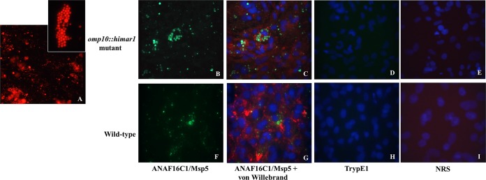
Infection of RF/6A endothelial cells at 10 days postinfection with the omp10::himar1 mutant and wild-type Anaplasma marginale strains. (A) RF/6A endothelial cells infected with the A. marginale omp10::himar1 mutant expressing mCherry red fluorescent protein, with the inset showing a single endothelial cell infected with multiple colonies. The A. marginale (green) omp10::himar1 mutant (B and C) and the wild type (F and G) in endothelial cells (red). In panels C and G, host cell nuclei were counterstained with DAPI (blue). Similar fluorescence was not detected in infected cells stained with control antibodies Tryp1E1 or NRS for cells infected with the omp10::himar1 mutant (D and E) or in cells infected with the wild type (H and I).
Figures 4B and C show the immunofluorescence results of the specific binding of the antibody ANAF16C1 (anti-Msp5) to omp10::himar1 mutant A. marginale (green) and antibody against human von Willebrand factor to RF/6A cells (red). Juxtaposition of the green and red fluorescence signals showed colocalization of omp10::himar mutant A. marginale (green) within endothelial cells (red), confirming that the entire monolayer was formed by endothelial cells. Fluorescence similar to that emitted using these two specific antibodies was not detected in RF/6A endothelial cells infected with omp10::himar1 mutant A. marginale labeled with the negative-control monoclonal antibody TrypE1 or NRS (Fig. 4D and E). Similar results were obtained from RF/6A cells infected with wild-type A. marginale (Fig. 4F to I).
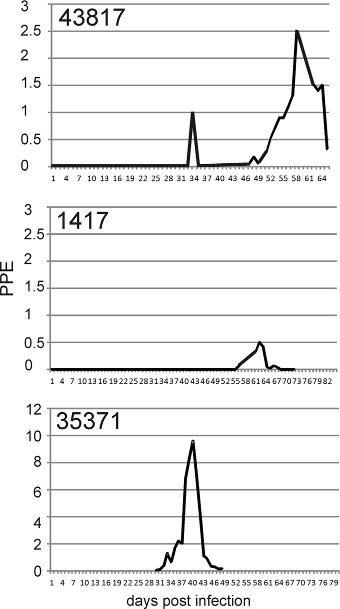
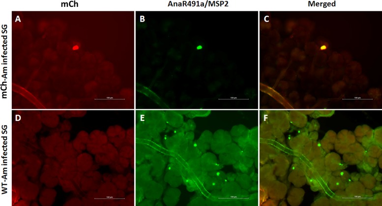
To determine if the A. marginale omp10::himar1 mutant infects cattle, we attempted infection of 2 spleen-intact calves and 1 splenectomized calf. Inoculation of a spleen-intact calf with 105 mutant organisms failed to establish infection, as shown by negative Giemsa-stained blood smears, PCR, Southern blotting, and competitive enzyme-linked immunosorbent (cELISA) assays. Because of this, a second attempt was made to infect the same calf at day 71 after the initial inoculation, delivering approximately 108 omp10::himar1 mutant A. marginale organisms. This animal was monitored for a further 46 days with no signs of infection, suggesting that this mutant had a reduced infectivity in cattle compared to that of the wild type (data not shown). Splenectomized calves are known to be less resistant to A. marginale infection than spleen-intact calves and adults (9, 33). Typically, spleen-intact calves control the first (acute) bacteremia and then become persistently infected, with additional cyclic peaks of bacteremia (9). In contrast, splenectomized calves generally fail to control the first peak of bacteremia, which can increase to >50% PPE, and then they succumb to infection. Administration of 1010 organisms to a splenectomized calf resulted in a patent erythrocytic bacteremia in 34 days and a second peak on day 58 (Fig. 5). Feeding of D. andersoni ticks on this infected calf resulted in 100% acquisition of omp10::himar1 mutant organisms in both the midgut and salivary glands, showing that although infectivity of the mutant was reduced, the mutant was acquired by ticks at rates and levels similar to those of the wild type. Anaplasma marginale burdens in the tick were determined by qPCR for msp5 (on the genome backbone) and mcherry (on the transposed insert) and ranged for the mutant from 103 to 105 (Table 3). This is similar to tick burdens determined previously for wild-type A. marginale (25). The positive ticks were fed on a naive, spleen-intact calf which had a peak parasitemia of 0.5 PPE at day 62 (Fig. 5). Ticks were collected from the transmission calf, dissected to collect salivary glands, and subjected to immunofluorescence microscopy (Fig. 6). The sections were stained with antibody against Msp2. The yellow staining in Fig. 6C indicates colocalization of Msp2 and the omp10::himar1 mutant.
FIG 5.
Infection profile of the omp10::himar1 mutant strain compared to wild-type A. marginale in cattle. Bacteremia (left axis) is reported as percent parasitized erythrocytes (PPE) as determined by microscopic evaluation of Giemsa-stained blood smears. Animal number is indicated in the top left of each graph. Animal number 43817 was a splenectomized animal infected with the omp10::himar1 mutant strain by injection of tissue culture material; animal number 1417 was a spleen-intact animal infected with the omp10::himar1 mutant strain by tick inoculation, while spleen-intact animal number 35371 was infected with wild-type A. marginale by tick inoculation.
TABLE 3.
Quantitation of the omp10::himar1 mutant in tick tissues
| Tick no. | GEa (log10 ± SD) |
|||
|---|---|---|---|---|
| SG |
MG |
|||
| msp5 | mcherry | msp5 | mcherry | |
| 1 | 5.12 ± 0.02 | 4.88 ± 0.02 | 5.38 ± 0.04 | 4.83 ± 0.02 |
| 2 | 5.42 ± 0.03 | 4.49 ± 0.01 | 4.33 ± 0.03 | 4.93 ± 0.02 |
| 3 | 4.36 ± 0.03 | 5.69 ± 0.01 | 4.07 ± 0.05 | 4.87 ± 0.01 |
| 4 | 4.95 ± 0.02 | 4.82 ± 0.03 | 4.74 ± 0.04 | 4.91 ± 0.03 |
| 5 | 4.09 ± 0.03 | 5.78 ± 0.02 | 4.68 ± 0.01 | 4.56 ± 0.03 |
| 6 | 4.97 ± 0.05 | 6.18 ± 0.01 | 4.51 ± 0.05 | 4.94 ± 0.01 |
| 7 | 4.92 ± 0.03 | 5.15 ± 0.02 | 5.55 ± 0.01 | 4.83 ± 0.03 |
| 8 | 5.30 ± 0.04 | 6.07 ± 0.02 | 5.66 ± 0.04 | 4.88 ± 0.02 |
| 9 | 5.02 ± 0.04 | 4.98 ± 0.03 | 4.94 ± 0.05 | 4.84 ± 0.02 |
| 10 | 5.19 ± 0.02 | 5.21 ± 0.05 | 5.40 ± 0.01 | 4.96 ± 0.01 |
GE, genome equivalents per salivary gland pair (SG) or per tick midgut (MG).
FIG 6.
The A. marginale omp10::himar1 mutant infects ticks. Immunofluorescence images showing infection of D. andersoni salivary gland (SG) acinar cells with the omp10::himar1 mutant (mCh-Am) (top) and wild-type A. marginale (WT-Am) control (bottom). (A to C) Salivary glands from tick acquisition fed on a calf infected with mCh-Am (red) were colabeled with AnaR4901a primary antibodies against Anaplasma marginale MSP2 and a conjugated Alexa Fluor 488 goat anti-rabbit IgG-H&L secondary antibody (green). (D to F) Salivary glands from control ticks infected with wild-type A. marginale showing multiple colonies of mCherry-negative, MSP2-positive A. marginale.
DISCUSSION
The ability of A. marginale to cause disease often depends on its ability to invade and replicate within different hosts. During this work, we investigated the phenotype of omp10::himar1 mutant A. marginale that carries the Himar1 transposon integrated within the omp10 gene.
The typical developmental cycle of A. marginale in tick cells involves two stages: a reticulate or vegetative form (RF) and the infective dense-core (DC) form. Upon internalization, A. marginale is enclosed within a parasitophorous vacuole, where it transitions into a reticulate form that divides by binary fission to produce multiple organisms that subsequently will change into the infective dense forms that will be released to infect adjacent cells (33–35). Electron microscopy analysis shows that A. marginale omp10::himar1 mutants develop in a fashion similar to the wild type in tick cell culture (20, 34–36) and suggests that the gross structure of the outer membrane of these organisms was not affected by disruption in the expression of omp10, omp9, omp8, and omp7. Growth curves support these findings since they show that A. marginale omp10::himar1 mutant growth is comparable to that of the wild type during infection of tick cell cultures. Stability of the Himar1 insertion under non-antibiotic-selectable conditions has been demonstrated in several pathogenic bacterial species (37, 38), and this is also true for the A. marginale omp10::himar1 mutant. Stability of transposon insertion in these mutants was monitored during seven serial passages over 3 months in cultures of infected ISE6 cells maintained without antibiotic selection. Quantitative PCR shows similar levels of the spectinomycin resistance gene (from the transposon) and the opag2 gene (from the genome backbone). Maintenance of the red fluorescent phenotype for the length of the culture period confirms these results, since the mcherry reporter gene is also found in the transposon. Hence, loss of the red fluorescence phenotype due to the lack of antibiotic selection pressure will correlate with the loss of the transposon insertion.
Previous work has indicated a possible important role of omp10, omp9, omp8, and omp7 in the pathogenesis of A. marginale. For example, these genes are expressed at lower levels in tick cells than in erythrocytes, and outer membrane proteome analysis and cross-linking experiments showed that although expressed, Omp9, Omp8, and Omp7 proteins are not detected at the surface of A. marginale-infecting tick cell culture (15, 17). On the other hand, in infected erythrocytes, these proteins are localized at the surface of A. marginale, suggesting a possible rearrangement in surface topology during the transition from tick to mammalian cell infection (17). Therefore, with the putative inability to upregulate the expression of these proteins on the surface of the pathogen, we hypothesized that the A. marginale omp10::himar1 mutant would not be infective to mammalian cells. To test this, infectivity of the A. marginale omp10::himar1 mutant in RF/6A endothelial cell cultures and in cattle was assessed. Surprisingly, the A. marginale omp10::himar1 mutant invaded and replicated within RF/6A endothelial cells similar to the wild type, demonstrating that disruption of omp10 and downstream genes did not affect infectivity of these mutants in endothelial cell cultures in vitro. Although it took three attempts, we were able to establish the A. marginale omp10::himar1 mutant in cattle. A splenectomized calf had a detectable PPE of 1% 34 days postinoculation. However, the infection was controlled, and a subsequent peak parasitemia of 2.5% occurred at day 58 and was again controlled. Typically, wild-type A. marginale infections in splenectomized animals will result in consistently increasing parasitemias to high levels until the animal dies or is euthanized. Ticks were able to acquire and transmit the omp10::himar1 mutant to a spleen-intact calf. A. marginale burden within the ticks ranged from 103 to 105 cells in the midgut and the salivary gland, which is typical for transmission-fed ticks (25).
A. marginale has been transformed only twice. The first transformant was by homologous recombination with a green fluorescent protein (GFP) marker into the St. Maries strain (32), while the second transformant is the one used in the current study (13). Interestingly, both transformed strains behave similarly when used to infect bovine hosts, producing very low PPE levels compared to those of wild-type infections (Fig. 5) (39, 40). Transcriptional profiling of the GFP mutant demonstrated alterations in gene transcriptional patterns for nucleotide biosynthesis, translation, and translation elongation (41). The omp10::himar1 mutant was found to have altered transcription of the omp10 operon (13). These two mutations have very different effects on the transcriptional patterns on the cell, and yet both have reduced vigor during mammalian infection. These transformed organisms are candidates for development as vaccine strains (40).
In summary, the work presented here shows that disruption of the omp10 operon does not result in altered morphology or altered in vitro growth of A. marginale. Neither is expression of the omp10 operon essential for the growth of A. marginale in ticks or cattle. However, this A. marginale omp10::himar1 mutant showed reduced infectivity and growth in its bovine host. In the past, naturally attenuated strains have been used in vaccines despite their empirical derivation and drawbacks associated with live blood vaccines. These data suggest that a rational strategy employing transposon mutagenesis to attenuate A. marginale for growth in cattle is feasible. If the efficiency of transposon mutagenesis can be increased, this would provide a means to define virulence mechanisms and identify protective antigens in A. marginale and other Anaplasmataceae.
ACKNOWLEDGMENTS
We thank Xiaoya Cheng (Washington State University, Pullman, WA, USA), Heather L. Wamsley (University of Florida, Gainesville, FL, USA), Melanie G. Pate (University of Florida, Gainesville, FL, USA), and Anna M. Lundgren (University of Florida, Gainesville, FL, USA) for their expert technical assistance.
This work was supported by The Wellcome Trust, grant number GR075800M.
REFERENCES
- 1.Kocan KM, de la Fuente J, Blouin EF, Coetzee JF, Ewing SA. 2010. The natural history of Anaplasma marginale. Vet Parasitol 167:95–107. doi: 10.1016/j.vetpar.2009.09.012. [DOI] [PubMed] [Google Scholar]
- 2.Brayton KA, Dark MJ, Palmer GH. 2009. Anaplasma, p 85–117. In Nene V, Kole C (ed), Genome mapping and genomics in animal-associated microbes. Springer-Verlag, Berlin, Germany. [Google Scholar]
- 3.Kuttler KL. 1984. Anaplasma infections in wild and domestic ruminants: a review. J Wildl Dis 20:12–20. doi: 10.7589/0090-3558-20.1.12. [DOI] [PubMed] [Google Scholar]
- 4.Kazimirova M, Stibraniova I. 2013. Tick salivary compounds: their role in modulation of host defences and pathogen transmission. Front Cell Infect Microbiol 3:43. doi: 10.3389/fcimb.2013.00043. [DOI] [PMC free article] [PubMed] [Google Scholar]
- 5.Singhgh SK, Girschick HJ. 2003. Tick-host interactions and their immunological implications in tick-borne diseases. Curr Sci 85:1284–1298. doi: 10.3389/fmicb.2013.00337. [DOI] [Google Scholar]
- 6.Kocan KM, De La Fuente J, Blouin EF, Garcia-Garcia JC. 2002. Adaptations of the tick-borne pathogen, Anaplasma marginale, for survival in cattle and ticks. Exp Appl Acarol 28:9–25. doi: 10.1023/A:1025329728269. [DOI] [PubMed] [Google Scholar]
- 7.Hajdusek O, Sima R, Ayllon N, Jalovecka M, Perner J, de la Fuente J, Kopacek P. 2013. Interaction of the tick immune system with transmitted pathogens. Front Cell Infect Microbiol 3:26. doi: 10.3389/fcimb.2013.00026. [DOI] [PMC free article] [PubMed] [Google Scholar]
- 8.Kocan KM, Goff WL, Stiller D, Claypool PL, Edwards W, Ewing SA, Hair JA, Barron SJ. 1992. Persistence of Anaplasma marginale (Rickettsiales: Anaplasmataceae) in male Dermacentor andersoni (Acari: Ixodidae) transferred successively from infected to susceptible calves. J Med Entomol 29:657–668. [DOI] [PubMed] [Google Scholar]
- 9.Palmer GH, Brown WC, Rurangirwa FR. 2000. Antigenic variation in the persistence and transmission of the ehrlichia Anaplasma marginale. Microbes Infect 2:167–176. doi: 10.1016/S1286-4579(00)00271-9. [DOI] [PubMed] [Google Scholar]
- 10.Meeus PF, Brayton KA, Palmer GH, Barbet AF. 2003. Conservation of a gene conversion mechanism in two distantly related paralogues of Anaplasma marginale. Mol Microbiol 47:633–643. doi: 10.1046/j.1365-2958.2003.03331.x. [DOI] [PubMed] [Google Scholar]
- 11.Meeus PF, Barbet AF. 2001. Ingenious gene generation. Trends Microbiol 9:353–355, discussion 355–356. doi: 10.1016/S0966-842X(01)02112-6. [DOI] [PubMed] [Google Scholar]
- 12.Hammac GK, Pierle SA, Cheng X, Scoles GA, Brayton KA. 2014. Global transcriptional analysis reveals surface remodeling of Anaplasma marginale in the tick vector. Parasit Vectors 7:193. doi: 10.1186/1756-3305-7-193. [DOI] [PMC free article] [PubMed] [Google Scholar]
- 13.Crosby FL, Wamsley HL, Pate MG, Lundgren AM, Noh SM, Munderloh UG, Barbet AF. 2014. Knockout of an outer membrane protein operon of Anaplasma marginale by transposon mutagenesis. BMC Genomics 15:278. doi: 10.1186/1471-2164-15-278. [DOI] [PMC free article] [PubMed] [Google Scholar]
- 14.Brayton KA, Kappmeyer LS, Herndon DR, Dark MJ, Tibbals DL, Palmer GH, McGuire TC, Knowles DP Jr. 2005. Complete genome sequencing of Anaplasma marginale reveals that the surface is skewed to two superfamilies of outer membrane proteins. Proc Natl Acad Sci U S A 102:844–849. doi: 10.1073/pnas.0406656102. [DOI] [PMC free article] [PubMed] [Google Scholar]
- 15.Noh SM, Brayton KA, Knowles DP, Agnes JT, Dark MJ, Brown WC, Baszler TV, Palmer GH. 2006. Differential expression and sequence conservation of the Anaplasma marginale msp2 gene superfamily outer membrane proteins. Infect Immun 74:3471–3479. doi: 10.1128/IAI.01843-05. [DOI] [PMC free article] [PubMed] [Google Scholar]
- 16.Pierle SA, Dark MJ, Dahmen D, Palmer GH, Brayton KA. 2012. Comparative genomics and transcriptomics of trait-gene association. BMC Genomics 13:669. doi: 10.1186/1471-2164-13-669. [DOI] [PMC free article] [PubMed] [Google Scholar]
- 17.Noh SM, Brayton KA, Brown WC, Norimine J, Munske GR, Davitt CM, Palmer GH. 2008. Composition of the surface proteome of Anaplasma marginale and its role in protective immunity induced by outer membrane immunization. Infect Immun 76:2219–2226. doi: 10.1128/IAI.00008-08. [DOI] [PMC free article] [PubMed] [Google Scholar]
- 18.Lopez JE, Siems WF, Palmer GH, Brayton KA, McGuire TC, Norimine J, Brown WC. 2005. Identification of novel antigenic proteins in a complex Anaplasma marginale outer membrane immunogen by mass spectrometry and genomic mapping. Infect Immun 73:8109–8118. doi: 10.1128/IAI.73.12.8109-8118.2005. [DOI] [PMC free article] [PubMed] [Google Scholar]
- 19.Palmer GH, Brown WC, Noh SM, Brayton KA. 2012. Genome-wide screening and identification of antigens for rickettsial vaccine development. FEMS Immunol Med Microbiol 64:115–119. doi: 10.1111/j.1574-695X.2011.00878.x. [DOI] [PMC free article] [PubMed] [Google Scholar]
- 20.Munderloh UG, Blouin EF, Kocan KM, Ge NL, Edwards WL, Kurtti TJ. 1996. Establishment of the tick (Acari:Ixodidae)-borne cattle pathogen Anaplasma marginale (Rickettsiales:Anaplasmataceae) in tick cell culture. J Med Entomol 33:656–664. [DOI] [PubMed] [Google Scholar]
- 21.Munderloh UG, Lynch MJ, Herron MJ, Palmer AT, Kurtti TJ, Nelson RD, Goodman JL. 2004. Infection of endothelial cells with Anaplasma marginale and Anaplasma phagocytophilum. Vet Microbiol 101:53–64. doi: 10.1016/j.vetmic.2004.02.011. [DOI] [PubMed] [Google Scholar]
- 22.Palmer GH, Oberle SM, Barbet AF, Goff WL, Davis WC, McGuire TC. 1988. Immunization of cattle with a 36-kilodalton surface protein induces protection against homologous and heterologous Anaplasma marginale challenge. Infect Immun 56:1526–1531. [DOI] [PMC free article] [PubMed] [Google Scholar]
- 23.Noh SM, Turse JE, Brown WC, Norimine J, Palmer GH. 2013. Linkage between Anaplasma marginale outer membrane proteins enhances immunogenicity but is not required for protection from challenge. Clin Vaccine Immunol 20:651–656. doi: 10.1128/CVI.00600-12. [DOI] [PMC free article] [PubMed] [Google Scholar]
- 24.Agnes JT, Brayton KA, LaFollett M, Norimine J, Brown WC, Palmer GH. 2011. Identification of Anaplasma marginale outer membrane protein antigens conserved between A. marginale sensu stricto strains and the live A. marginale subsp. centrale vaccine. Infect Immun 79:1311–1318. doi: 10.1128/IAI.01174-10. [DOI] [PMC free article] [PubMed] [Google Scholar]
- 25.Futse JE, Ueti MW, Knowles DP Jr, Palmer GH. 2003. Transmission of Anaplasma marginale by Boophilus microplus: retention of vector competence in the absence of vector-pathogen interaction. J Clin Microbiol 41:3829–3834. doi: 10.1128/JCM.41.8.3829-3834.2003. [DOI] [PMC free article] [PubMed] [Google Scholar]
- 26.Torioni de Echaide S, Knowles DP, McGuire TC, Palmer GH, Suarez CE, McElwain TF. 1998. Detection of cattle naturally infected with Anaplasma marginale in a region of endemicity by nested PCR and a competitive enzyme-linked immunosorbent assay using recombinant major surface protein 5. J Clin Microbiol 36:777–782. [DOI] [PMC free article] [PubMed] [Google Scholar]
- 27.Knowles D, Torioni de Echaide S, Palmer G, McGuire T, Stiller D, McElwain T. 1996. Antibody against an Anaplasma marginale MSP5 epitope common to tick and erythrocyte stages identifies persistently infected cattle. J Clin Microbiol 34:2225–2230. [DOI] [PMC free article] [PubMed] [Google Scholar]
- 28.Carreno AD, Alleman AR, Barbet AF, Palmer GH, Noh SM, Johnson CM. 2007. In vivo endothelial cell infection by Anaplasma marginale. Vet Pathol 44:116–118. doi: 10.1354/vp.44-1-116. [DOI] [PubMed] [Google Scholar]
- 29.Wamsley HL, Alleman AR, Johnson CM, Barbet AF, Abbott JR. 2011. Investigation of endothelial cells as an in vivo nidus of Anaplasma marginale infection in cattle. Vet Microbiol 153:264–273. doi: 10.1016/j.vetmic.2011.05.035. [DOI] [PubMed] [Google Scholar]
- 30.Wamsley HL, Barbet AF. 2008. In situ detection of Anaplasma spp. by DNA target-primed rolling-circle amplification of a padlock probe and intracellular colocalization with immunofluorescently labeled host cell von Willebrand factor. J Clin Microbiol 46:2314–2319. doi: 10.1128/JCM.02197-07. [DOI] [PMC free article] [PubMed] [Google Scholar]
- 31.Barbet AF, Blentlinger R, Yi J, Lundgren AM, Blouin EF, Kocan KM. 1999. Comparison of surface proteins of Anaplasma marginale grown in tick cell culture, tick salivary glands, and cattle. Infect Immun 67:102–107. [DOI] [PMC free article] [PubMed] [Google Scholar]
- 32.Felsheim RF, Chavez AS, Palmer GH, Crosby L, Barbet AF, Kurtti TJ, Munderloh UG. 2010. Transformation of Anaplasma marginale. Vet Parasitol 167:167–174. doi: 10.1016/j.vetpar.2009.09.018. [DOI] [PMC free article] [PubMed] [Google Scholar]
- 33.Kocan KM, de la Fuente J, Guglielmone AA, Melendez RD. 2003. Antigens and alternatives for control of Anaplasma marginale infection in cattle. Clin Microbiol Rev 16:698–712. doi: 10.1128/CMR.16.4.698-712.2003. [DOI] [PMC free article] [PubMed] [Google Scholar]
- 34.Kocan KM. 1986. Development of Anaplasma marginale Theiler in ixodid ticks: coordinated development of a rickettsial organism and its tick host, p 472–505. In Sauer JR, Hair JA (ed), Morphology, physiology, and behavioral biology of ticks. John Wiley & Sons, New York, NY. [Google Scholar]
- 35.Blouin EF, Kocan KM. 1998. Morphology and development of Anaplasma marginale (Rickettsiales: Anaplasmataceae) in cultured Ixodes scapularis (Acari: Ixodidae) cells. J Med Entomol 35:788–797. [DOI] [PubMed] [Google Scholar]
- 36.Kocan KM. 1992. Recent advances in the biology of Anaplasma spp. in Dermacentor andersoni ticks. Ann N Y Acad Sci 653:26–32. doi: 10.1111/j.1749-6632.1992.tb19626.x. [DOI] [PubMed] [Google Scholar]
- 37.Rholl DA, Trunck LA, Schweizer HP. 2008. In vivo Himar1 transposon mutagenesis of Burkholderia pseudomallei. Appl Environ Microbiol 74:7529–7535. doi: 10.1128/AEM.01973-08. [DOI] [PMC free article] [PubMed] [Google Scholar]
- 38.Pelicic V, Morelle S, Lampe D, Nassif X. 2000. Mutagenesis of Neisseria meningitidis by in vitro transposition of Himar1 mariner. J Bacteriol 182:5391–5398. doi: 10.1128/JB.182.19.5391-5398.2000. [DOI] [PMC free article] [PubMed] [Google Scholar]
- 39.Noh SM, Ueti MW, Palmer GH, Munderloh UG, Felsheim RF, Brayton KA. 2011. Stability and tick transmission phenotype of GFP-transformed Anaplasma marginale through a complete in vivo infection cycle. Appl Environ Microbiol 77:330–334. doi: 10.1128/AEM.02096-10. [DOI] [PMC free article] [PubMed] [Google Scholar]
- 40.Hammac GK, Ku PS, Galletti MF, Noh SM, Scoles GA, Palmer GH, Brayton KA. 2013. Protective immunity induced by immunization with a live, cultured Anaplasma marginale strain. Vaccine 31:3617–3622. doi: 10.1016/j.vaccine.2013.04.069. [DOI] [PMC free article] [PubMed] [Google Scholar]
- 41.Pierle SA, Hammac GK, Palmer GH, Brayton KA. 2013. Transcriptional pathways associated with the slow growth phenotype of transformed Anaplasma marginale. BMC Genomics 14:272. doi: 10.1186/1471-2164-14-272. [DOI] [PMC free article] [PubMed] [Google Scholar]


