ABSTRACT
Results of stereotactic radiotherapy (SRT) for spinal intradural metastases developing inside or adjacent to the previous external-beam radiation therapy (EBRT) field are shown in 3 cases. One case of spinal intramedullary metastasis and two cases of intradural extramedullary metastases were treated using a Novalis shaped-beam SRT. Case 1 developed an intramedullary metastatic tumor in the C1 spinal medulla inside the previous whole brain EBRT field and another lesion adjacent to the field in the C2 spinal medulla. Case 2 developed intradural extramedullary metastasis around C6-8 inside the previous EBRT field for the primary lung adenocarcinoma. Case 3 developed multiple spinal intradural extramedullary metastatic deposits after surgical resection and following whole brain EBRT for brain metastasis. We delivered 24 to 36 Gy in 5 to 12 fractions. The treated tumors were stable or decreased in size until the patients’ death from the primary cancer (10, 22, and 5 months). Neurological symptoms were stable or improved in all 3 patients. Palliative SRT using Novalis is expected to be safe and effective even if the patient develops spinal intradural metastases within or adjacent to the previous irradiation field.
Key Words: IMRT, Intradural, Metastasis, Spine, Stereotactic radiotherapy
INTRODUCTION
With the progress of recent multimodality cancer treatments and advancement of imaging to detect newly developing lesions, retreatment of late relapse or a second neoplasm has become more common in the management of patients with cancers. The spinal cord is considered a major dose-limiting organ in radiotherapy because radiation myelopathy can result in severe functional deficits. The low tolerance of the spinal cord to radiation1) often limits the treatment dose to a level far below the optimal tumor treatment dose, especially in retreatment of tumors located within or immediately adjacent to the previous external beam radiation therapy (EBRT) field. Conventional EBRT lacks the precision to allow delivery of large doses of radiation near radiosensitive structures, such as the spinal cord. If the radiation dose could be confined more precisely to the treatment volume, the likelihood of successful tumor control should increase at the same time that the risk of spinal cord injury is minimized. Stereotactic radiotherapy (SRT) or radiosurgery (SRS) delivers a highly conformal and large radiation dose to a localized tumor by means of stereotactic approach. We report 3 cases of spinal intradural metastatic tumors, developing within or immediately adjacent to the previous EBRT field, which were treated with SRT and the results were favorable.
PATIENTS AND METHODS
Patients
Three patients (Cases 1, 2, and 3) with spinal intradural metastases were treated by SRT using the Novalis System (BrainLAB, Tokyo, Japan). All patients signed Institutional Review-Board informed consent forms. Spinal intradural metastases developed within or just adjacent to the previous EBRT field. The intervals between the previous EBRT and the Novalis SRT were 3, 14, and 6 months, respectively. Case 1 had C (cervical) 1 and C2 intramedullary metastasis. Case 2 had C6-8 extramedullary metastasis. Case 3 had C1-2 and L (lumbar) 1 extramedullary metastases.
Stereotactic radiotherapy technique
The Novalis SRT system consists of several stereotactic components. Infrared passive reflectors attached non-invasively to the patient’s body shell surface were used for positioning of the target close to the isocenter of a linear accelerator. X-ray image guidance was used for fine positioning adjustments based on internal anatomy, such as the spinal bone. The infrared guidance is also used to monitor external patient motion during treatment. A head and neck thermoplastic shell for cervical spinal lesions, or a vacuum couch and a thermoplastic body shell for thoracic, lumbar, and sacral lesions (Fig. 1), minimize the patient’s movement during treatment. Spinal lesions are exactly targeted after localization of spinal bone structures during Novalis SRT as described above.
Fig. 1.
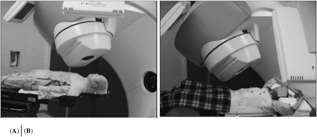
Two types of shells for patient fixation during Novalis stereotactic radiation therapy (SRT). One is for cervical spinal lesions (A) and the other for thoracic, lumbar, and sacral lesions (B).
RESULTS
Total doses of 24 to 36 Gy in 5 to 12 fractions were delivered to spinal intradural metastases within or adjacent to the previous irradiation field. Chemotherapy was continued after Novalis SRT in all 3 patients. In Case 1, the C1 and C2 intramedullary tumors were stable until the patient’s death 10 months after SRT. In Case 2, the C6-8 tumor disappeared and no relapse occurred until the patient’s death 22 months after SRT. In Case 3, the intradural tumors were stable until the patient’s death 5 months after SRT. Neurological symptoms were stable in Case 1. Symptoms were improved in Case 2 and Case 3 after SRT.
Case presentation
Case 1 67-year-old male, Fig. 2
Fig. 2.
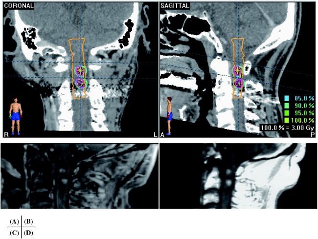
Case 1. Coronal view (A) and sagittal view (B) of CT with contrast enhancement during treatment plan of Novalis SRT for C1 and C2 intramedullary metastases on BrainSCAN (BrainLAB, Tokyo, Japan). Sagittal views of MRI before (C) and 4 months after SRT (D). The treated lesions were stable for 10 months after SRT until the patient died from primary lung adenocarcinoma.
The patient suffered from lung adenocarcinoma for 15 years. He underwent resection of the lung tumor initially and then received chemotherapy for relapse of lung cancer 14 years later. In addition, he received whole brain EBRT (WB-EBRT) of 40 Gy in 20 fractions, following surgical resection of the left frontal brain metastasis. The field of WB-EBRT covered the cervical spinal medulla of C1. He then developed gait disturbance due to spinal intramedullary metastases of C1 and C2. The tumor in C1 was in the field of the previous WB-EBRT, and that of C2 was just outside the field. SRT by circular multi-arc of a cone collimator using Novalis was performed for both spinal lesions. A total dose of 24 Gy (at 100% isodose) in 8 fractions was delivered for the C1 lesion (planned target volume, PTV=0.23 ml) and 36 Gy in 12 fractions was delivered to the C2 lesion (0.07 ml) using a multi-circular cone collimator. D95 (rate of PTV volume covered in 95% dose) was 96% in both tumors. Both tumors were stable until death from primary lung carcinoma 10 months after the Novalis SRT. The patient developed mild ataxia before treatment but the symptoms were stable until his death.
Case 2 44-year-old male, Fig. 3
Fig. 3.
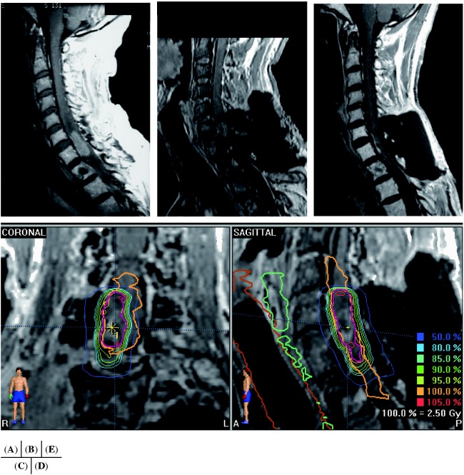
Case 2. Sagittal MRI before (A) and after (B) the second surgical resection of recurrent intradural extramedullary metastasis around C6-8. Coronal view (C) and sagittal view (D) of MRI with gadolinium (Gd) enhancement during treatment plan of Novalis SRT for residual tumor on the surface of the spinal medulla. The tumor almost disappeared within 3 months after SRT (E) and no relapse occurred prior to the patient’s death 22 months after SRT.
The patient developed intradural extramedullary metastasis around C6-8 originating initially along the C8 nerve root, which was inside the previous irradiation field for the primary right upper lung adenocarcinoma. The patient developed left-sided hemi-dysesthesia. The spinal tumor was once resected subtotally, but recurred 3 months later. The site of the spinal tumor was included in the field of 42 Gy out of a total 60 Gy of the previous irradiation for right upper lung adenocarcinoma. We treated the residual tumor (PTV=1.74 ml) by SRT with 25 Gy (at 100% isodose) in 10 fractions using a coplanar intensity-modulated multi-beam after a second surgical subtotal resection. D95 was 96%. The tumor disappeared and no relapse occurred prior to the patient’s death 22 months after Novalis SRT and the patient’s neurological symptoms had partially improved.
Case 3 55-year-old male, Fig. 4, 5
Fig. 4.
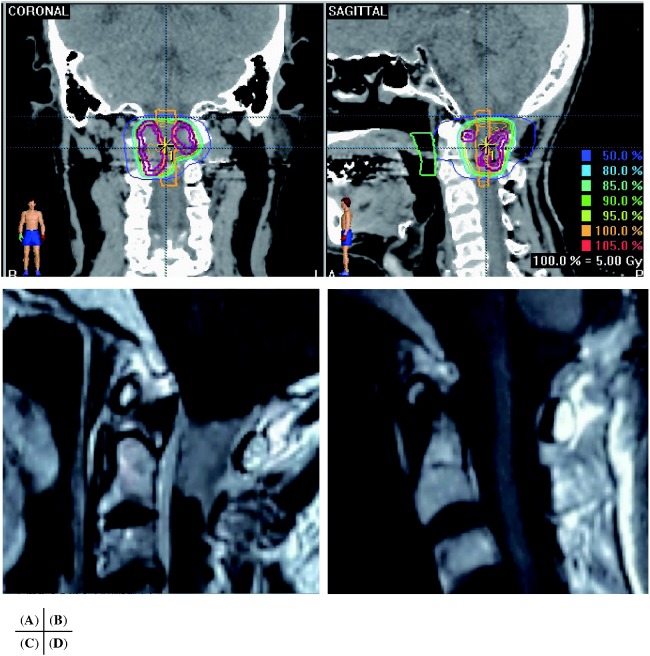
Case 3. Coronal (A) and sagittal (B) view of treatment planning of SRT on CT. The tumor around C1-2 (C: sagittal MRI with gadolinium enhancement before SRT) disappeared within 3 months after the treatment (D, sagittal MRI with Gd enhancement). No relapse occurred prior to the patient’s death from primary cancer 5 months after the Novalis treatment.
Fig. 5.
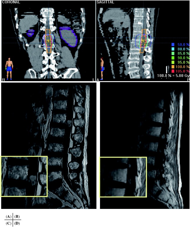
Case 3. Coronal (A) and sagittal (B) view of treatment planning of SRT on CT. The tumor around L1 (C: sagittal MRI with Gd enhancement before SRT) was decreased in size within 8 months after SRT (D, sagittal MRI with Gd enhancement).
The patient received chemoradiation therapy for lung adenocarcinoma, and developed headache and nausea due to cerebellar metastasis 10 months later. The cerebellar tumor was surgically resected, then treated with WB-EBRT of 40 Gy. The patient developed occipital pain 6 months later. Spinal magnetic resonance imaging (MRI) revealed multiple spinal intradural metastases thought to be cerebrospinal fluid seeding. A C1-2 tumor (11.25 ml) caused occipital pain and an L1 tumor (15.27 ml) caused lumbar pain. Both tumors were treated with Novalis SRT for palliation to relieve the pain. A total dose of 25 Gy (at 100% isodose) in 5 fractions was delivered to both tumors using a coplanar conformal multi-beam. D95 was 96% in both tumors. Both tumors remarkably shrunk within 2 months after SRT. No relapse occurred until death from primary lung carcinoma 5 months after Novalis SRT. Pain was partially relieved a week after the treatment.
DISCUSSION
Novalis stereotactic radiotherapy is a precise and useful technique to safely deliver radiation to lesions involving the spinal cord. Since the Novalis system uses X-ray image analysis to correct patient position before each treatment session, spinal lesions are exactly targeted after localization of spinal bone structures.2, 3) The spinal cord is spared as much as possible, while the tumor receives a higher dose than possible with conventional EBRT. The versatility of the Novalis system in precisely confirming radiation to lesions in close relationship to the spine provides an important palliative treatment for patients with malignancies threatening their QOL. We obtained preferable results by Novalis SRT in 3 cases of spinal intradural metastases in or just adjacent to the field of previous irradiation. In all 3 cases, the treated tumors were controlled until the patients’ death.
There are some reports on successful results of SRT or SRS for spinal metastases,2-7) but most describe only spinal bone metastases. There are few reports on the results of SRT or SRS for spinal intradural metastases. Endo et al. 8) reviewed reports of conventional EBRT for intramedullary spinal cord metastases, and found that a total dose of 25 to 40 Gy improved patients symptoms in 84.2% (116 out of 191). Shin et al. 9) reported treatment results of spine SRS for intradural and intramedullary metastases in 9 patients (11 tumors). The median treatment dose was 13.8 Gy (range, 10–16 Gy). They observed that only 1 case (11%) developed tumor progression, and no adverse effects were noted.
The clinical radiation tolerance dose of the spinal cord is important because radiation myelopathy is one of the most feared complications of radiotherapy. The tolerance dose (TD) to the spinal cord has been defined based on retrospective analysis of patients who developed myelopathy from treatment errors or overlapping fields. It is usually quoted as 45 Gy to 50 Gy in 2-Gy fractions, which is known to be TD 5/5, 5% severe complication probability in 5 years.10) However, more recent studies that included large numbers of patients have shown that a more realistic TD 5/5 could be up to 60 Gy.11) Animal studies have also shown a similar tolerance dose of the monkey spinal cord.12)
The dose-volume effect of the spinal medulla against radiation is not established.6, 13. 14) Concerning single-session radiosurgery for spinal bone metastasis, Ryu et al. 6) considered that the partial volume tolerance of the human spinal cord is at least 10 Gy to 10% of the spinal cord volume defined as 6 mm above and below the radiosurgery target. Gerszten et al. 15) calculated the volume of the entire spinal canal receiving more than 800 cGy during treatment planning in order to estimate the safety of SRS. Sahgal et al. 16) investigated 5 cases of radiation myelopathy and indicated that SRS 10 Gy to a maximum point dose for the thecal sac is safe. Milano et al. 17) reviewed a reported series and mentioned that the volume of the thecal sac receiving more than 14 Gy and 10 Gy must be considered. Other centers using intensity-modulated, near-simultaneous, CT-image-guided SRT techniques have employed doses of 6–30 Gy in 1–5 fractions.5)
Retreatment of the spinal cord generally has been contraindicated. This concept was based on the assumption that long-term recovery from radiation damage does not occur in nerve tissue and, hence, a normal spinal cord would not tolerate re-irradiation. Recent retreatment studies with animals using limb paralysis as an endpoint have shown substantial recovery of spinal cord injury when sufficient time had elapsed after the initial treatment.13) However, a certain interval is necessary for recovery. Ang et al. 18) observed that 2 years was apparently a sufficient time for recovery, allowing full-dose re-irradiation in monkeys. Ryu et al. 10) reported a successful result of ‘full-dose’ re-irradiation for glial tumor in the C1-Th1 spinal medulla. The patient had received 46.8 Gy in 26 fractions at the initial EBRT but the tumor recurred locally 12 years later. A total dose of 45 Gy in 25 fractions were given for relapse. The patient was reportedly doing well 5 years after the second EBRT. Sahgal et al. 19) found that a dose of approximately 70 Gy or less, in a total maximum point dose normalized to a 2 Gy equivalent dose, was safe.
As the recurrent spinal tumors in our cases were located inside or just adjacent to the spinal medulla, a certain amount of spinal medulla next to the tumor would be irradiated together with the tumor itself. The tumor was also inside or just adjacent to the previous irradiation field. In these cases, the interval between the previous irradiation and the Novalis SRT was not long, at 3, 14, and 6 months, respectively. We considered that the best way to reduce the irradiated volume as little as possible is with the stereotactic radiation technique. In Case 1 (C1 lesion) and Case 2 we reduced the volume of the re-irradiated volume by a steep dose fall-off between the tumor and the surrounding spinal parenchyma by SRT. In Case 1 (C2 lesion) and Case 3 we spared the previously irradiated area using SRT. A circular beam with a cone collimator was used in Case 1 because the tumors were spherical and surrounded all the way by spinal parenchyma, though a micro-multi-leaf collimator was employed in Case 2 and Case 3. Though the follow-up time was short in all 3 cases as a result of the patients’ death from the primary lesions, symptoms did not develop due to side effects on the spinal re-irradiation at 10, 22, and 5 months, respectively.
We usually use around 30 to 32.5 Gy in 5 to 6 fractions or 30 to 40 Gy in 10 fractions at 100% isodose for spinal bone metastases if the patient has not received prior EBRT in the area. For intradural metastases, 25 Gy in 5 fractions to 36 Gy in 12 fractions is delivered. In Cases 1 (C1 lesion) and Case 2 we gave reduced doses due to concern of previous radiation. Neither developed side effects of SRT, though the follow-up time was short.
CONCLUSIONS
Palliative SRT is expected to be a safe and effective treatment if the patient develops spinal intradural metastases within or adjacent to the previous irradiation field. We obtained preferable results for SRT using the Novalis system in 3 cases of spinal intradural metastases within or just adjacent to the field of previous irradiation. SRT is a precise and useful technique for delivering radiation safely to lesions involving the spinal cord and simultaneously reducing the dose to the surrounding structures. SRT may delay neurological deterioration and improve quality of life without complications.
Conflict of interest
none
REFERENCES
- 1).Wara WM, Phillips TL, Sheline GE, Schwade JG. Radiation tolerance of the spinal cord. Cancer. 1975; 35: 1558–1562. [DOI] [PubMed]
- 2).De Salles AAF, Pedroso AG, Medin P, Agazaryan N, Solberg T, Cabatan-Awang C, Espinoza DM, Ford J, Selch MT. Spinal lesions treated with Novalis shaped beam intensity-modulated radiosurgery and stereotactic radiotherapy. J Neurosurg. 2004; 101 (Suppl 3) 435–440. [PubMed]
- 3).Ryu S, Jin R, Jin JY, Chen Q, Rock J, Anderson J, Movsas B. Pain control by image-guided radiosurgery for solitary spinal metastasis. J Pain Symptom Manage. 2008; 35: 292–298. [DOI] [PubMed]
- 4).Benzil DL, Saboori M, Mogilner AY, Rocchio R, Moorthy C. Safety and efficacy of stereotactic radiosurgery for tumors of the spine. J Neurosurg. 2004; 101 (Suppl 3): 413–418. [PubMed]
- 5).Gerszten PC, Burton SA, Ozhasoglu C, Welch WC. Radiosurgery for spinal metastases. Clinical experience in 500 cases from a single institution. Spine. 007; 32: 193–199. [DOI] [PubMed]
- 6).Ryu S, Jin J-Y, Jin R, Rock J, Ajlouni M, Movsas B, Rosenblum M, Kim JH. Partial volume tolerance of the spinal cord and complications of single-dose radiosurgery. Cancer. 2007; 109: 626–628. [DOI] [PubMed]
- 7).Ryu SI, Chang SD, Kim DH, Murphy MJ, Le QT, Martin DP, Adler JR Jr. Image-guided hypo-fractionated stereotactic radiosurgery to spinal lesions. Neurosurgery. 2001; 49: 838–846. [DOI] [PubMed]
- 8).Endo S, Hida K, Yano S, Ito M, Yamaguchi S, Kashiwazaki D, Kinoshita R, Shirato H, Iwasaki Y. Intramedullary spinal cord metastasis treated with radiation therapy: report of 3 cases. No Shinkei Geka. 2008; 36: 345–349. [PubMed]
- 9).Shin DA, Huh R, Chung SS, Rock J, Ryu S. Stereotactic spine Radiosurgery for intradural and intramedullary metastasis. Neurosurg Focus. 2009; 27: E10. [DOI] [PubMed]
- 10).Ryu S, Gorty S, Kazee AM, Bogart J, Hahn SS, Dalal PS, Chung CT, Sagermann RH. ‘Full dose’ reirradiation of human cervical spinal cord. Am J Clin Oncol (CCT). 2000; 23: 29–31 [DOI] [PubMed]
- 11).Schultheiss TE. Spinal cord radiation "tolerance": doctorine versus data. Int J Radiat Oncol Biol Phys. 2000; 19: 219–221. [DOI] [PubMed]
- 12).Wong CS, Poon JK, Hill RP. Re-irradiation tolerance in the rat spinal cord: influence of level of initial damage. Radiother Oncol. 1993; 26: 132–138. [DOI] [PubMed]
- 13).Hashii H, Mizumoto M, Kanemoto A, Harada H, Asakura H, Hashimoto T, Furutani K, Katagiri H, NakasuY, Nishimura T. Radiotherapy for patients with symptomatic intramedullary spinal cord metastasis. J Radiat Res. 2011; 52: 641–645. [DOI] [PubMed]
- 14).Mizumoto M, Harada H, Asakura H, Hashimoto T, Furutani K, Hashii H, Murata H, Takagi T, Katagiri H, Takahashi M, Nishimura T. Radiotherapy for patients with metastases to te spinal column: a review of 603 patients at Shizuoka Cancer Center Hospital. Int J Radiat Oncol Biol Phys. 2011; 79: 208–213. [DOI] [PubMed]
- 15).Gerszten PC, Ozhasoglu C, Burton SA, Vogel WJ, Atkins BA, Kalnicki S, Welch WC. CyberKnife frameless stereotactic radiosurgery for spinal lesions: clinical experience in 125 cases. Neurosurgery. 2004; 55: 89–99. [PubMed]
- 16).Sahgal A, Ma L, Gibbs I, Gerszten PC, Ryu S, Soltys S, Weinberg V, Wong S, Chang E, Fowler J, Larson DA. Spinal cord tolerance for stereotactic body radiotherapy. Int J Radiat Oncol Biol Phys. 2010; 77: 548–553. [DOI] [PubMed]
- 17).Milano MT, Usuki KY, Walter KA, Clark D, Schell MC. Stereotactic radiosurgery and hypofractionated stereotactic radiotherapy: Normal tissue dose constraints of the central nervous system. Cancer Treat Rev. 2011; 37: 567–578. [DOI] [PubMed]
- 18).Ang KK, Stephens LC. Prevention and management of radiation myelopathy. Oncology. 1994; 8: 71–76 [PubMed]
- 19).Sahgal A, Ma L, Weinberg V, Gibbs IC, Chao s, Chang U-K, Werner-Wasik M, Angelov L, Chang EL, Sohn M-J, Soltys SG, Letourneau D, Ryu S, Gerszten PC, Fowler J, Wong CS, Larson DA. Reirradiation human spinal cord tolerance for stereotactic body radiotherapy. Int J Radiat Oncol Biol Phys. 2012; 82: 107–116. [DOI] [PubMed]


