Abstract
Introduction
Automatic fire suppression systems use hydrofluorocarbons (HF) to extinguish fires chemically. At high temperatures, HF can release hydrofluoric acid (HFA), a toxic, potentially lethal gas.
Case report
A 52-year-old male visited our Pulmonary Division with dyspnea of 8-months duration. He had been working at a facility that manufactured fire extinguishers. Bronchoscopy was performed and a transbronchial lung biopsy was taken from the right lower lobe. After the patient was diagnosed with pulmonary alveolar proteinosis (PAP), whole-lung lavage was performed. In this case, fire extinguisher gas induced pulmonary alveolar proteinosis. This material should be used with care and investigated further.
Discussion
HFA is corrosive and penetrates organic materials, including body tissues. Depending on the mode of exposure, skin ulceration, pulmonary injury, or even systemic shock can result. This report describes PAP that developed after chronic, repeated exposure to fire extinguisher spray. Hydrofluoric acid can induce pulmonary disorders such as PAP.
Keywords: Hydrofluorocarbons (HF), Hydrofluoric acid (HFA), Pulmonary alveolar proteinosis (PAP)
Background
Hydrofluorocarbons (HF) are used as extinguishing agents [1]. The most frequently used HF is 1,1,1,2,3,3,3-hepatofluoropropane, which is generally non-toxic and stable [1]. However, it can decompose over time, when exposed to high temperature or under certain environmental conditions [1]. Hydrofluoric acid (HFA), an aqueous form of HF, is highly water-soluble and a weak acid [2]. Exposure to HFA might not cause symptoms initially. Over time, however, fluoride ions bind to intracellular calcium or magnesium, causing liquefactive tissue necrosis or systemic electrolyte disturbance [3,4]. Tracheobronchitis, pulmonary edema, pneumonia, bronchospasm, and acute respiratory failure syndrome have been reported after HFA exposure [1,4-6]. However, there are no reports of pulmonary alveolar proteinosis (PAP) secondary to HF extinguishing agents. We report the diagnosis and successful treatment of PAP that developed after fire extinguisher testing. PAP has been reported in workers exposed to aluminum dust, paint, sawdust, silica, synthetic plastic fumes, and indium-tin oxide. However, there are no reports of PAP after repeated fire extinguisher use.
Case presentation
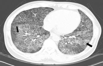
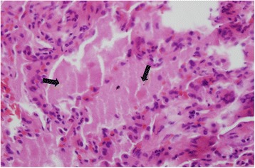
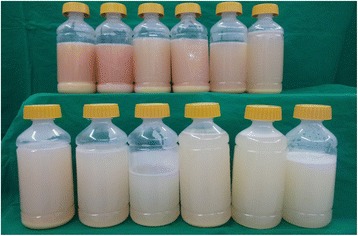
A 52-year-old male visited our Pulmonary Division with dyspnea of 8-months duration. He had smoked for 30 years and had a 5-year history of hypertension for which he was prescribed medication. For 20 years, he had a desk job at a facility that manufactured fire extinguishers. Beginning 8 months ago, he was exposed to gas from fire extinguishers repeatedly, for less than 10 s each time. He complained of an intermittent cough that was relieved within a few hours. Two months before visiting our hospital, he was exposed to the gas over an extended period of time. The severity of his symptoms increased rapidly. His symptoms included dyspnea with a cough on exertion and eye irritation. He was exposed to the fire extinguisher spray roughly three times per week. On physical examination, he looked ill. His blood pressure and body temperature were normal. His lips were cyanotic. Rales were heard in both lower lungs. The initial oxygen saturation on room air was 88%. Arterial blood gas analysis showed a pH of 7.469, a PCO2 of 29.3 mmHg, PO2 of 79.7 mmHg, HCO3 21.4 mEq/L, and saturation of 96.6% with a nasal cannula receiving supplemental oxygen at 3 L/min. Laboratory tests including blood and sputum were normal. Pulmonary function tests showed FEV1 3.14 L (96%), FVC 3.45 L (77%), FEV1/FVC 91%, and DLCO 42%. A simple chest x-ray revealed a bilateral pulmonary infiltration, while contrast-enhanced chest computed tomography showed ground glass opacities with a crazy paving appearance (Figure 1). Bronchoscopy with bronchial alveolar lavage (BAL) and a transbronchial lung biopsy of the right lower lobe were performed. Bronchoscopy showed proteinaceous secretions in both bronchial trees. The BAL fluid was milky and opaque. Microscopically, the transbronchial lung biopsy showed complete filling of alveoli with periodic-acid-Schiff-positive granular material in the preserved alveolar architecture (Figure 2). The patient was diagnosed with pulmonary alveolar proteinosis. Whole-lung lavage was performed under general anesthesia (Figure 3). On discharge, a simple chest x-ray showed improved lung haziness bilaterally. His respiratory symptoms improved. Sixteen months later, pulmonary function tests showed FEV1 3.57 L (115%), FVC 4.34 L (104%), FEV1/FVC 82%, and DLCO 81%.
Figure 1.

Contrast enhanced computed tomography revealing Crazy paving patterned diffuse ground glass attenuation with inter/intralobular septal thickening, representing diffuse alveolar damage, both lungs (black arrow).
Figure 2.

Histology of the left lung, pulmonary alveolar proteinosis high magnification photomicrograph showing complete filling of alveoli with periodic-acid-Schiff-positive granular material in preserved alveolar architecture (black arrow) (PAS, x400).
Figure 3.

Whole lung lavage fluid, first bottle (left and upper) is milky, opaque fluid and thick sediment layer. Last bottle (right lower) is fade compared to previously bottle and no sediment.
Discussion
Pulmonary alveolar proteinosis is a rare pulmonary disorder in which surfactant proteins and lipids accumulate within the intra-alveolar spaces [7,8]. PAP is classified into primary and secondary forms: secondary PAP is caused by hematological agents, infection, or the inhalation of dust or fumes [7-9]. PAP has been reported in workers exposed to aluminum dust, paint, sawdust, silica, synthetic plastic fumes, and indium-tin oxide [8]. However, there are no reports on PAP after fire extinguisher use, as occurred in our patient. The hydrocarbon used in fire extinguishers is 1,1,1,2,3,3,3-hepatofluoropropane, which is non-toxic under stable conditions, but can decompose into HF at high temperatures, particularly in combination with extended exposure to humidity, contaminants, certain metals, metal surfaces, or contact with certain liquids or vapors [1]. HFA is corrosive and penetrates organic materials, including body tissues. Depending on the mode of exposure, skin ulceration, pulmonary injury, or even systemic shock can result [2-5]. Wu et al. investigated HFA exposures occurring from 1991 to 2010: dermal exposure was the most frequent (83.6%), but inhalation only (7.1%), combined dermal and inhalation (1.9%), and combined ocular and inhalation (0.3%) exposure were also reported [4]. Kono et al. and Kawaura et al. also reported pulmonary injury accompanied by acute respiratory distress syndrome after HFA inhalation [5-7]. Zierold et al. reported that three US military personnel died of acute respiratory failure after using a fire extinguisher in their vehicle following a rocket-propelled grenade attack. They presumably inhaled the HFA generated from HF by the high temperature within the vehicle [1].
In our case, a high-temperature garbage incinerator was located in the same room where the patient performed the fire extinguisher spray test. Most likely, the HF was converted to HFA, which was then inhaled. Notably, we did not measure the concentration of autoantibodies to granulocyte–macrophage colony-stimulating factor (GM-CSF) in this patient. However, Cummings et al. asserted that inhalational exposure cannot be excluded when primary PAP is suspected [10,11]. In addition, Inoue et al. reported a dust inhalation history in 23% of primary PAP cases [11,12]. In our case, we surmise that the GM-CSF autoantibody status is less important. The limitation of this paper is a single case. Therefore we needed further studies to determine if there is an association with HFA.
Conclusions
In conclusion, this report describing PAP that developed after chronic, repeated fire extinguisher spray exposure reveals that hydrofluoric acid can induce pulmonary disorders such as PAP in addition to pneumonitis and acute respiratory failure. These findings highlight the need for caution and greater attention to this risk.
Consent
Written informed consent was obtained from the patient for publication of this case report. A copy of the written consent is available for review by the Editor–in–chief of this journal.
Footnotes
Competing interests
The authors declare that they have no competing interests.
Authors’ contributions
YJK has written the manuscript and contribution with treatment when the patient was admitted. JYS has contribution with analysis and interpretation of data. SMK has contribution with whole larvage. SYK, JWP, SPL and SML has discussed about this manuscript. SHJ treated the patient and has revised the manuscript. All authors have read and approved the final manuscript.
Contributor Information
YuJin Kim, Email: gene2001@gilhospital.com.
JiYoung Shin, Email: biochem98@daum.net.
ShinMyung Kang, Email: shinm.kang@gmail.com.
SunYoung Kyung, Email: light@gilhospital.com.
Jeong-Woong Park, Email: jwpark@gilhospital.com.
SangPyo Lee, Email: drsplee@gilhospital.com.
SangMin Lee, Email: allergylung@gilhospital.com.
Sung Hwan Jeong, Email: jsw@gilhospital.com.
References
- 1.Zierold D, Chauvier M. Hydrogen fluoride inhalation injury from a fire suppression system. Mil Med. 2012;177(1):108–12. doi: 10.7205/MILMED-D-11-00165. [DOI] [PubMed] [Google Scholar]
- 2.Ozcan M, Allahbeickaraghi A, Dundar M. Possible hazardous effects of hydrofluoric acid and recommendations for treatment approach: a review. Clin Oral Invest. 2012;16:15–23. doi: 10.1007/s00784-011-0636-6. [DOI] [PubMed] [Google Scholar]
- 3.Imanishi M, Dote T, Tsuji H, Tanida E, Yamadori E, Kono K. Time-dependent changes of blood parameters and fluoride kinetics in rats after acute exposure to subtoxic hydrofluoric acid. J Occup Health. 2009;51:287–93. doi: 10.1539/joh.M8016. [DOI] [PubMed] [Google Scholar]
- 4.Wu ML, Yang CC, Ger J, Tsai WJ, Deng JF. Acute hydrofluoric acid exposure reported to Taiwan poison control center, 1991–2010. Hum Exp Toxicol. 2014;33(5):449–54. doi: 10.1177/0960327113499165. [DOI] [PubMed] [Google Scholar]
- 5.Kono K, Watanabe T, Dote T, Usuda K, Nishiura H, Tagawa T, et al. Successful treatments of lung injury and skin burn due to hydrofluoric acid exposure. Int Arch Occup Environ Health. 2000;73:S93–7. doi: 10.1007/PL00014634. [DOI] [PubMed] [Google Scholar]
- 6.Kawaura F, Fukuoka M, Aragane N, Hayashi S. Acute respiratory distress syndrome induced by hydrogen fluoride gas inhalation. Nihon Kokyuki Gakkai Zasshi. 2009;47(11):991–5. [PubMed] [Google Scholar]
- 7.Dote T, Kono K, Usuda K, Shimizu H, Kawasaki T, Dote E. Lethal inhalation exposure during maintenance operation of a hydroge fluoride liquefying tank. Toxicol Ind Health. 2003;19:51–4. doi: 10.1191/0748233703th174oa. [DOI] [PubMed] [Google Scholar]
- 8.Trapnell BC, Whitsett JA, Nakata K. Pulmonary alveolar proteinosis. N Engl J Med. 2003;349:2527–39. doi: 10.1056/NEJMra023226. [DOI] [PubMed] [Google Scholar]
- 9.Bonella F, Bauer PC, Griese M, Ohshimo S, Guzman J, Costabel U. Pulmonary alveolar proteinosis: New insights from a single-center cohort of 70 patients. Respir Med. 2011;105:1908–16. doi: 10.1016/j.rmed.2011.08.018. [DOI] [PubMed] [Google Scholar]
- 10.Cummings KJ, Donat WE, Ettensohn DB, Roggli VL, Ingram P, Kreiss K. Pulmonary alveolar proteinosis in workers at an indium processing facility. Am J Respir Crit Care Med. 2010;181:458–64. doi: 10.1164/rccm.200907-1022CR. [DOI] [PMC free article] [PubMed] [Google Scholar]
- 11.Costabel U, Nakata K. Pulmonary alveolar proteinosis associated with dust inhalation. Am J Respir Crit Care Med. 2010;181:427–8. doi: 10.1164/rccm.200912-1800ED. [DOI] [PubMed] [Google Scholar]
- 12.Inoue Y, Trapnell BC, Tazawa R, Arai T, Takada T, Hizawa N, et al. Japanese center of the rare lung diseases consortium: Characteristics of a large cohort of autoimmune pulmonary alveolar proteinosis patients in Japan. Am J Respir Crit Care Med. 2008;177:752–62. doi: 10.1164/rccm.200708-1271OC. [DOI] [PMC free article] [PubMed] [Google Scholar]


