Abstract
We analyzed the clinicopathological features of angiomatoid fibrous histiocytoma (AFH) in 21 cases with emphasis on variant morphology. In our series, ten patients were male and eleven were female. The patients’ mean age was 26.9 years old. Tumors were located on the lower limbs in eight cases, upper limbs in three, trunk in five, head and neck in four, and trachea in one. Microscopically, thirteen cases were characterized by typical AFH. Tumor cells showed marked tumor pleomorphism with giant hyperchromatic nuclei in two cases. Mitotic figures (2-3/10HPF) were found in two cases. Focal necrosis was found in one case. A number of multinucleated giant cells were found in two cases. Two cases showed obvious myxoid change in the stromal. Prominent sclerosing changes in the stromal component were found in two cases. Immunohistochemistry staining showed tumor cells were positive for EMA, desmin, and CD68. Five cases demonstrated the presence of rearrangement of the EWSR1 gene by FISH detection. Only two patients had tumor recurrence at 3 and 6 months after tumor resection, respectively. In conclusion, AFH has variant histological patterns. The differential diagnosis includes inflammatory myofibroblastic tumor, aneurysmal fibrous histiocytoma, follicular dendritic cell tumor, and metastatic tumor of lymph node.
Keywords: Angiomatoid fibrous histiocytoma, clinicopathological features, variant morphology
Introduction
Angiomatoid fibrous histiocytoma (AFH) is a rare tumor of intermediate biological potential, typically arising in extremity soft tissue of children and young adults. However, it has been also reported to occur in nonsomatic soft tissue sites such as hard palate, mediastinum, vulva, retroperitoneum, ovary, and lung [1,2]. Histologically, typical AFHs are characterized by varying proportions of spindled or oval histiocytoid cells with syncytial growth, arranged in a nodular pattern, pseudovascular spaces, and prominent lymphoplasmacytic rim. Recently, variants of AFH have been reported in the literature [3-5]. Herein, we collected 21 cases of AFH and analyzed their clinicopathological features with emphasis on morphology variants and differential diagnosis.
Materials and methods
Patients
We retrospectively reviewed the medical records including consult files of 21 cases diagnosed as angiomatoid fibrous histiocytoma in the Department of Pathology, The First Affiliated Hospital, Sun Yat-Sen University, China, between January 2008 and May 2014. The histopathology of tumour was determined by two pathologists according to the histopathological criteria of WHO classification of tumours of soft tissue and bone [6]. For the research purposes of these clinical materials, prior patient’s consents and approval from the Institutional Research Ethics Committee were obtained. Clinical information including age, gender, tumor site, tumor size, and follow-up information was obtained (Table 1).
Table 1.
Clinicopathological features of 21 cases of angiomatoid fibrous histiocytoma
| Case No. | Sex/age | Site | Size (cm) | Tumor histological features | EWSR1 rearrangement |
|---|---|---|---|---|---|
| 1 | F/64 | Right thigh | 4.3 | Classical type | NA |
| 2 | F/30 | Left knee | 2.1 | Classical type | NA |
| 3 | F/17 | Left thigh | 2.0 | Classical type | NA |
| 4 | F/23 | Left lower mandible | 1.5 | Classical type | NA |
| 5 | F/83 | Left upper arm | 2.0 | Classical type | NA |
| 6 | M/8 | Right leg | 1.0 | Classical type | NA |
| 7 | F/19 | Left knee | 2.0 | Classical type | NA |
| 8 | F/16 | Left abdominal wall | 10.0 | Classical type | NA |
| 9 | M/23 | Scalp | 2.0 | No pseudoangiomatoid spaces | NA |
| 10 | M/27 | Trachea | 3.0 | No pseudoangiomatoid spaces and lymphoid rim; myxoid change and scattered multinuclear giant cells in the stromal | + |
| 11 | M/12 | Right shoulder | 2.0 | No pseudoangiomatoid spaces; tumor giant cells; remarkable myxoid change in the stromal | + |
| 12 | F/15 | Left buttock | 4.0 | No pseudoangiomatoid spaces; active mitotic figures (2/10HPF) | NA |
| 13 | F/55 | Left upper arm | 2.0 | No pseudoangiomatoid spaces; prominent sclerosing stromal | NA |
| 14 | F/38 | Right thigh | 1.8 | Classical type | NA |
| 15 | M/33 | Right neck | 2.0 | No pseudoangiomatoid spaces; tumor giant cells; numerous capillary blood vessels in the stromal | NA |
| 16 | M/14 | Left humeral back | 2.0 | Classical type | + |
| 17 | F/16 | Right forearm | 2.0 | Classical type | + |
| 18 | M/18 | Left popliteal space | 2.5 | Classical type | - |
| 19 | M/12 | Neck | 4.0 | Classical type; active mitotic figures (3/10HPF) | NA |
| 20 | M/25 | Retroperitoneum | 8.0 | No pseudoangiomatoid spaces; focal tumor necrosis; prominent sclerosing stromal; scattered multinuclear giant cells in the stromal | + |
| 21 | M/16 | Left thigh | 2.0 | No pseudoangiomatoid spaces | NA |
NA: not available.
Immunohistochemical staining
Immunohistochemical staining was carried out on formalin-fixed, paraffin-embedded tissue using an EnVision kit (Dako, Carpinteria, CA). The following primary antibodies purchased from Dako Corporation were used: cytokeratin, epithelial membrane antigen (EMA), vimentin, smooth muscle actin (SMA), desmin, S-100 protein, CD21, CD35, CD34, CD31, CD68, CD56, neuron specific enolase, chromogranin A, synaptophysin, CD31, MyoD1, myogenin, HMB-45, Melan-A, anaplastic lymphoma kinase (ALK), and Ki-67. Positive and negative control slides were employed.
Fluorescence in situ hybridization
Fluorescence in situ hybridization (FISH) detection for EWSR1 breakpoints was performed according to the manufacturer’s protocols (Beijing GP Medical Technologies, China). The result showed more than 15% tumour cells had separate red and green signals, indicating presence of rearrangement of the EWSR1 gene.
Results
Clinical features
Of 21 AFH patients, ten patients were male and eleven were female. The patients’ age ranged from 8 to 83 years old with mean age 26.9 and median age 19 years old. The majority of AFH patients presented with a painless, slowly-growing, superficial soft tissue mass. Only one patient with endotracheal AFH complained of persistent cough for 1 year with sputum production, hemoptysis and dyspnea on exertion. Tumors were located on the lower limbs in eight cases, upper limbs in three, trunk in five, head and neck in four, and trachea in one. Simple tumor excision was performed in all patients without adjuvant radiotherapy or chemotherapy. Follow-up was available in all patients, and only two patients (9.5%, 2/21) had tumor recurrence at 3 months and 6 months after tumor resection, respectively. The other patients were well and alive without recurrence or metastasis after a median duration of 48 months (range 4-148 months).
Pathological findings
Grossly, mean tumor size was 3.0 cm with a range of 1.0-10.0 cm and median 2.0 cm in the greatest diameter. The majority of tumors were typically well circumscribed and firm, simulating a lymph node. In our series, tumors showed red-brown haemorrhagic cystic spaces of variable sizes in twelve cases on cut surface. The tumors in other nine cases were gray-white. Of 21 cases, one case (case 11) showed prominent myxoid change with gelatinous consistency. One case (case 20) had focal necrosis on cut surface.
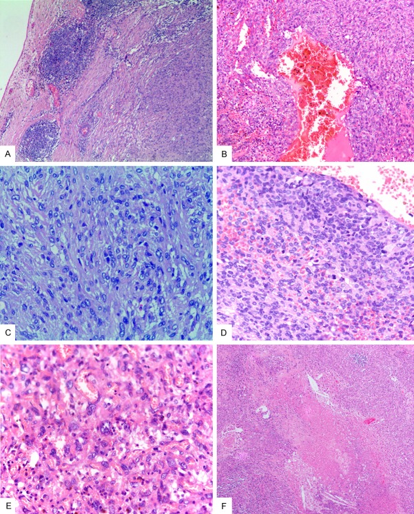
Microscopically, thirteen cases was characterized by varying proportions of spindled or oval histiocytoid cells arranged in a nodular pattern, pseudoangiomatous spaces filled with blood and surrounded by tumor cells and prominent lymphoplasmacytic rim with occasional germinal centers mimicking a lymph node metastatic tumor. However, no pseudoangiomatous space was found in other 8 cases. In the nodular area, the tumor cells showed syncytial appearance and were largely plump or ovoid, and more focally epithelioid with abundant cytoplasm. There was slight nuclear atypia. Few mitotic figures were found in 19 cases. No atypical mitotic figures were found. In our series, tumor cells showed marked tumor pleomorphism with giant hyperchromatic nuclei in two cases. Mitotic figures (2-3/10HPF) were found in two cases. Focal necrosis was found in one case. A number of multinucleated giant cells were found within the tumor stromal in two cases. Some multinuclear giant cells were similar to Langhans’ giant cells or Touton giant cells. Two cases showed obvious myxoid change in the stromal of tumor. Prominent sclerosing changes in the stromal component were found in two cases. Numerous capillary blood vessels were found in one case (Figures 1, 2).
Figure 1.
A. The tumor was surrounded by dense lymphoplasmatic infiltrate and thick fibrous pseudocapsule, HE×100; B. Pseudoangiomatoid space in the center of tumor, HE×200; C. Tumor cells were plump and epitheloid cells with indistinct cell borders and nuclei, HE×400; D. Mitotic figures were found in this case, HE×200; E. Numerous capillary blood vessels and tumor giant cells were shown in this case, HE×400; F. Focal tumor necrosis was shown in this case, HE×40.
Figure 2.

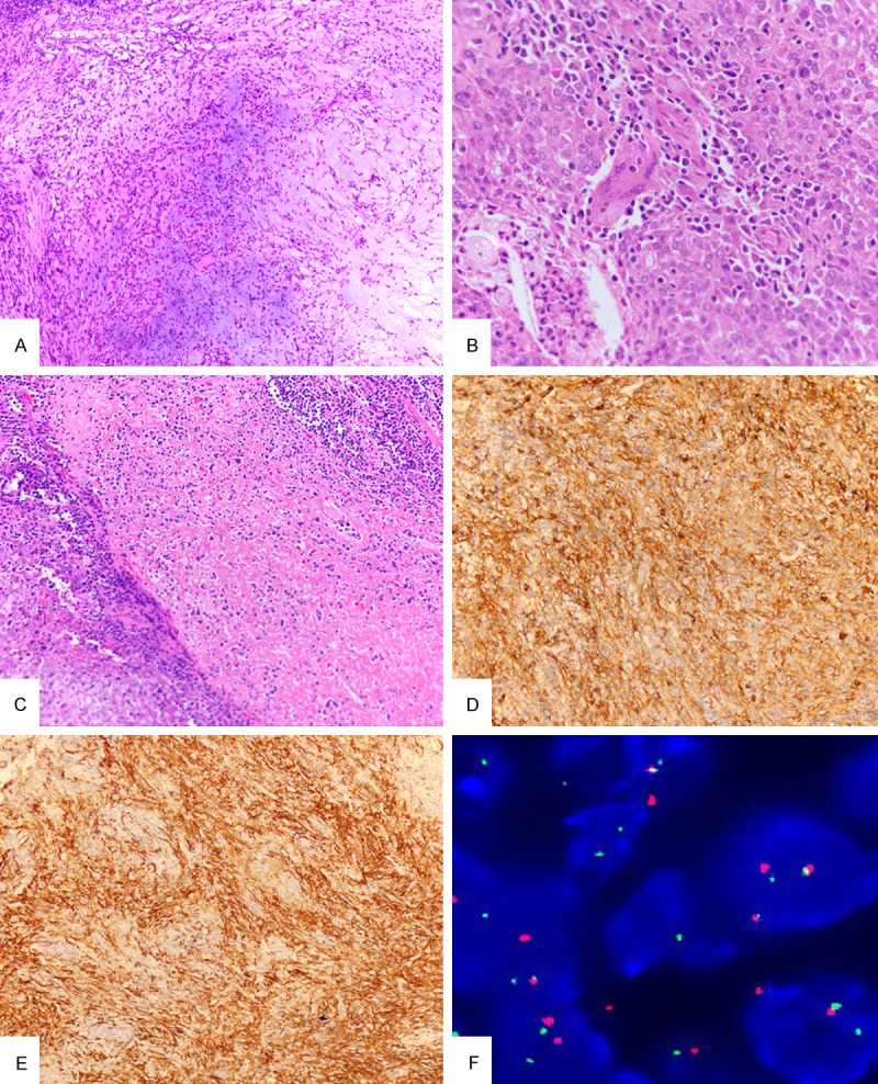
(A) Remarkable myxoid change in the stromal, HE×100; (B) Multinuclear giant cell in the stromal, HE×200; (C) Prominent sclerosing in the stromal, HE×100; (D-E) The tumor cells showed strong positive for EMA (D) and desmin (E), IHC×200; (F) Tumor cell showed separate red and green signals, indicating presence of rearrangement of the EWSR1 gene, Fluorescence in situ hybridization, ×400.
As shown in Figure 2, immunohistochemistry staining showed tumor cells were positive for vimentin (21/21, 100%), EMA (11/21, 52%), desmin (14/21, 66%), CD68 (17/21, 88%). Focal cytokeratin and SMA expression was found in 2 cases, respectively. Ki-67 positive rate was approximately 1-2 percent of tumor cells. However, the tumor cells were negative for S-100 protein, CD56, neuron specific enolase, chromogranin A, synaptophysin, CD21, CD35, CD34, CD31, MyoD1, myogenin, HMB-45, Melan-A, and ALK. FISH detection for EWSR1 breakpoints was performed in 6 cases of AFH, 5 cases (83%) showed the presence of rearrangement of the EWSR1 gene.
Discussion
AFH was first described by Enzinger and termed as angiomatoid malignant fibrous histiocytoma [7]. Because of their relatively rarity of metastasis and the overall excellent clinical course, it was classified as an intermediate tumor in WHO classification of tumours of soft tissue and bone. AFH usually occurs in the soft tissue forming a well circumscribed subcutaneous nodule on the extremities, head and neck and trunk. Recently, AFH has been reported in unusual sites including the lung, mediastinum, vulva, retroperitoneum, ovary, pulmonary artery, kidney, and intracranial [1,2,8,9].
Microscopically, the tumor characteristically has pseudoangiomatoid spaces. The tumor cell is a bland histiocytoid or fibroblast-like which shows few mitotic figures and is surrounded by a dense lymphoid tissue and thick fibrous pseudocapsule. However, variant morphology of AFH has been reported in the literature. Chen et al. have reported that unusual features were found focally in some AFH cases, including clear cells, rhabdomyoblast-like cells, pulmonary edema-like pattern and tumor cell cords lying in a myxoid stroma [2]. Bohman et al. have reported 27 cases of AFH and found ten (37%) displayed significant areas of sclerosis; three of these ten had areas with a perineurioma-like pattern. Nine displayed at least moderate pleomorphism. Eight had eosinophils in the stroma. One had a reticulated pattern of cells in a myxoid stroma [3]. Kao et al. have reported 13 cases of AFH and found four cases showed solid histology without pseudoangiomatoid spaces and another one lacked peripheral lymphoid infiltrates. Nuclear pleomorphism was striking in two cases, moderate in seven, with osteoclasts seen in two cases. In three AFHs with sclerotic matrix, one exhibited perivascular hyalinisation and nuclear palisading. Varyingly myxoid stromal was found in three cases [5]. Most recently, Schaefer and Fletcher have reported myxoid variant of AFH in a series of 21 cases [4]. In our series, no pseudoangiomatous space was found in 8 cases. Tumor cells showed remarkable tumor pleomorphism with giant hyperchromatic nuclei in two cases. Mitotic figures (2-3/10HPF) were found in two cases. A number of multinuclear giant cells were found within the tumor stromal in two cases. Two cases showed obvious myxoid change in the stromal of tumor. Prominent sclerosing changes in the stromal component were found in two cases. We firstly reported numerous capillary blood vessels were found in one AFH case. Tumor cells of AFH are usually positive for CD68, EMA, desmin, and CD99, but negative for S-100 protein, CD21, CD35, and CD34 by immunohistochemistry staining. In our series, tumor cells were positive for vimentin (21/21, 100%), EMA (11/21, 52%), desmin (14/21, 66%), and CD68 (17/21, 88%).
AFH has a variety of morphologic features and lacks specific immunophenotypic markers. It is important to differentiate from other tumors including inflammatory myofibroblastic tumor, aneurysmal fibrous histiocytoma, follicular dendritic cell tumor, and metastatic tumors of lymph node. The main difference is that the tumor cells of inflammatory myofibroblastic tumor, which are myofibroblasts, have distinct cell borders and eosinophilic cytoplasm, and often vesicular nuclei with distinct nucleoli. The second point is the distribution of the inflammatory cells intimately intermingled with the neoplastic cells without forming a peritumoral rim. The third point is that they show variable staining for actin, desmin, and ALK although AFH is positive for desmin in about 50% of cases. Aneurysmal fibrous histiocytoma which is an uncommon pathologic variant of dermatofibroma and has blood spaces is easily confused with AFH. However, in addition to the features of a typical dermatofibroma, aneurysmal fibrous histiocytoma has large cleft-like or cavernous blood-filled spaces with numerous hemosiderin pigments. Furthermore, aneurysmal fibrous histiocytoma is negative for factor XIIIa, CD34, CD31, CD68, and desmin, but strongly positive for vimentin by immunohistochemistry staining. Follicular dendritic cell tumor usually has rich inflammatory cells infiltration and the spindle tumor cells are arranged in nodular or whirl structure. However, the main difference from AFH is that follicular dendritic cell tumor is composed of a polymorphic population of inflammatory cells including B-lymphocytes, T-lymphocytes, plasma cells and spindle cells in addition to the expression of follicular dendritic cell markers including CD21, CD23, CD35 and EBV expression. Finally, typical AFH is surrounded by a dense lymphoid tissue and thick fibrous pseudocapsule, which is easily misdiagnosed as metastatic tumor of lymph node including malignant melanoma, spindle cell carcinoma, and other metastatic soft tissue tumors. The most important difference is that the true lymph node has marginal sinus. But AFH has absence of such structure. Moreover, histological features, immunohistochemistry staining, and clinical history are helpful for differentiating AFH from metastatic tumors.
To our knowledge, three types of gene translocations in AFH including t (2:22) (q33:q12) EWSR1-CREB1, t (12:16) (q13:p11) FUS-ATF1, and t (12:22) (q13:q12) EWSR1-ATF1 have been reported [10,11]. In our series, 5 cases (83%, 5/6) showed the presence of rearrangement of the EWSR1 gene by FISH detection for EWSR1 breakpoints. Although molecular investigations are crucial in final diagnosis of AFH, these gene fusions is not specific for AFH because they can also occur in other soft tissue tumors and carcinoma such as clear cell sarcoma (of tendons and aponeuroses) and hyalinizing clear cell carcinoma of the salivary gland [12,13].
AFH is rather indolent with 2%-11% local recurrences and less than 1% metastasis. In our series, only two patients (9.5%, 2/21) had tumor recurrence at 3 months and 6 months after tumor resection, respectively. The other patients were well and alive without recurrence or metastasis after a median duration of 48 months (range 4-148 months). The results suggest that variant morphology of AFH has no significant prognostic implications.
Disclosure of conflict of interest
None.
References
- 1.Song JY, Lee SK, Kim SG, Rotaru H, Baciut M, Dinu C. Angiomatoid fibrous histiocytoma on the hard palate: case report. Oral Maxillofac Surg. 2012;16:237–42. doi: 10.1007/s10006-011-0297-2. [DOI] [PubMed] [Google Scholar]
- 2.Chen G, Folpe AL, Colby TV, Sittampalam K, Patey M, Chen MG, Chan JK. Angiomatoid fibrous histiocytoma: unusual sites and unusual morphology. Mod Pathol. 2011;24:1560–70. doi: 10.1038/modpathol.2011.126. [DOI] [PubMed] [Google Scholar]
- 3.Bohman SL, Goldblum JR, Rubin BP, Tanas MR, Billings SD. Angiomatoid fibrous histiocytoma: an expansion of the clinical and histological spectrum. Pathology. 2014;46:199–204. doi: 10.1097/PAT.0000000000000073. [DOI] [PubMed] [Google Scholar]
- 4.Schaefer IM, Fletcher CD. Myxoid variant of so-called angiomatoid “malignant fibrous histiocytoma”: clinicopathologic characterization in a series of 21 cases. Am J Surg Pathol. 2014;38:816–23. doi: 10.1097/PAS.0000000000000172. [DOI] [PubMed] [Google Scholar]
- 5.Kao YC, Lan J, Tai HC, Li CF, Liu KW, Tsai JW, Fang FM, Yu SC, Huang HY. Angiomatoid fibrous histiocytoma: clinicopathological and molecular characterisation with emphasis on variant histomorphology. J Clin Pathol. 2014;67:210–15. doi: 10.1136/jclinpath-2013-201857. [DOI] [PubMed] [Google Scholar]
- 6.Fletcher CD, Bridge JA, Hogendoorn PC, Mertens F. WHO classification of tumours of soft tissue and bone. Lyon: IARC Press; 2013. pp. 306–309. [Google Scholar]
- 7.Enzinger FM. Angiomatoid malignant fibrous histiocytoma: a distinct fibrohistiocytic tumor of children and young adults simulating a vascular neoplasm. Cancer. 1979;44:2147–57. doi: 10.1002/1097-0142(197912)44:6<2147::aid-cncr2820440627>3.0.co;2-8. [DOI] [PubMed] [Google Scholar]
- 8.Ghigna MR, Hamdi S, Petitpretz P, Rohnean A, Florea V, Mussot S, Dartevelle P, Dorfmuller P, Tu L, Thuillet R, Guignabert C, Thomas-de-Montpreville V. Angiomatoid fibrous histiocytoma of the pulmonary artery: a multidisciplinary discussion. Histopathology. 2014;65:278–82. doi: 10.1111/his.12429. [DOI] [PubMed] [Google Scholar]
- 9.Ochalski PG, Edinger JT, Horowitz MB, Stetler WR, Murdoch GH, Kassam AB, Engh JA. Intracranial angiomatoid fibrous histiocytoma presenting as recurrent multifocal intraparenchymal hemorrhage. J Neurosurg. 2010;112:978–82. doi: 10.3171/2009.8.JNS081518. [DOI] [PubMed] [Google Scholar]
- 10.Rossi S, Szuhai K, Ijszenga M, Tanke HJ, Zanatta L, Sciot R, Fletcher CD, Dei Tos AP, Hogendoorn PC. EWSR1-CREB1 and EWSR1-ATF1 fusion genes in angiomatoid fibrous histiocytoma. Clin Cancer Res. 2007;13:7322–28. doi: 10.1158/1078-0432.CCR-07-1744. [DOI] [PubMed] [Google Scholar]
- 11.Raddaoui E, Donner LR, Panagopoulos I. Fusion of the FUS and ATF1 genes in a large, deep-seated angiomatoid fibrous histiocytoma. Diagn Mol Pathol. 2002;11:157–62. doi: 10.1097/00019606-200209000-00006. [DOI] [PubMed] [Google Scholar]
- 12.Flucke U, Mentzel T, Verdijk MA, Slootweg PJ, Creytens DH, Suurmeijer AJ, Tops BB. EWSR1-ATF1 chimeric transcript in a myoepithelial tumor of soft tissue: a case report. Hum Pathol. 2012;43:764–68. doi: 10.1016/j.humpath.2011.08.004. [DOI] [PubMed] [Google Scholar]
- 13.Antonescu CR, Katabi N, Zhang L, Sung YS, Seethala RR, Jordan RC, Perez-Ordoñez B, Have C, Asa SL, Leong IT, Bradley G, Klieb H, Weinreb I. EWSR1-ATF1 fusion is a novel and consistent finding in hyalinizing clear-cell carcinoma of salivary gland. Genes Chromosomes Cancer. 2011;50:559–70. doi: 10.1002/gcc.20881. [DOI] [PubMed] [Google Scholar]


