Abstract
Tooth enamel is the hardest substance in the human body and has a unique combination of hardness and fracture toughness that protects teeth from dental caries, the most common chronic disease worldwide. In addition to a high mineral content, tooth enamel comprises organic material that is important for mechanical performance and influences the initiation and progression of caries; however, the protein composition of tooth enamel has not been fully characterized. Here, we determined that epithelial hair keratins, which are crucial for maintaining the integrity of the sheaths that support the hair shaft, are expressed in the enamel organ and are essential organic components of mature enamel. Using genetic and intraoral examination data from 386 children and 706 adults, we found that individuals harboring known hair disorder–associated polymorphisms in the gene encoding keratin 75 (KRT75), KRT75A161T and KRT75E337K, are prone to increased dental caries. Analysis of teeth from individuals carrying the KRT75A161T variant revealed an altered enamel structure and a marked reduction of enamel hardness, suggesting that a functional keratin network is required for the mechanical stability of tooth enamel. Taken together, our results identify a genetic locus that influences enamel structure and establish a connection between hair disorders and susceptibility to dental caries.
Introduction
Dental caries are initiated at the surface of the tooth by microorganisms metabolizing food residues and releasing acids that dissolve enamel minerals (1). Their development is influenced by nutrition, hygiene, and genetic factors affecting the structure of tooth enamel (1–5).
During tooth development, amelogenin and other matrix components (enamelin, ameloblastin) are deposited by enamel-secreting ameloblasts in a highly structured manner to form enamel rods that are fully mineralized during maturation, with degradation of protein components and deposition of hydroxyapatite minerals (6). Mature enamel consists of approximately 96% minerals, 3% water, and 1% organic material by weight. Most of the organic material is located in the inner enamel where it forms very characteristic structures called enamel tufts due to their appearance at the dentin-enamel junction. Tuft material extends throughout the enamel thickness as thin enamel rod sheaths surrounding each individual rod (7). Enamel tufts and rod sheaths play an essential role in the mechanical performance of enamel (8, 9) and influence the initiation and progression of caries (10, 11). It has been suggested that they contain keratin-like material (12, 13), but a high degree of crosslinking has precluded full characterization of their protein composition (14).
While tooth and hair morphogenesis utilize common molecular pathways, the final structures produced are very different and involve specific structural components (15, 16). In the hair, hair keratins form filaments that are highly crosslinked and are major components forming the different layers of the hair shaft and supporting tissues, such as the inner root sheath, the companion layer, and outer root sheath. Here, we show that epithelial hair keratins are crucial structural components of human enamel and that mutations in these keratins increase the susceptibility to dental caries.
Results and Discussion
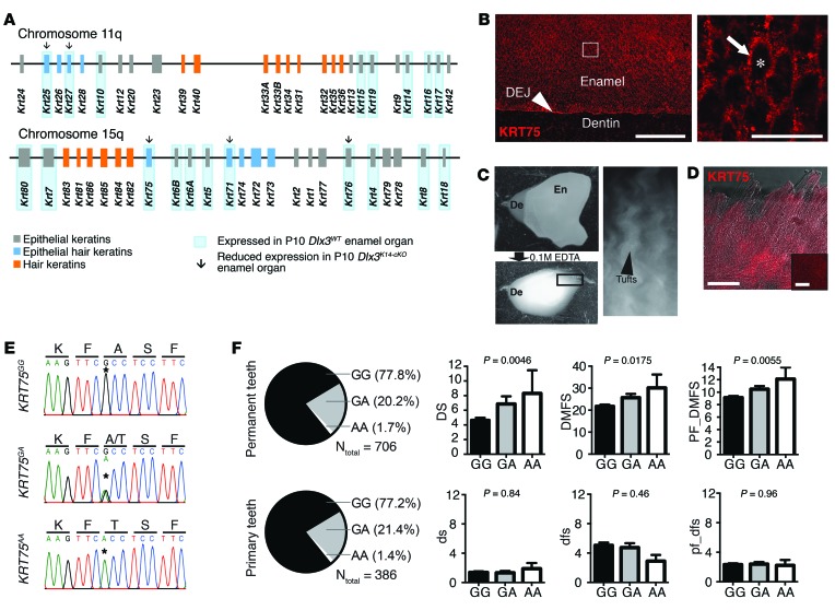
In a previous study, we showed that the transcription factor DLX3 directly controls the expression of hair keratins during hair follicle development (17). Performing transcriptome analysis on murine enamel organs lacking DLX3 (Krt14-Cre Dlx3fl/fl, herein referred to as Dlx3K14-cKO mice at P10), we found that several epithelial hair keratins (Krt25, Krt27, Krt71, Krt75, and Krt76), expressed in the supporting tissues surrounding the hair shaft (inner root sheath and companion layer), were transcribed in the enamel organ and exhibited significantly downregulated expression in Dlx3K14-cKO mice (Figure 1A and Supplemental Figure 1; supplemental material available online with this article; doi:10.1172/JCI78272DS1). In addition to previously detected KRT5 and KRT14, which are expressed in most epithelia, we detected the mRNA of other epithelial keratins in the enamel organ: Krt6a, Krt6b, Krt7, Krt10, Krt15, Krt16, Krt17, Krt19, and Krt80, none of which were significantly affected by Dlx3 deletion (Figure 1A and Supplemental Figure 1).
Figure 1. Epithelial hair keratins in enamel and association of polymorphism in KRT75 with increased susceptibility to caries in humans.
(A) Diagram representing the 2 keratin clusters on chromosomes 11q and 15q and highlighting the keratins expressed in mouse enamel organ and those that are downregulated in Dlx3K14-cKO mice at P10 (RNA-seq). (B) Detection of KRT75 on polished and etched section of mature human tooth showing the dentin-enamel junction (DEJ) and cross sections of enamel rods. Magnification on the right shows enamel rods (asterisk) and interrods (arrow). Scale bars: 100 μm (left); 20 μm (right).(C) Isolation of organic material (enamel tufts and rod sheaths) from human enamel (En) after demineralization. Enamel tufts are visualized at the surface of the dentin (De). (D) Detection of KRT75 in isolated organic material. Scale bars: 100 μm; 20 μm (inset). (E) Sequencing of the human KRT75 gene harboring a missense polymorphism (rs2232387, asterisks). Three genotypes are shown: KRT75GG, KRT75GA, and KRT75AA. (F) Genetic association between the rs2232387 polymorphism and caries experience in primary and permanent dentition. Pie charts show allelic distribution of the rs2232387 polymorphism measured on 706 adults and 386 children. Graphs show measure of caries experience for each genotype: left, number of tooth surfaces with untreated decay (DS and ds); center, number of decayed, missing due to decay, and filled surfaces (DMFS and dfs); right, partial DMFS and dfs indices limited to the molars and premolars pit and fissure (PF and pf) surfaces.
We investigated the expression pattern of KRT75 in mouse enamel organ and in mature human enamel. KRT75 was expressed by mouse ameloblasts at the secretory stage (Supplemental Figure 2A) and detected in the tufts and rod sheaths of mature human enamel (Figure 1, B and C). Interestingly, until enamel matrix proteins were identified, enamel was thought to be a keratinized structure, like hair and nail, with an extreme degree of mineralization (18, 19). Here, we show that a subset of hair keratins is expressed by ameloblasts, and we demonstrate that KRT75 is incorporated into the tufts and rod sheaths in mature enamel. Since keratins are not secreted proteins, their deposition in enamel may be related to the retention of cell fragments at the periphery of the rods (20).
Mutations in hair keratins have been reported in various human pathological conditions (21, 22). Among them, pseudofolliculitis barbae, a hair disorder characterized by the formation of ingrown hair in the facial area upon shaving, is associated with a polymorphism (rs2232387) in the KRT75 gene and more prevalent in African-Americans due to the curly nature of their hair (23). This G-to-A missense mutation (KRT75GA or KRT75AA genotypes) leads to an alanine-to-threonine substitution at position 161 (A161T) in a highly conserved region of the 1A α-helical segment of KRT75 (Figure 1E). To determine whether the KRT75A161T mutation had clinical impact on enamel function, we tested the genetic association between the KRT75 polymorphism (rs2232387) and tooth decay assessed as dental caries experience in the primary dentition of 386 children (mixed European descent, 6–12 years) and permanent dentition of 706 adults (mixed European descent, 25–50 years). The missense A allele of rs2232387, which was observed in 23% of the samples, was associated with a higher number of carious tooth surfaces in adults but not children (Figure 1F and Supplemental Table 1).
Association with a second rarer missense polymorphism in KRT75 (rs2232398) leading to a glutamate-to-lysine substitution at position 337 (E337K) was shown previously for loose anagen hair syndrome (24). In our cohort, this polymorphism was observed in 1% of the samples and was associated with increased caries in children only (Supplemental Table 2). The 2 missense polymorphisms were not in linkage disequilibrium, and in adults, there was modest evidence of a statistical interaction between them (Supplemental Table 3), with one participant having both risk variants and 30 decayed tooth surfaces. Results were unchanged after adjusting for tooth-brushing frequency and fluoride exposure. Altogether, these results indicate that genetic variation in KRT75 increases the risk of dental caries and that the effects of specific polymorphisms may differ across dentitions and by genotype at other loci. KRT75 can therefore be added to the list of enamel formation genes (amelogenin, ameloblastin, enamelin, tuftelin) in which polymorphisms have been previously associated with caries experience (25–27).
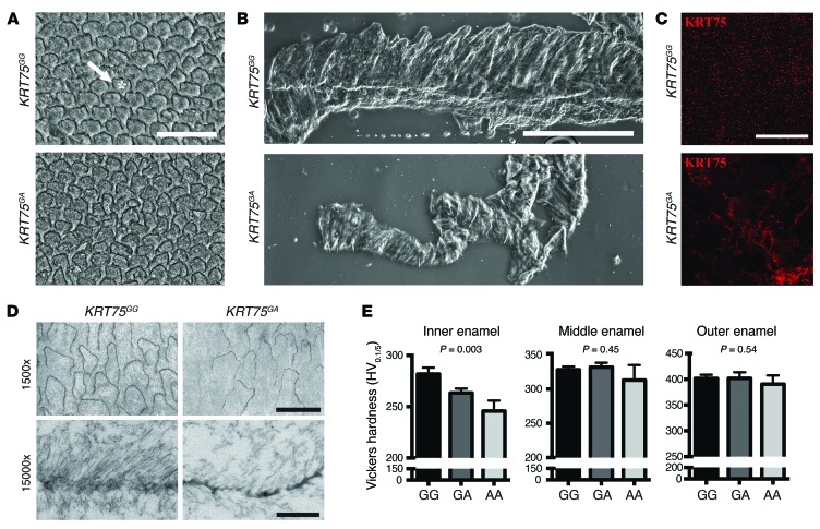
To determine the functional effects of the missense polymorphism in KRT75 on enamel, we conducted structural analysis of human enamel from individuals carrying the rs2232387 polymorphism. Scanning electron microscopy analysis of polished and etched tooth samples sectioned in the plane transecting the enamel rods revealed that the distribution and characteristic keyhole shape of the rods were altered in individuals carrying the variant A allele (Figure 2A). Tufts and rod sheaths isolated from patients with the KRT75GA or KRT75AA genotype appeared disorganized (Figure 2B), and the distribution pattern of KRT75 was highly disrupted (Figure 2C). This was further corroborated by transmission electron microscopy analysis (Figure 2D). These results suggest that epithelial hair keratins stabilize enamel tufts and rod sheaths to support enamel rods during their formation, which is similar to their function in supporting the hair shaft (23, 28, 29). The functional role of KRT75 in teeth was further supported by a mouse model (Krt75tm1Der) carrying a Krt75 mutation leading to hair defects (30) as well as defects in the assembly of enamel rods (Supplemental Figure 2, B–E).
Figure 2. Effects of polymorphism in KRT75 on enamel and tufts structure and on enamel hardness.
(A) Scanning electron microscopy analysis of ground, polished, and etched human molars from patients with (KRT75GA) or without (KRT75GG) a missense A allele at rs2232387. Asterisk, rod; arrow, interrod. Scale bar: 20 μm. (B) Differential interference contrast imaging of tufts isolated from molars from individuals with KRT75GG or KRT75GA genotype. Scale bar: 500 μm. (C) Detection of KRT75 protein in tufts from individuals with KRT75GG or KRT75GA genotype. Scale bar: 20 μm. (D) Transmission electron microscopy of organic material isolated after demineralization of enamel from individuals with KRT75GG or KRT75GA genotype. Lower panels show high resolution images of enamel rod sheaths. Scale bars: 10 μm (upper panels); 500 nm (lower panels). (E) Vickers enamel hardness (adjusted for race) measured on molar sections from individuals with KRT75GG, KRT75GA, or KRT75GA genotype. n = 6 (3 of mixed European descent and 3 African-Americans) for each group.
To evaluate the mechanical consequences of these enamel defects, we performed microhardness testing on molars from 6 individuals (3 of mixed European descent and 3 African-Americans) of each genotype (KRT75GG, KRT75GA, and KRT75AA). The missense variant in KRT75 led to a dose-dependent decrease in the hardness of inner enamel, where organic material is more abundant (Figure 2E and Supplemental Table 4). Interestingly, we observed significant differences in inner enamel hardness between participants of mixed European descent and African-Americans for all 3 genotypes considered (Supplemental Figure 3), and KRT75 genotype and race together accounted for 71% of variation in the inner enamel hardness (Supplemental Table 4). However, in contrast to pseudofolliculitis barbae (23), the clinical outcome of the rs2232387 polymorphism in terms of enamel defects and susceptibility to caries does not depend on race.
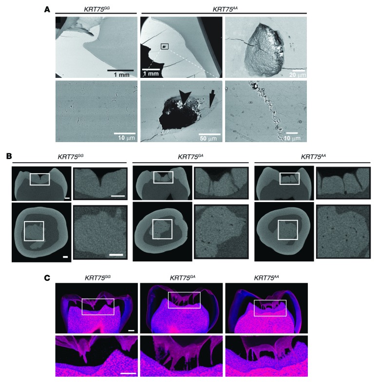
Scanning electron microscopy analysis of the polished specimens used for microindentation revealed the presence of cracks and holes (20–100 μm in diameter) in the enamel from several individuals carrying the rs2232387 polymorphism (Figure 3A). The area surrounding the holes was hypomineralized, as indicated by the reduction in density (Figure 3A, arrow), and spherical particles observed in these holes suggested the presence of bacteria (Figure 3A, arrowhead), which was confirmed by Gram staining (Supplemental Figure 4). Micro-computed tomography (micro-CT) analysis of molars revealed the presence of tubular projections of the pit and fissure area into the enamel. These structures were shallow in individuals with KRT75GG; however, they penetrated through the enamel in individuals carrying the missense A allele (Figure 3, B and C). In 3 of the carriers analyzed, the number of these tubules was very high (10 to 20), and most of them reached the dentin (Figure 3C and Supplemental Videos 1–6). These tubular lesions, which possibly form along a group of prisms (31), may have serious clinical implications, such as delayed diagnosis of decay, due to lack of visible signs on the tooth surface and a fast progression of the disease into the dentin. Enamel rod boundaries have been acknowledged as the main pathway for acid attack and demineralization, and the proteins located at the rod sheath were shown to be more chemically stable in enamel from patients with healthy teeth than in sound enamel areas from patients who are prone to develop caries (10, 11). Moreover, the pit and fissure area of mature permanent human teeth has a very convoluted prism arrangement with high concentration of tufts (7) and is normally resistant to acid/carious attack (32), suggesting that tufts offer some protection against caries. Our results suggest that tufts and rod sheaths destabilized by the presence of KRT75A161T mutant protein have a reduced capacity to protect against caries.
Figure 3. Tubular projections of molar fissures forming thin holes deep into the enamel.
(A) Backscattered scanning electron microscopy of polished sections of tooth crowns from individuals with common G alleles (KRT75GG) or with missense A alleles (KRT75AA) at rs2232387. Note for KRT75AA the presence of cracks and holes surrounded by partially demineralized enamel (arrow) and containing particles resembling bacteria (arrowhead). Scale bars: 1 mm (upper left and central panels); 20 μm (upper right panel); 50 μm (lower central panel); 10 μm (lower left and right panels). (B) Micro-CT analysis of molars showing the presence of thin holes going from the enamel surface and extending into the dentin detected in patients with the missense A allele. Scale bars: 5 mm. (C) Micro-CT reconstructions of molar sections showing that the thin holes in molars from individuals with a KRT75GA or KRT75AA genotype are tubular projections of the fissures that run throughout the enamel. Enamel is transparent, while dentin is colored in red, based on density. A thin layer at the surface of the enamel appears red due to the transition between air density and enamel density. Scale bars: 1 mm.
This study provides the first characterization, to our knowledge, of specific epithelial hair keratins present in mature enamel and links hair disorder–associated polymorphisms in the KRT75 gene to increased susceptibility to dental defects and caries. We anticipate that other mutations in epithelial hair keratins will show a similar association.
Methods
Further information can be found in Supplemental Methods.
Statistics.
Linear regression was used to test the association of genetic polymorphisms with dental caries experience and enamel hardness. A P value of less than 0.05 was considered significant. Data are expressed as mean ± SEM, and error bars denote SEM.
Study approval.
Informed consent was provided by all participants or their guardians. All procedures, forms, and protocols were approved by NIAMS, the Institutional Review Boards of the University of Pittsburgh, West Virginia University, and the Walter Reed National Military Medical Center. All animal work was approved by the NIAMS Animal Care and Use Committee.
Accession numbers.
All original completed microarray data and RNA-Seq data were deposited in the NCBI’s Gene Expression Omnibus (GSE57984). The genotype and phenotype data from the Center for Oral Health Research in Appalachia (COHRA) study cohort are available in the NCBI’s database of Genotypes and Phenotypes (dbGaP) (phs000095.v2.p1).
Supplementary Material
Acknowledgments
This work was supported by the Intramural Research Program of NIAMS (ZIA AR041171-07 to M.I. Morasso) and by grants from the National Institute of Dental and Craniofacial Research (R01-DE014899 and U01-DE018903 to M.L. Marazita and R56-DE016703 to E. Beniash). We thank Paul Bible, Meghan Kellett, Julie Erthal, and Juliane Lessard from the Laboratory of Skin Biology. We thank members of NIAMS: Martha Somerman, Leon Nesti, Gustavo Gutierrez-Cruz, Hong-Wei Sun, and the Light Imaging Facility. We thank Glen Imamura and the residents at the Walter Reed National Military Medical Center; Yang Xu, Toshiki Soejima, and Robert J. Weyant from the University of Pittsburgh; Daniel W. McNeil and Richard Crout from West Virginia University; Jiang Chen and Dennis Roop from the University of Colorado Denver. We would like to express our gratitude to the COHRA participants and field research teams whose contributions made this work possible.
Footnotes
Conflict of interest: The authors have declared that no conflict of interest exists.
Reference information:J Clin Invest. 2014;124(12):5219–5224. doi:10.1172/JCI78272.
References
- 1.Shaw JH. Causes and control of dental caries. N Engl J Med. 1987;317(16):996–1004. doi: 10.1056/NEJM198710153171605. [DOI] [PubMed] [Google Scholar]
- 2.[No authors listed]. The problem of dental caries. Nature. 1932;129(3269):926–927. doi: 10.1038/129926a0. [DOI] [Google Scholar]
- 3.Neumann HH. Dental caries. Lancet. 1947;1(6458):806. doi: 10.1016/s0140-6736(47)91560-2. [DOI] [PubMed] [Google Scholar]
- 4.Crabb HS, Mortimer KV. Dental caries and enamel structure. Nature. 1966;209(5023):611–612. doi: 10.1038/209611a0. [DOI] [PubMed] [Google Scholar]
- 5.Werneck RI, Mira MT, Trevilatto PC. A critical review: an overview of genetic influence on dental caries. Oral Dis. 2010;16(7):613–623. doi: 10.1111/j.1601-0825.2010.01675.x. [DOI] [PubMed] [Google Scholar]
- 6.Simmer JP, et al. Regulation of dental enamel shape and hardness. J Dent Res. 2010;89(10):1024–1038. doi: 10.1177/0022034510375829. [DOI] [PMC free article] [PubMed] [Google Scholar]
- 7.Robinson C, Lowe NR, Weatherell JA. Amino acid composition, distribution and origin of “tuft” protein in human and bovine dental enamel. Arch Oral Biol. 1975;20(1):29–42. doi: 10.1016/0003-9969(75)90149-1. [DOI] [PubMed] [Google Scholar]
- 8.Baldassarri M, Margolis HC, Beniash E. Compositional determinants of mechanical properties of enamel. J Dent Res. 2008;87(7):645–649. doi: 10.1177/154405910808700711. [DOI] [PMC free article] [PubMed] [Google Scholar]
- 9.Chai H, Lee JJ, Constantino PJ, Lucas PW, Lawn BR. Remarkable resilience of teeth. Proc Natl Acad Sci U S A. 2009;106(18):7289–7293. doi: 10.1073/pnas.0902466106. [DOI] [PMC free article] [PubMed] [Google Scholar]
- 10.Pincus P. Relation of enamel protein to dental caries. Nature. 1948;161(4104):1014. doi: 10.1038/1611014a0. [DOI] [PubMed] [Google Scholar]
- 11.Little K. Caries-prone and caries-resistant teeth. Nature. 1962;193:388–389. doi: 10.1038/193388a0. [DOI] [PubMed] [Google Scholar]
- 12.Lesot H, Smith AJ, Matthews JB, Ruch JV. An extracellular matrix protein of dentine, enamel, and bone shares common antigenic determinants with keratins. Calcif Tissue Int. 1988;42(1):53–57. doi: 10.1007/BF02555839. [DOI] [PubMed] [Google Scholar]
- 13.Robinson C, Shore RC, Kirkham J. Tuft protein: its relationship with the keratins and the developing enamel matrix. Calcif Tissue Int. 1989;44(6):393–398. doi: 10.1007/BF02555967. [DOI] [PubMed] [Google Scholar]
- 14.Robinson C, Hudson J. Tuft protein: protein cross-linking in enamel development. Eur J Oral Sci. 2011;119(suppl 1):50–54. doi: 10.1111/j.1600-0722.2011.00906.x. [DOI] [PubMed] [Google Scholar]
- 15.Pispa J, Thesleff I. Mechanisms of ectodermal organogenesis. Dev Biol. 2003;262(2):195–205. doi: 10.1016/S0012-1606(03)00325-7. [DOI] [PubMed] [Google Scholar]
- 16.Biggs LC, Mikkola ML. Early inductive events in ectodermal appendage morphogenesis. Semin Cell Dev Biol. 2014;25–26C:11–21. doi: 10.1016/j.semcdb.2014.01.007. [DOI] [PubMed] [Google Scholar]
- 17.Hwang J, Mehrani T, Millar SE, Morasso MI. Dlx3 is a crucial regulator of hair follicle differentiation and cycling. Development. 2008;135(18):3149–3159. doi: 10.1242/dev.022202. [DOI] [PMC free article] [PubMed] [Google Scholar]
- 18.Pincus P. Enamel protein. Nature. 1936;138:970. doi: 10.1038/138970a0. [DOI] [Google Scholar]
- 19.Pautard FG. Mineralization of keratin and its comparison with the enamel matrix. Nature. 1963;199:531–535. doi: 10.1038/199531a0. [DOI] [PubMed] [Google Scholar]
- 20.Warshawsky H, Josephsen K. The behavior of substances labeled with 3H-proline and 3H-fucose in the cellular processes of odontoblasts and ameloblasts. Anat Rec. 1981;200(1):1–10. doi: 10.1002/ar.1092000102. [DOI] [PubMed] [Google Scholar]
- 21.McLean WH, Irvine AD. Disorders of keratinisation: from rare to common genetic diseases of skin and other epithelial tissues. Ulster Med J. 2007;76(2):72–82. [PMC free article] [PubMed] [Google Scholar]
- 22.Pan X, Hobbs RP, Coulombe PA. The expanding significance of keratin intermediate filaments in normal and diseased epithelia. Curr Opin Cell Biol. 2013;25(1):47–56. doi: 10.1016/j.ceb.2012.10.018. [DOI] [PMC free article] [PubMed] [Google Scholar]
- 23.Winter H, et al. An unusual Ala12Thr polymorphism in the 1A α-helical segment of the companion layer-specific keratin K6hf: evidence for a risk factor in the etiology of the common hair disorder pseudofolliculitis barbae. J Invest Dermatol. 2004;122(3):652–657. doi: 10.1111/j.0022-202X.2004.22309.x. [DOI] [PubMed] [Google Scholar]
- 24.Chapalain V, et al. Is the loose anagen hair syndrome a keratin disorder? A clinical and molecular study. Arch Dermatol. 2002;138(4):501–506. doi: 10.1001/archderm.138.4.501. [DOI] [PubMed] [Google Scholar]
- 25.Slayton RL, Cooper ME, Marazita ML. Tuftelin, mutans streptococci, and dental caries susceptibility. J Dent Res. 2005;84(8):711–714. doi: 10.1177/154405910508400805. [DOI] [PubMed] [Google Scholar]
- 26.Patir A, et al. Enamel formation genes are associated with high caries experience in Turkish children. Caries Res. 2008;42(5):394–400. doi: 10.1159/000154785. [DOI] [PMC free article] [PubMed] [Google Scholar]
- 27.Shimizu T, et al. Enamel formation genes influence enamel microhardness before and after cariogenic challenge. PLoS One. 2012;7(9):e45022. doi: 10.1371/journal.pone.0045022. [DOI] [PMC free article] [PubMed] [Google Scholar]
- 28.Harel S, Christiano AM. Genetics of structural hair disorders. J Invest Dermatol. 2012;132(E1):E22–E26. doi: 10.1038/skinbio.2012.7. [DOI] [PubMed] [Google Scholar]
- 29.Duverger O, Morasso MI. To grow or not to grow: hair morphogenesis and human genetic hair disorders. Semin Cell Dev Biol. 2014;25–26:22–33. doi: 10.1016/j.semcdb.2013.12.006. [DOI] [PMC free article] [PubMed] [Google Scholar]
- 30.Chen J, Jaeger K, Den Z, Koch PJ, Sundberg JP, Roop DR. Mice expressing a mutant Krt75 (K6hf) allele develop hair and nail defects resembling pachyonychia congenita. J Invest Dermatol. 2008;128(2):270–279. doi: 10.1038/sj.jid.5701038. [DOI] [PubMed] [Google Scholar]
- 31. Robinson C, Kirkham J, Shore RC, Brookes SJ, Wood SR. Enamel matrix function and the tuft enigma: a role in directing enamel tissue-architecture: a partial sequence of human ameloblastin. In: Goldberg M, Boskey A, Robinson C, ed. Chemistry And Biology Of Mineralised Tissues. Proceedings Of The Sixth International Conference. Vittel, France: American Academy of Orthopaedic Surgeons; 2000:209–213. [Google Scholar]
- 32. Robinson C, Weatherell JA, Hallsworth AS. Alterations in the composition of permanent human enamel during carious attack. In: Leach SA, Edgar WM, eds. Demineralization and Remineralization of the Teeth. Oxford, United Kingdom: IRL Press; 1982:209–223. [Google Scholar]
Associated Data
This section collects any data citations, data availability statements, or supplementary materials included in this article.


