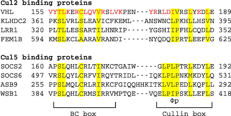
Figure 1. Sequence alignment of VHL BC box and cullin box to other Cul2 and Cul5 binding proteins.
The regions for the BC box and cullin box are marked. Conserved residues are highlighted and the Φp (Φ indicates a hydrophobic residue) motif is shown. VHL missense mutations associated with VHL disease are shown in red.

