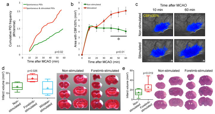
Figure 7. Tactile stimulation-induced PIDs worsen ischemic tissue perfusion and outcome.
a. Average cumulative induced and spontaneous PID frequency in mice receiving tactile stimulation starting 10 minutes after MCAO (red, n=14), and spontaneous PID frequency in non-stimulated controls (green, n=17). None of the mice in these cohorts were subjected to transient hypoxia or hypotension as part of the experimental protocol.
b. Area of perfusion defect with ≤30% residual CBF increased in proportion with PID occurrence in the stimulated group over time. Two-way ANOVA for repeated measures.
c. Representative laser speckle flowmetry images showing the expansion of perfusion defect (blue pixels with ≤30% residual CBF) between 10 and 60 minutes after MCAO in a mouse that received tactile stimulation, and the stable perfusion defect in a mouse without tactile stimulation.
d. Infarct volumes measured using TTC staining were larger in the group receiving forepaw but not whisker stimulation starting 10 minutes after MCAO (n=6, 4 and 4 in control, forepaw and whisker stimulation groups; p<0.05; t-test). These experiments were performed in a separate cohort of mice without endotracheal intubation or mechanical ventilation in order to minimize surgical morbidity and mortality. The experimental protocol was otherwise identical to other tactile stimulation experiments above. There were a total of 2.25±0.25 PIDs in the forepaw-stimulated group, whereas no spontaneous or induced PID was detected in non-stimulated and whisker stimulated cohorts, respectively.
e. Infarct volumes measured 72 hours after stroke onset using H&E-stained coronal brain sections were larger in the group receiving forepaw stimulation starting 10 minutes after dMCAO (n=9 each; t-test). In this cohort, we initially studied n=5 mice per group. The data showed higher than expected coefficient of variation. We then performed a power analysis, determined n=10 mice per group as the appropriate sample size, and added 5 more mice in each group. One mouse was excluded in each group based on a priori criteria (1 in non-stimulated group due to total absence of an infarct; 1 in stimulated group due to clip-related trauma precluding reliable infarct measurement). Therefore, final sample sizes were n=9 per group. As with the 24-hour assessment group above, these experiments were performed in a separate cohort of mice without endotracheal intubation or mechanical ventilation in order to minimize surgical morbidity and mortality. In this experimental cohort, inhalation gas included 70%N2/30%O2 mixture. By doing this, we also reproduced the effect of stimulation-induced PIDs on tissue outcome in the absence of N2O. The experimental protocol was otherwise identical to other tactile stimulation experiments above. Representative sections show the infarct at each slice level in one non-stimulated and forepaw and shoulder stimulated mouse each.

