Abstract
Proliferating mammalian stem and cancer cells express telomerase (TERT) in an effort to extend chromosomal G-overhangs and maintain telomere ends. Telomerase-expressing cells also have higher levels of the single-stranded DNA binding protein SSB1, which has a critical role in DNA double-strand break repair. Here we report that SSB1 binds specifically to G-strand telomeric DNA in vitro and associates with telomeres in vivo. SSB1 interacted with the TERT catalytic subunit and regulates its interaction with telomeres. Deletion of SSB1 reduced TERT interaction with telomeres and lead to G-overhang loss. While SSB1 was recruited to DSB sites, we found no corresponding change in TERT levels at these sites, implying that SSB1-TERT interaction relied upon a specific chromatin structure or context. Our findings offer an explanation for how telomerase is recruited to telomeres to facilitate G-strand DNA extension, a critical step in maintaining telomere ends and cell viability in all cancer cells.
Keywords: SSB1, G-overhang, telomerase, TERT, DNA damage response
Introduction
The telomeric ends of all eukaryotic chromosomes are capped with nucleoprotein complexes that prevent chromosome degradation or the formation of chromosome end-to-end fusions. Capping by the multi-protein shelterin complex, through binding to telomere-specific DNA sequences also prevents aberrant recognition of the free DNA ends as a DNA double-strand break (DSB), which would typically activates the DNA damage response. Three protein subunits (TRF1, TRF2, and POT1) of the shelterin complex are involved in direct recognition of the TTAGGG repeats found in telomeres. Three additional sheletrin components (TIN2, TPP1 and Rap1) function in tandem to distinguish telomeres from interstitial DNA double-strand breaks. Both TPP1 and POT1 have also been implicated in the regulation of telomerase recruitment to telomeres (1).
The catalytic unit of telomerase is TERT and its activity is necessary for the immortality of many cancers and is mostly inactive in somatic cells, suggesting that telomerase inhibition could selectively repress cancer cell growth with minimal side effects on normal tissue. Mammalian telomeres maintained by telomerase consist of long tracks of G-rich double-stranded DNA repeats that end in a G-rich, single-stranded DNA (ssDNA) overhang. The mammalian telomerase consists of a reverse transcriptase (the catalytic subunit called TERT) and a functional RNA template (called TR or TERC) and is a unique ribonuleoprotien enzyme that is responsible for adding the telomeric repeats onto the 3′ ends of chromosomes during S-phase DNA synthesis. Telomerase is required in order to maintain a cells ‘regenerative’ (i.e., stem cell) or proliferative (i.e., transformed cell) capacity. While the enzymatic propertied of telomerase are well described, the mechanism by which mammalian telomerase is loaded onto the G-rich single-stranded telomeric overhang is largely unknown. Telomeric overhangs are normally bound by the single strand-binding protein, POT1 (2) while the TPP1 oligosaccharide-oligonucleotide (OB)-fold binding domain of sheletrin TPP1 is sufficient for telomerase recruitment to telomeres (1). As several DNA repair-associated proteins, including single-stranded DNA binding protein 1 (SSB1), also have OB-fold domains, their role in telomerase recruitment to telomeres requires further elucidation.
Similar to other single-stranded DNA binding proteins (e.g., replication protein A, RPA), SSB is an essential component of the DNA repair machinery in eukaryotes. Human ssDNA-binding proteins 1 and 2 (hSSB1 and hSSB2, also known as NABP2 and NABP1, respectively), together with the integrator complex subunit 3 (INTS3) and C9orf80, form a heterotrimeric protein complex that participates in DNA damage responses and in the maintenance of genome stability (3). Recent studies have shown that SSB1 and SSB2 also appear to protect newly replicated leading- and lagging-strand DNA of telomeres (3); however, their specific function(s) at the telomere remains largely unknown.
Gu and coworkers reported that deletion of murine Ssb1 (mSsb1) resulted in increased chromatid-type fusions involving both leading- and lagging-strand telomeric DNA (3). Their observation suggests, but does not unequivocally prove, that SSB1 is required for the protection of G-overhangs. In addition, both mSsb1 and mSsb2 localize to a subset of telomeres and are required to repair TRF2-deficient telomeres (3). The localization of mSsb1 to damaged DNA requires interaction with INTS3 (4), while its association with telomeric ssDNA is dependent on interaction with Pot1a (3), indicating these functions and interactions are mediated differently. To investigate this possibility, we generated mouse embryonic fibroblast cells from mSSB1 conditional knockout mice and used this model system to demonstrate here that SSB1 interacts with TERT and is required for telomerase recruitment to telomeres. Moreover, depletion of SSB1 results in the loss of G-overhangs, suggesting that SSB1 has a crucial role in maintaining the structure of telomere ends. However, while SSB1 is also recruited to DNA DSB sites, under this circumstance telomerase is not co-recruited, indicating SSB1 participates in two mechanistically distinct cellular processes.
Materials and Methods
Cells
The culture conditions for human HEK293, and HCT116 cells have been described previously (5). The conditional RosaCreERT2 Ssb1flox/flox mice were generated as described previously (6). Mouse embryonic fibroblasts (MEFs) were generated from E13.5 embryos and Cre expression was induced by treatment with tamoxifen at a concentration of 0.2 µM for the indicated time.
Chromosomal aberration analysis
Chromosomal aberrations analysis was carried out in exponentially growing cells as described previously (5). Cell cycle measurements were performed by flow cytometric analysis (7). Treatment of cell lines with siRNA was performed as described (8). Generation of mutant ΔhSSB1, siRNA transfection, immunofluorescence, and protein retention assay were carried out as previously described (8–12). Cells in different cell cycle phases were enriched by serum starvation and thymidine block. Endogenous hSSB1 was depleted by 3’ UTR-specific hSSB1-siRNA in cells expressing GFP-OB-hSSB1.
Detection of telomeres, centromeres and telomerase assay
Telomeres and centromeres in metaphase spreads were detected by fluorescent in situ hybridization (FISH) with a telomere- or centromere specific probe as described previously (5). Nondenaturing in-gel hybridization to determine relative amounts of telomeric single-stranded DNA (G tails) was performed as described previously (5, 13, 14). Telomerase activity was determined as previously described (15, 16).
Electrophoretic mobility shift assay (EMSA)
The interaction of recombinant hSSB1 with G-rich strand (TTAGGG)5, C-rich strand (AATCCC)5 and GC-duplex oligonucleotides was investigated using native acrylamide electrophoretic mobility shift analysis. Increasing concentrations of hSSB1 were incubated with 32P-labelled oligonucleotide ssDNA (50 pmol) in buffer (20mM HEPES, pH 7.3, 100mM KCl and 1mM MgCl2, 1 mg µl−1 bovine serum albumin) at 200 C for 30 min in 10 µL total volume. Reactions were resolved on 10% native acrylamide/TBE gel described previously (17). Gels were exposed to a phosphorimage plate and visualized with a Fuji FLA-5000 Phosphoimager.
Western blot analysis, immunoprecipitation (IP) and chromatin-immunoprecipitation (ChIP)
Western blot analysis, IP and ChIP were performed by standard procedures as previously described (5, 12). In brief, hSSB1 or Flag antibody immunoprecipitated human DNA was membrane bound by slot-blotting and then hybridized with 32P-labeled DNA probe. The quantitation of the DNA signals was achieved by phosphorimaging. Antibodies used in this study were supplied by Sigma (M2-Flag) Calbiochem (Mre11, Rad51), Upstate (γ-H2AX), Roche (BrdU), Calbiochem (hTERT) and Invitrogen (GFP and Alexa secondary antibodies). Sheep antiserum to hSSB1 was raised against full-length recombinant His-tagged hSSB1 and have been described previously (12, 17, 18).
Detection of hSSB1 and hTERT proteins at I-SceI induced DSB sites by ChIP in different phases of the cell cycle
DR95 cells expressing Flag-hTERT treated with 2 mM thymidine for 14 h then washed with 1X PBS and released into fresh media as described previously (19). The cell cycle distribution was determined using flow cytometry to measure DNA content. The percentage of cells in different phases was calculated from summation of G1 + S+ S/G2 phase cells (Supplemental Table 1), eliminating M phase cells which were quantitated by metaphase scoring. The closest PCR product to the DSB site is 94–378 (19, 20).
Results
Preferential binding of hSSB1 to the telomere G-strand
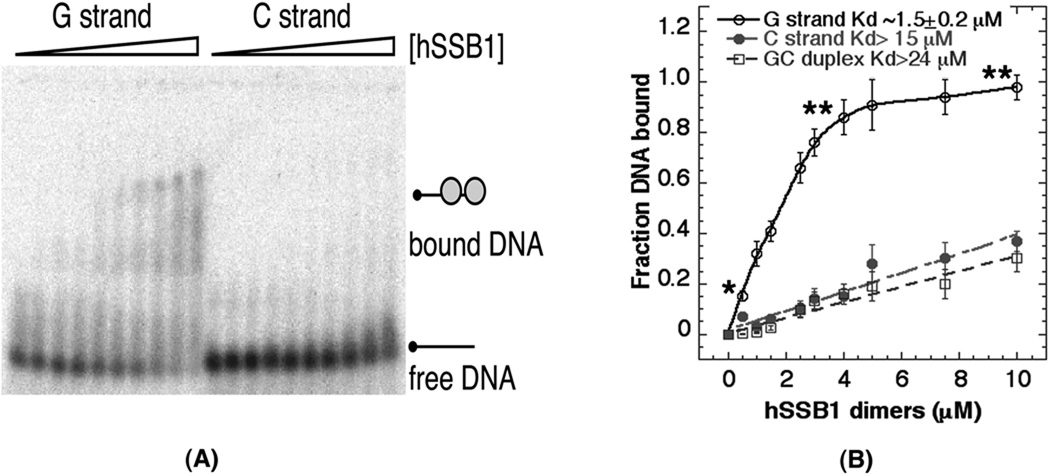
Many proteins of the DNA DSB repair machinery are constitutively expressed, as are some proteins involved in telomere maintenance, and demonstrate increased chromatin retention in cells following irradiation (21–23). Indeed, some such proteins even play dual roles. Similar to RPA and ATR, SSB1 is involved in DNA DSB repair by homologous recombination (24, 25) and helps protect newly replicated telomeres (3). Although recombinant SSB1 is thought to exhibit a non-specific ssDNA-binding activity, we have previously shown that hSSB1 prefers binding to polypyrimidine or d(GT) ssDNA substrates and that increasing dA content was inhibitory (17). This is also true for SSBs found in other organisms; a marked decrease in binding to polyadenine sequences has been reported for Escherichia coli SSB (26). In fact, the structure of a hSSB, such as RPA, bound to ssDNA (27) shows only two DNA bases that efficiently base-stack with aromatic amino acids in the protein’s ssDNA-binding cleft. The identity of these DNA bases is likely to be important for binding capacity; the larger adenine base is not as thermodynamically favorable for binding (as pyrimidines) (17, 26) due to stearic hindrance and/or inefficient base stacking with the aromatic side chains. We therefore determined, by electrophoretic mobility shift analysis, whether SSB1 can specifically bind to mammalian DNA containing G- or C-rich telomere-like sequences. Results support binding of recombinant SSB1 to single-stranded G-rich telomeric oligonucleotides (TTAGGG)5, but not to C-rich telomeric oligonucleotides (AATCCC) or GC duplex oligonucleotides (Fig. 1A & B). The non-preference for the C strand may be due, at least in part, to the adjacent adenine bases in the repeated telomeric motif, which likely mimics the effect of poly-A sequences. Without a structure, we can only postulate this reason for the inability of hSSB1 to efficiently position itself on this C-rich sequence.
Fig. 1. hSSB1 interaction with DNA.
(A) Electrophoretic mobility shift analysis showing preferential binding of recombinant SSB1 to single stranded DNA substrates which are either G- or C-rich telomere oligonucleotides. (B) The data is from triplicate gel shift experiments showing binding curves of hSSB1 using G- and C-rich oligonucleotides. hSSB1 preferentially binds G-rich oligonucleotides with minimum binding to C-rich oligonucleotides. The difference in hSSB1 binding with G-rich vs. C-rich oligonucleotides is statistically significantly (*P < 0.05 and **P < 0.01 as determined by the chi-square test).
Depletion of SSB1 results in the G-overhang loss from telomeres
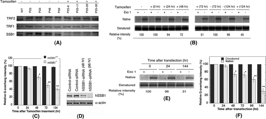
Given that hSSB1 binds the G-rich the sequences of telomeres, we next explored whether SSB1 binding plays a direct role in regulating and maintaining the length of G-overhangs. We constructed a Ssb1 (Ssb1 flox/flox allele (6)) conditional deletion mutant using tamoxifen-induced Cre recombinase expression (Cre-ERT2) in mouse embryonic fibroblasts (MEFs) and demonstrated efficient deletion of mSsb1 without affecting the levels of TRF1 or TRF2 (Fig. 2A). Using a sensitive double strand-specific nuclease technique (28, 29) we measured G-overhang at different time points following tamoxifen-induced mSsb1 deletion. As shown in Figure 2B, the level of native single-stranded G-overhang DNA decreased with increasing post-induction times in CreERT2; Ssb1fl/fl cells (Fig. 2B & C). Similarly, depletion of hSSB1 in human HEK293 cells with hSSB1-specific siRNA (Fig. 2D) also resulted in loss of G-overhangs (Fig. 2E, F; Supplementary Fig. 1) (17, 28). Taken together, these results support the argument that SSB1 regulates the stability of telomeric G-overhangs in mouse and human cells. To determine whether the G-overhang reduction was due to breaks near the telomeres, we next conducted telomeric FISH analysis to detect any loss of telomeric signals in metaphase cells (Supplementary Fig. 2). We reasoned that G-overhang loss would not result in a total loss of telomere signals, unless the loss led to extensive telomere degradation or a subtelomeric break. Concurrent with the loss of G-overhangs, we observed telomere end-to-end associations but without telomere signal loss (Supplementary Fig, 2A), a situation also observed in cells defective in ATM function (16, 30). In addition, a higher frequency of Robertsonian fusions was observed in Ssb1-deleted cells (P<0.01) (Supplementary Fig. 2B–D), suggesting that G-overhang loss was accompanied by breaks at or near the telomere that led to telomere fusions. A similar situation was observed in hSSB1 depleted human cells, where dicentric chromosome formation was detected (Supplementary Fig. 2E, F).
Fig. 2. Effect of SSB1 depletion on G-overhang.
(A) Western blot of Ssb1, TRF1 and TRF2 in Rosa CreERT2: Ssb1flox/flox MEF cells at different population (P) doublings with and without tamoxifen treatment. (B, E) In-gel hybridization of telomeres. For native, C probe for G-overhang was used. (B, C) MEFs with and without depletion of Ssb1 (B) and quantitation from three independent experiments (C) The difference in loss of G-overhang are statistically significantly (*P < 0.05 and **P < 0.01 as determined by the chi-square test). (D) Western blot of hSSB1 in HEK293 cells with and without knock down of hSSB1. (E, F) In gel hybridization analysis of G-overhang in 293 cells with and without depletion of hSSB1 (E) and quantitation from three independent experiments (F). The difference in G-overhang loss is statistically significantly (*P < 0.05 and **P < 0.01 as determined by the chi-square test).
SSB1 is required for TERT recruitment to telomeres
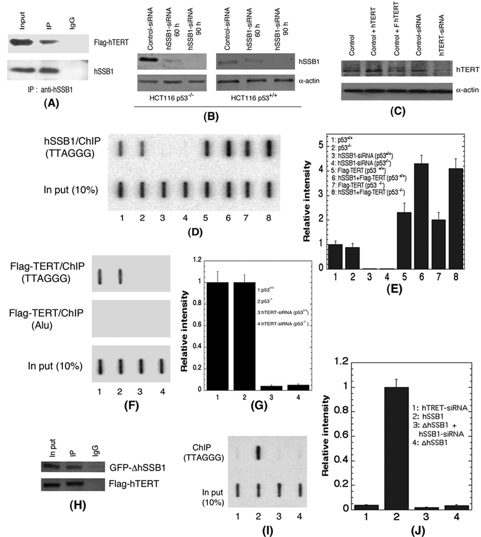
A possible mechanism by which SSB1 facilitates the protection and/or maintenance of G-overhangs is through recruitment of telomerase, perhaps via direct binding of TERT to the G-rich single-stranded overhangs during DNA replication. To investigate this possibility, we performed SSB1 and hTERT co-immunoprecipitation experiments, immunoprecipitating endogenous hSSB1 from cells expressing Flag-hTERT (Supplementary Fig. 3A) followed by immunoblotting with an anti-Flag antibody. Similarly we expressed GFP-hSSB1 and Flag-hTERT, then immunoprecipitated TERT with Flag antibody followed by immunoblotting with an anti-GFP antibody (Supplementary Fig. 3B). In both cases hSSB1 and hTERT associated together in a complex (Fig. 3A).
Fig. 3. Interaction of hSSB1 with telomeres.
(A) Flag-tagged hTERT was expressed in 293 cells and immunoprecipitated with hSSB1 antibody. hTERT was detected by Flag antibody. (B) Detection of hSSB1 in HCT116 cells with and without p53. hSSB1 was depleted with specific siRNA. (C) Depletion of hTERT by UTR-specific siRNA and detection by western blots. (D, E) Detection (D) and quantification (E) of hSSB1 at the telomeres by chromatin-immunoprecipitation analysis using an hSSB1 antibody. Lanes 1, 2: control; lanes 3, 4: hSSB1 depleted by specific siRNA; lane 5: p53+/+ cells with over-expression of hTERT; lane 6: p53+/+ cells with over-expression of hTERT and hSSB1; lane 7: p53−/− cells with over-expression of hTERT; lane 8: p53−/− cells with over-expression of hTERT and hSSB1. (F, G) ChIP analysis by using Flag-antibody to analyze Flag-hTERT at the telomeres; lane 1 : p53+/+ cells; lane 2 : p53−/− cells; lane 3: p53+/+ cells with depleted hTERT and lane 4: p53−/− cells with depleted hTERT (H) Interaction of ΔOB−hSSB1 with hTERT was determined by immunoprecipitation. (I, J) Effect of hTERT deletion or expression of hSSB1 with OB-depletion (GFP-???????????ΔOB−hSSB1) on the interaction of hSSB1 with telomeres determined by ChIP. Lane 1: HEK293 cells with hTERT depletion by specific siRNA and ChIP with hSSB1 antibody; lane 2: 293 cells ChIP with hSSB1 antibody; lane 3: 293 cells with depletion of endogenous hSSB1 and expression of GFP-ΔOB−hSSB1, ChIP with GFP antibody and; lane 4: 293 cells with expression of GFP-ΔOB−hSSB1 and ChIP with GFP antibody.
Since hTERT is known to localize to telomeres, chromatin immunoprecipitation (ChIP) was performed to determine whether hSSB1 also co-localizes to telomeric DNA. Irrespective of p53 status, hSSB1 is present in cells (Fig. 3B) and readily detected in association with telomeric DNA (Fig. 3D & 3E). Depletion with siRNAs of either hSSB1 or hTERT (Fig. 3B & 3C) resulted in a corresponding loss of both hSSB1 and hTERT binding to telomeric DNA (Fig. 3 D–G), while over-expression of Flag-hTERT (Fig. 3C) increased telomere-associated hSSB1 levels independent of p53 status (Fig. 3D & 3E). To determine whether depletion of hSSB1 (Fig. 3B) affects the association of hTERT with telomeres, ChIP was performed by using an anti-Flag antibody to detect telomeric Flag-hTERT in hSSB1-depleted cells. Consistent with other results, the levels of telomere-bound hTERT were reduced as a consequence of hSSB1 depletion (Fig. 3D & E). In contrast, overexpression of hSSB1 and hTERT resulted in enhanced association of both proteins at the telomeres (Fig. 3 D & E; Supplementary Fig. C). Conversely, when hTERT was depleted with specific siRNA (Fig. 3C), the levels of telomere-associated hSSB1 were reduced (Fig. 3F & G). While over-expression of proteins can lead to misinterpretations, these results are consistent with experiments using native cells (31) and imply that hTERT and hSSB1 first interact with one another before the protein complex recognizes single-stranded G-overhangs of telomeres.
The OB-fold of TPP1 (a shelterin protein) is required for recruitment of hTERT to telomeres. We next assessed whether the OB domain of SSB1 is similarly associated with hTERT recruitment. A GFP tagged hSSB1 mutant protein tagged missing OB binding domain (ΔOB-hSSB1), expressed in 293 cells, still immunoprecipitated with hTERT (Fig. 3H). However, the ΔOB-hSSB1 mutant protein displayed decreased telomere association (Fig. 3I & 3J) and corresponding reduction in telomere-associated hTERT (Supplementary Fig 3C). Full length hSSB1 tagged with GFP (GFP-hSSB1) showed no reduction in telomere DNA binding (Supplementary Fig. 3D). These results imply that while the loss of the hSSB1 OB domain does not inhibit its interaction with hTERT, it does interfere with hSSB1/hTERT complex loading onto telomeric DNA.
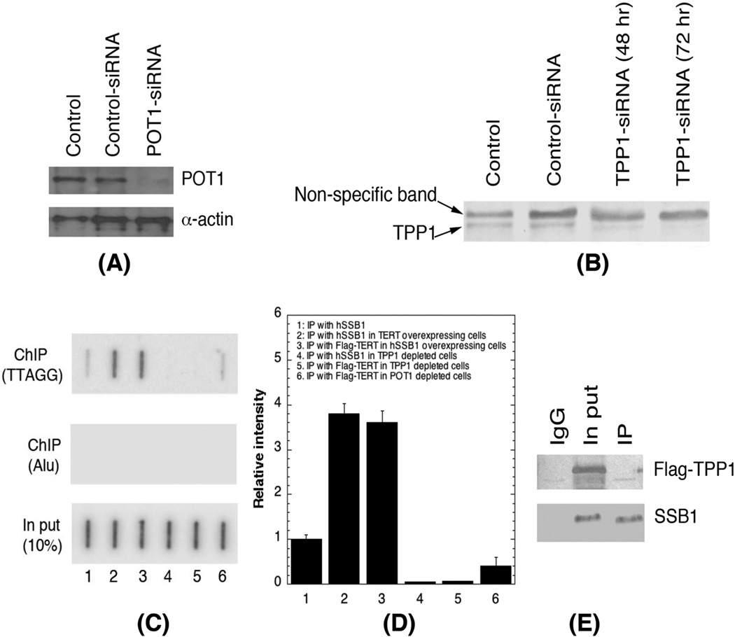
SSB1 has been reported to interact with another shelterin component POT1 (3), which when paired with TPP1 in a POT1-TPP1 heterodimer, has a higher affinity for telomeric single-stranded DNA compared to POT1 alone (32). Thus, we explored whether depletion of POT1 (Fig. 4A) or TPP1 (Fig. 4B) altered SSB1 association with telomere DNA. Depletion of TPP1, as did POT1 depletion, significantly reduced TERT and SSB1 association with telomeres (Fig. 4C & D). Although depletion of TPP1 essentially eliminated the association of telomeres with hTERT and hSSB1, we did not detect any interaction between SSB1 and TERT (Fig. 4E), suggesting that these two proteins independently regulate the recruitment of telomerase to the telomeres.
Fig. 4. Effect of POT1 and TPP1 depletion on hSSB1 and hTERT association with telomeric DNA.
(A, B) Knockdown of POT1 (A) and TPP1 (B) with specific si-RNA. (C, D) Lane 1 represents ChIP with hSSB1 antibody: Lane 2 represents ChIP with hSSB1 antibody from hTERT over-expressing cells: Lane 3 represents with ChIP with Flag-hTERT antibody in hSSB1 overexpressing cells: Lane 4 represents ChIP with hSSB1 antibody in TPP1 depleted cells: Lane 5 represents ChIP with Flag-hTERT antibody in TPP1 depleted cells and : Lane 6 represents ChIP with Flag-hTERT antibody in POT1 depleted cells. (E) Flag-tagged TPP1 was expressed in 293 cells and immunoprecipitated with hSSB1 antibody. TPP1 was detected by Flag antibody.
TRF1, but not TRF2, affects binding of hSSB1 to telomeric DNA
POT1 binds specifically to single-stranded 5′-(T)TAGGGT TAG-3′ sites at either the 3′ end or within the recognition sequence (33). In contrast, homodimeric TRF1 and TRF2 can each directly recognize and interact with telomere double-stranded DNA containing TTAGGG repeats. TRF1 is known to regulate telomere length; its overexpression results in progressive shortening of telomere length, whereas a telomere non-binding TRF1 mutant exhibits increased telomere elongation. Thus we explored the possibility that TRF1 may interact with hSSB1 and/or alter hSSB1 recruitment to telomeres. Co-immunoprecipitation studies did not detect any interaction between, hSSB1 and either TRF1 or TRF2 (Supplementary Fig. 4A & B). However, depletion of cellular TRF1 (Supplementary Fig. 4C) increased the amount of telomere-associated hSSB1 (Supplementary Fig. 4E), while depletion of TRF2 (Supplementary Fig. 4D) had no effect (Supplementary Fig. 4E). These results suggest that although TRF1 does not interact with hSSB1, its cellular levels may influence the recruitment of hSSB1 to telomeres. TRF2 depletion did not seem to play a role in hSSB1 recruitment to telomeres (Supplementary Fig. 4D & E).
S-phase specific interaction of SSB1 with telomeres
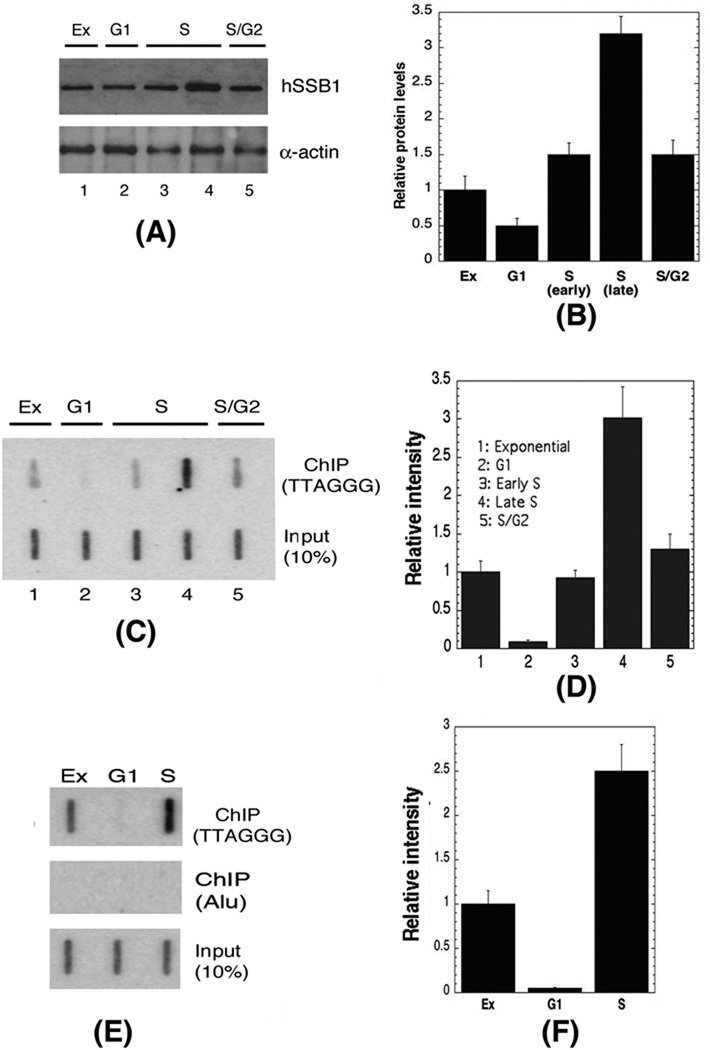
Telomeres are synthesized throughout DNA replication in mammalian cells (34). As such, we sought to determine whether SSB1 levels and telomere binding a function of cell cycle position. Cells were enriched in different phases by serum deprivation as well as by thymidine block. For serum deprivation, cells were incubated until confluence, washed, and incubated for 48 hr in serum-free medium. Cell-cycle analysis by flow cytometry revealed that more than 95% of cells were in G1-phase of the cell cycle. Western blot analysis indicated hSSB1 protein levels peaked during S-phase (Fig. 5A & B). Furthermore, ChIP analysis indicated that telomere-associated hSSB1 levels peaked in mid to late S-phase cells then declined in S/G2 phase cells before reaching minimum in G1-phase cells (Fig. 5C & D). We similarly examined ectopically expressed Flag-hTERT binding to telomeres as a function of cell cycle position by ChIP analysis and found binding to telomeric DNA in S-phase, but not G1-phase cells (Fig. 5E & F). Together, these results indicate that SSB1 recruitment, as does hTERT, occurs specifically during telomere replication.
Fig. 5. Interaction of hSSB1 and hTERT with telomeres as a function of cell cycle position.
(A, B) hSSB1 levels in different phases of the cell cycle. (C, D) Interaction of hSSB1 with telomeres in different phases of the cell cycle by ChIP using anti-hSSB1 antibody: results are from three independent experiments. (E, F) Interaction of hTERT with telomeres in different phases of cell cycle by ChIP using Flag-hTERT antibody.
hSSB1, but not hTERT, associates directly with DNA DSBs
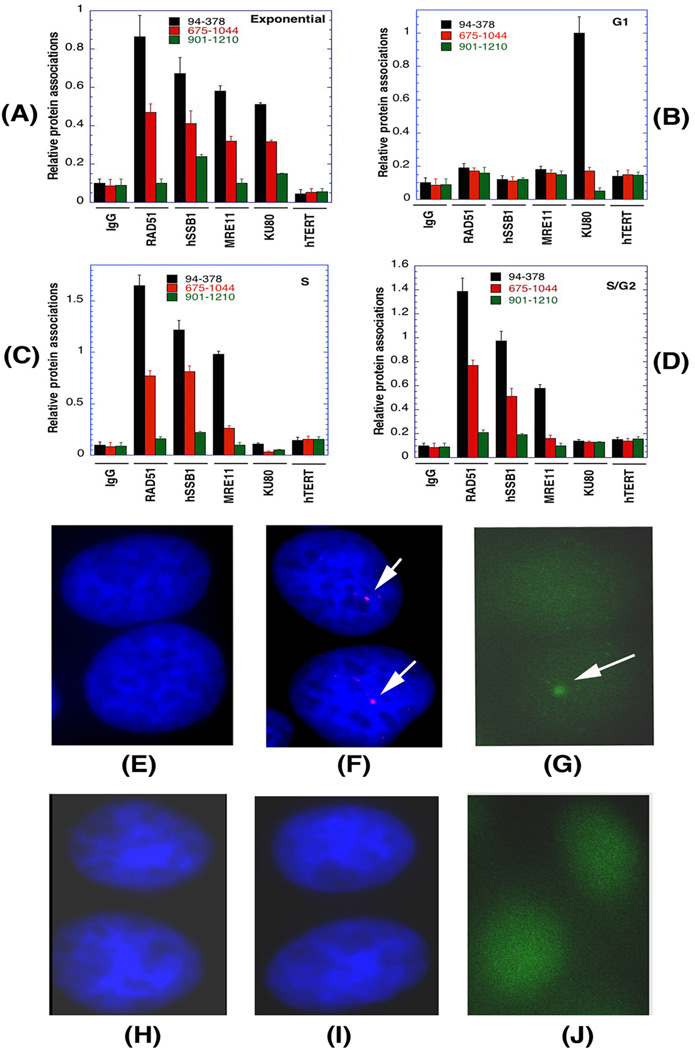
Expression of hTERT enhances DNA DSB repair and suppresses genomic instability (35). Since hSSB1 interacts with hTERT and hSSB1 has an increased presence at DSB sites, we assessed whether hTERT levels also increase at DNA DSBs. To determine recruitment of hSSB1 and hTERT to interstitial DNA DSBs, we compared the loading of each protein at different distances from an l-Scel induced DSB site (19). Cells enriched in G1, S or S/G2 phase (Supplementary Table 1), were induced for l-Scel site cleavage and analyzed by ChIP, using RAD51, hSSB1, MRE11 KU80, and hTERT specific antibodies in combination with site specific PCR primer pairs. In exponentially growing asynchronous cells, RAD51, hSSB1, MRE11 and KU80 appeared elevated in closest proximity to the DNA DSB and declined as the distance from the DSB increased. In contrast, the level of hTERT at the DSB site was similar to that of the control and both did not fluctuate with distance (Fig. 6A). In G1-phase cells, only KU80 levels were high in close proximity to the DSB break and declined with distance whereas levels of RAD51, hSSB1, MRE11 and hTERT were essentially background and constant across the break site (Figure 6B). However, in S- and S/G2-phase cells, high RAD51 or MRE11 levels localized near the DNA break and declined with distance whereas there was no change in hTERT or Ku80 recruitment (Fig. 6C & 6D). Moreover, SSB1 foci formation at the l-Scel DSB site could be detected by immunofluorescence of (Fig. 6E–G), but not hTERT foci formation (Fig. 6H–J), corroborating the ChIP protein mapping results.
Fig. 6. Localization of repair proteins and hTERT to a DSB site as measured by ChIP and immunofluorescence.
Cell synchronization, cell-cycle analysis, I-SceI-induced DSB, and ChIP analysis were done according to the described procedure (19, 20). The closest PCR product to the DSB site is 94–378. (A–D) Exponentially growing asynchronous cells (A); G1 phase cells (B); S phase cells (C) and; S/G2 phase cells (D). (E–H) Detection of repair proteins and hTERT at I-Sce1 site by immunofluorescence. Control cells with out I-Sec1 digestion (E); hSSB1 antibody detected as red by Alexa fluor 568 (F) and green by Alexa fluor 488 (G); control cells without digestion (H); hTERT antibody detected as red by Alexa fluor 568 (I) and green by Alexa fluor 488 (J).
Association of hSSB1 with telomeres requires hTERT
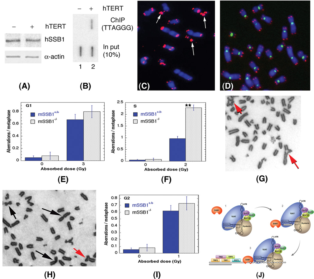
Cells called ALT (alternative lengthening of telomeres) lack detectable expression of endogenous hTERT but exhibit a telomerase-independent mechanism of telomere maintenance. The ALT cells lacking hTERT expressed hSSB1 levels similar to that of isogenic ALT cells ectopically expressing hTERT (Fig. 7A), however, hSSB1 association with telomeres was only detected in hTERT-expressing ALT cells (Fig. 7B). Telomere fluorescent in situ hybridization (FISH) analysis indicated that ALT cells presented with variable telomere signals (Fig. 7C), which was unaffected by hSSB1 over-expression. In contrast, ectopic expression of hTERT resulted in gradual stabilization of the telomere size (Fig. 7D), emphasizing the need for both hSSB1 and hTERT for normal telomere maintenance.
Fig. 7. Effect of hSSB1 loss on genomic stability.
(A) Protein levels hSSB1 in ALT cells with and without expression of hTERT. (B) Interaction of hSSB1 with telomeres as determined by ChIP. Lane 1: ALT cells without expression of hTERT; Lane 2: with expression of hTERT. (C, D) Telomeres detected by telomere specific FISH. (C) Metaphase of ALT cell showing large variation in telomere signals and (D) expression of hTERT and regaining telomerase activity show uniformity of the telomere signals. (E–I) Chromosomal aberrations in mSsb1+/+ and mSsb1−/− cells after IR exposure. For analysis of G1-phase specific chromosome aberrations, cells were irradiated (3 Gy), incubated for 12 h, and then treated for 3 h with colcemid, followed by hypotonic treatment and fixation for scoring metaphase chromosome aberrations. Categories of asymmetric chromosome aberrations scored included Robertsonian fusions, dicentrics, centric rings, interstitial deletions-acentric rings, and terminal deletions (E). For S-phase-specific chromosome aberrations, cells were irradiated with 2 Gy, incubated for 6 h, and metaphases were harvested after 3 h of colcemid treatment (F–H). S-phase specific aberration observed in the first round of metaphases included tri- quadri-radials, breaks and gaps. When cells irradiated in S-phases were allowed to go for two or more cell divisions, the frequency of aberrations including Robertsonian fusion increased only in mSsb1−/− MEFs. The difference in chromosomal aberrations in S-phase between cells with and without SSB1 are statistically significantly (P < 0.01 as determined by the chi-square test). For G2-type chromosome aberrations, exponential-phase cells were irradiated with 1 Gy, incubated for 1 h, followed by 3 h of colcemid treatment and subsequent hypotonic treatment fixation to analyze chromosome aberrations at metaphases (H, I). Model depicting the interaction of SSB1 with TERT and their recruitment to the G-strand of single stranded telomeric DNA (J).
Loss of SSB1 increases Robertsonian fusions
Our results demonstrate a role for hSSB1 in the recruitment of TERT to telomeres and the extension of telomeric G-overhangs. To elucidate the role for hSSB1 in regulating telomerase activity, we performed an in vitro TRAP assay, using extracts from cells with and without SSB1 depletion of SSB1. Depletion of SSB1 had no apparent effect on in vitro telomerase activity, implying that the SSB1/TERT interaction functions mainly in telomerase recruitment to telomeres rather than regulation of telomerase activity (Supplementary Fig. 5).
Mouse cells deficient for SSB1 have been shown to exhibit higher frequencies of chromatid fusions due to nucleolytic degradation of G-overhangs (3). An unanswered question is whether SSB1 depletion-induced loss of G-overhangs also affects telomere fusions and loss of telomere signals. FISH analysis with a telomere specific probe (Supplementary Fig. 2) of mouse MEF cells containing a tamoxifen/Cre inducible mSsb1 gene deletion, detected frequent loss of telomere signals in SSB1-depleted cells and a gradual increase in the frequency of Robertsonian fusions with increasing drug treatment time. No metaphase cells with more than 5–7 Robertsonian fusions were observed, suggesting an upper limit to the number of fusions that a cell may have before its ability to enter metaphase is compromised.
SSB1 depletion leads to defective DNA damage response in S-phase
Both hSSB1 and mSsb1 have been implicated in the DNA damage response. Thus, depletion of mSsb1 in MEFs may cause spontaneous genomic instability (3, 17, 36). Measurements of the spontaneous chromosome aberration frequency in metaphase cells, with or without depletion of mSsb1, showed a higher frequency of chromosome aberrations in mSSB1-depleted MEFs (Supplementary Table. 2). Robertsonian translocations were one type aberration frequently observed, indicating the same genomic instability mechanism is triggered in both mouse and human cells by SSB1 deficiency (Supplementary Fig. 2).
To examine whether defects in repair induces chromosome aberrations at specific phases of the cell cycle, we analyzed chromosome- or chromatid-type aberrations at metaphase after exposure to ionizing radiation. We previously reported that G1-specific aberrations detected at metaphase are mostly of the chromosome type (12, 13), whereas S-phase-type aberrations detected at metaphase are chromosomal- and chromatid-type (12, 13). G2-type aberrations detected at metaphase are predominantly of the chromatid type (12).
To measure G1-specific chromosome damage caused by loss of Ssb1, we exposed the exponentially growing mouse MEF cells to 3 Gy of IR and scored aberrations at metaphase, as described previously (12, 13). The loss of mSsb1 did not appear to affect the frequency of chromosome aberrations in G1 cells (Fig. 7E). In S-phase cells, mSsb1 depletion induced higher frequencies of chromatid and chromosomal aberrations (Fig. 7F). In addition, irradiated S-phase and mSsb1-depleted cells exhibited higher frequencies of tri-and quadri-radials. We observed Robertsonian fusions and fragments once these cells had undergone one cell division, implicating a critical role for SSB1 in S-phase DNA repair (Fig. 7G & 7H). Upon evaluating G2-phase-specific chromosome repair, we concluded that the loss of mSsb1 had no effect on the frequency of chromosome aberrations (Fig. 7I), reinforcing the idea that mSsb1 participates in chromosome repair/or protection primarily in S phase.
Discussion
The OB-fold domain, a hallmark of single-stranded DNA binding proteins, facilitates the binding of these proteins to single-stranded DNA which is present at telomeric ends, as well as during DNA replication and DNA double-strand break repair (37, 38). Studies of the shelterin component TPP1 indicate that OB-fold proteins can also regulate recruitment of telomerase to telomeres (39), a process that is poorly understood. Previous studies have implicated a role for the OB-fold domain containing SSB1 and SSB2 proteins in the protection of newly replicated telomeres (3). In this series of investigations, we sought to elucidate the mechanisms through which SSB1 facilitates telomerase recruitment to telomeres. We demonstrated that SSB1 containing the OB-fold domain interacts with the G-overhangs of telomeres, helping in maintaining G-overhangs and telomere stability.
The shelterin complex includes the proteins TRF1, TRF2, TIN2, Rap1, TPP1, and POT1, but not SSB1, which is consistent with our results that show SSB1 is found at telomeres only during S-phase. Thus, the role of SSB1 in end-protection coincides with the role of telomerase as previously proposed (40). We also found that depletion of POT1 reduces hTERT as well as SSB1 recruitment, consistent with the model that removal or depletion of POT1 leads to telomere instability. In addition, TPP1 depletion resulted in the loss of hTERT recruitment to telomeres. Furthermore, we found that loss of TPP1 decreased association of SSB1 at the telomeres. We found telomere-associated SSB1 mostly in S-phase, underscoring the hypothesis that SSB1 plays a role in maintaining the function of telomerase at the telomeres. Our results support a model in which TPP1 is required for hTERT interaction with telomeres (1, 39), and SSB1 plays a critical role in aiding telomerase to maintain G-overhangs.
During homologous recombination-mediated DNA DSB repair, the MRN complex, in cooperation with Sae2/CtIP, resects the DNA DSB into single-stranded DNA that is then bound by RPA to facilitate ATR recruitment and the phosphorylation of downstream factors controlling DSB repair and cell cycle progression (41–44). In addition to RPA, SSB1 appears to also be involved in HR-mediated repair (36), that is consistent with our own observations. These results support the role of SSB1 in chromosome damage repair during S-phase. Although HR-mediated DNA DSB repair is also prevalent in the G2-phase of the cell cycle, cells deficient in SSB1 did not show any G2-specific defect in chromosome repair, implicating a particular role for SSB1 in S-phase when DNA synthesis is being coordinated with the maintenance of telomeres.
Our results align with those of Chan and Blackburn who described the role of telomerase in chromosome end-protection (40) as maintaining telomere capping. Gu and coworkers have also reported that mSSB1 protects the chromosome ends (3). Most of the sheletrin components are present at the telomeres in all phases of the cell cycle; however, SSB1 is present at telomeres only in S-phase, presumably to have a protective role during the period of G-overhang synthesis.
We showed that SSB1 interacts with TERT, and SSB1 is required for TERT to associate with telomeres, suggesting that SSB1 has a role in recruiting TERT to telomeres but not to the DNA DSB sites. Together with the observation that SSB1 forms detectable repairosome foci post-irradiation while hTERT does not, we conclude that the recruitment of hTERT through SSB1 is dependent on the different chromatin structure formed at telomeres versus those found within interstitial DNA (23).
Although TERT does not concentrate at DNA DSB sites, it is well documented that TERT expression enhances DNA DSB repair and suppresses genomic instability, underscoring a strong argument for telomerase having at least an indirect role in DNA repair (31, 35, 45). Gene expression is altered by TERT expression and, given that DNA repair factors, transcription regulators and chromatin modifying factors coordinate their functions depending on chromatin status (i.e., structure of the DNA), this supports the idea that SSB1 similarly plays multiple roles in these processes.
The fact that SSB1 depletion increased chromosomal aberrations (3, 17) remains consistent with a role for SSB1 in S-phase. We demonstrated that SSB1 depletion results in cell cycle-specific aberrations that correlate with loss of G-overhang synthesis in S-phase, again favoring the argument that SSB1 is critical for telomerase activity at the telomeres and in maintaining genomic stability. These results support a novel function for SSB1 in telomerase recruitment to telomeres during telomere replication (Fig. 7J). As in yeast, the telomere-binding protein, Cdc13p, positively regulates telomerase recruitment through the Est1 component of telomerase (46–48). Similarly, SSB1 interacts with TERT in an interaction that facilitates telomerase recruitment to telomeres.
In this study, we clearly established that SSB1 forms a complex with TERT and facilitates its binding to and extension of telomeres (i.e., maintaining G-overhangs). Conversely, SSB1 needs TERT in order to associate with telomeres, supported by the observation that TERT-depleted cells have very few telomere-associated SSB1. The lack of SSB1 at the telomeres in ALT cells that have normal expression of SSB1 but lack TERT expression, further confirms the critical role of the SSB1/telomerase complex. Furthermore, the gradual decrease in the amount of native telomeric DNA and subsequent increase in the frequency of Robertsonian fusions in cells deficient for SSB1, supports the argument that SSB1 plays a role in the extension of the telomeric DNA. Based on in vitro assays, SSB1 shows preferential binding to G-rich telomeric DNA sequences, which is consistent with the role of SSB1 in TERT mediated telomere maintenance. The observations that SSB1 interacts with the G-rich strand of telomeres in vitro; is enriched at telomeric DNA during S-phase as determined by ChIP; interacts with TERT as determined by co-immunoprecipitation; that depletion results in the loss of G-overhangs as determined in vivo; that depletion causes increased S-phase specific chromosome damage and reduction results in the gradual increase in Robertsonian fusions, overall demonstrate that SSB1 has a role in recruiting telomerase at the telomeres to maintain G-overhang length and in the maintenance of telomeres by telomerase, thus avoiding chromosome instability.
Supplementary Material
Acknowledgements
We thank members of Pandita laboratory and Arun Gupta for technical help. We thank Hanh Hoang and D. Singh for critical reading of the manuscript. This work has been supported by NIH grants CA129537, CA154320 and GM109768.
Footnotes
Author contributions: R.K.P., K.K.K., W.E.W., J.W.S., and T.K.P. designed research; R.K.P., T.C.T., D.U., Y.Z., A.B., L.C., C.R.H., W.S., N.H., Y.Z., performed experiments, and R.K.P., K.K.K., C.R.H. and T.K.P. wrote paper.
Competing financial interests:
The authors declare no competing financial interests and have no conflict of interest.
Uncategorized References
- 1.Nandakumar J, Bell CF, Weidenfeld I, Zaug AJ, Leinwand LA, Cech TR. The TEL patch of telomere protein TPP1 mediates telomerase recruitment and processivity. Nature. 2012;492(7428):285–289. doi: 10.1038/nature11648. Epub 2012/10/30. PubMed PMID: 23103865; PubMed Central PMCID: PMC3521872. [DOI] [PMC free article] [PubMed] [Google Scholar]
- 2.Baumann P, Cech TR. Pot1, the putative telomere end-binding protein in fission yeast and humans. Science. 2001;292(5519):1171–1175. doi: 10.1126/science.1060036. Epub 2001/05/12. PubMed PMID: 11349150. [DOI] [PubMed] [Google Scholar]
- 3.Gu P, Deng W, Lei M, Chang S. Single strand DNA binding proteins 1 and 2 protect newly replicated telomeres. Cell Res. 2013;23(5):705–719. doi: 10.1038/cr.2013.31. Epub 2013/03/06. PubMed PMID: 23459151; PubMed Central PMCID: PMC3641597. [DOI] [PMC free article] [PubMed] [Google Scholar]
- 4.Skaar JR, Richard DJ, Saraf A, Toschi A, Bolderson E, Florens L, et al. INTS3 controls the hSSB1-mediated DNA damage response. J Cell Biol. 2009;187(1):25–32. doi: 10.1083/jcb.200907026. Epub 2009/09/30. PubMed PMID: 19786574; PubMed Central PMCID: PMC2762097. [DOI] [PMC free article] [PubMed] [Google Scholar]
- 5.Pandita RK, Sharma GG, Laszlo A, Hopkins KM, Davey S, Chakhparonian M, et al. Mammalian Rad9 plays a role in telomere stability, S- and G2-phase-specific cell survival, and homologous recombinational repair. Mol Cell Biol. 2006;26(5):1850–1864. doi: 10.1128/MCB.26.5.1850-1864.2006. Epub 2006/02/16. PubMed PMID: 16479004. [DOI] [PMC free article] [PubMed] [Google Scholar]
- 6.Shi W, Bain AL, Schwer B, Al-Ejeh F, Smith C, Wong L, et al. Essential developmental, genomic stability, and tumour suppressor functions of the mouse orthologue of hSSB1/NABP2. PLoS genetics. 2013;9(2):e1003298. doi: 10.1371/journal.pgen.1003298. Epub 2013/02/15. PubMed PMID: 23408915; PubMed Central PMCID: PMC3567186. [DOI] [PMC free article] [PubMed] [Google Scholar]
- 7.Pandita TK, Hittelman WN. The contribution of DNA and chromosome repair deficiencies to the radiosensitivity of ataxia-telangiectasia. Radiat Res. 1992;131(2):214–223. Epub 1992/08/01. PubMed PMID: 1641475. [PubMed] [Google Scholar]
- 8.Pandita TK. Role of mammalian Rad9 in genomic stability and ionizing radiation response. Cell Cycle. 2006;5(12):1289–1291. doi: 10.4161/cc.5.12.2862. Epub 2006/06/09. PubMed PMID: 16760671. [DOI] [PubMed] [Google Scholar]
- 9.Hunt CR, Pandita RK, Laszlo A, Higashikubo R, Agarwal M, Kitamura T, et al. Hyperthermia activates a subset of ataxia-telangiectasia mutated effectors independent of DNA strand breaks and heat shock protein 70 status. Cancer Res. 2007;67(7):3010–3017. doi: 10.1158/0008-5472.CAN-06-4328. Epub 2007/04/06. PubMed PMID: 17409407. [DOI] [PubMed] [Google Scholar]
- 10.Sharma GG, So S, Gupta A, Kumar R, Cayrou C, Avvakumov N, et al. MOF and histone H4 acetylation at lysine 16 are critical for DNA damage response and double-strand break repair. Mol Cell Biol. 2010;30(14):3582–3595. doi: 10.1128/MCB.01476-09. Epub 2010/05/19. PubMed PMID: 20479123; PubMed Central PMCID: PMC2897562. [DOI] [PMC free article] [PubMed] [Google Scholar]
- 11.Kumar R, Hunt CR, Gupta A, Nannepaga S, Pandita RK, Shay JW, et al. Purkinje cell-specific males absent on the first (mMof) gene deletion results in an ataxia-telangiectasia-like neurological phenotype and backward walking in mice. Proc Natl Acad Sci U S A. 2011;108(9):3636–3641. doi: 10.1073/pnas.1016524108. Epub 2011/02/16. PubMed PMID: 21321203. [DOI] [PMC free article] [PubMed] [Google Scholar]
- 12.Singh M, Hunt CR, Pandita RK, Kumar R, Yang CR, Horikoshi N, et al. Lamin a/c depletion enhances DNA damage-induced stalled replication fork arrest. Molecular and cellular biology. 2013;33(6):1210–2122. doi: 10.1128/MCB.01676-12. Epub 2013/01/16. PubMed PMID: 23319047; PubMed Central PMCID: PMC3592031. [DOI] [PMC free article] [PubMed] [Google Scholar]
- 13.Dhar S, Squire JA, Hande MP, Wellinger RJ, Pandita TK. Inactivation of 14-3-3sigma influences telomere behavior and ionizing radiation-induced chromosomal instability. Mol Cell Biol. 2000;20(20):7764–7772. doi: 10.1128/mcb.20.20.7764-7772.2000. Epub 2000/09/26. PubMed PMID: 11003671. [DOI] [PMC free article] [PubMed] [Google Scholar]
- 14.Chow TT, Zhao Y, Mak SS, Shay JW, Wright WE. Early and late steps in telomere overhang processing in normal human cells: the position of the final RNA primer drives telomere shortening. Genes & development. 2012;26(11):1167–1178. doi: 10.1101/gad.187211.112. Epub 2012/06/05. PubMed PMID: 22661228; PubMed Central PMCID: PMC3371406. [DOI] [PMC free article] [PubMed] [Google Scholar]
- 15.Pandita TK, Hall EJ, Hei TK, Piatyszek MA, Wright WE, Piao CQ, et al. Chromosome end-to-end associations and telomerase activity during cancer progression in human cells after treatment with alpha-particles simulating radon progeny. Oncogene. 1996;13(7):1423–1430. Epub 1996/10/03. PubMed PMID: 8875980. [PubMed] [Google Scholar]
- 16.Pandita TK, Pathak S, Geard CR. Chromosome end associations, telomeres and telomerase activity in ataxia telangiectasia cells. Cytogenet Cell Genet. 1995;71(1):86–93. doi: 10.1159/000134069. Epub 1995/01/01. PubMed PMID: 7606935. [DOI] [PubMed] [Google Scholar]
- 17.Richard DJ, Bolderson E, Cubeddu L, Wadsworth RI, Savage K, Sharma GG, et al. Single-stranded DNA-binding protein hSSB1 is critical for genomic stability. Nature. 2008;453(7195):677–681. doi: 10.1038/nature06883. Epub 2008/05/02. PubMed PMID: 18449195. [DOI] [PubMed] [Google Scholar]
- 18.Gupta A, Hunt CR, Pandita RK, Pae J, Komal K, Singh M, et al. T-cell-specific deletion of Mof blocks their differentiation and results in genomic instability in mice. Mutagenesis. 2013;28(3):263–270. doi: 10.1093/mutage/ges080. Epub 2013/02/07. PubMed PMID: 23386701; PubMed Central PMCID: PMC3630520. [DOI] [PMC free article] [PubMed] [Google Scholar]
- 19.Rodrigue A, Lafrance M, Gauthier MC, McDonald D, Hendzel M, West SC, et al. Interplay between human DNA repair proteins at a unique double-strand break in vivo. EMBO J. 2006;25(1):222–231. doi: 10.1038/sj.emboj.7600914. Epub 2006/01/06. PubMed PMID: 16395335. [DOI] [PMC free article] [PubMed] [Google Scholar]
- 20.Gupta A, Hunt CR, Hegde ML, Chakraborty S, Udayakumar D, Horikoshi N, et al. MOF Phosphorylation by ATM Regulates 53BP1-Mediated Double-Strand Break Repair Pathway Choice. Cell reports. 2014;8(1):177–189. doi: 10.1016/j.celrep.2014.05.044. Epub 2014/06/24. PubMed PMID: 24953651. [DOI] [PMC free article] [PubMed] [Google Scholar]
- 21.Pandita TK, Dhar S. Influence of ATM function on interactions between telomeres and nuclear matrix. Radiat Res. 2000;154(2):133–139. doi: 10.1667/0033-7587(2000)154[0133:ioafoi]2.0.co;2. Epub 2000/08/10. PubMed PMID: 10931683. [DOI] [PubMed] [Google Scholar]
- 22.Kumar R, Horikoshi N, Singh M, Gupta A, Misra HS, Albuquerque K, et al. Chromatin modifications and the DNA damage response to ionizing radiation. Front Oncol. 2012;2:214. doi: 10.3389/fonc.2012.00214. Epub 2013/01/25. PubMed PMID: 23346550; PubMed Central PMCID: PMC3551241. [DOI] [PMC free article] [PubMed] [Google Scholar]
- 23.Smilenov LB, Dhar S, Pandita TK. Altered telomere nuclear matrix interactions and nucleosomal periodicity in ataxia telangiectasia cells before and after ionizing radiation treatment. Mol Cell Biol. 1999;19(10):6963–6971. doi: 10.1128/mcb.19.10.6963. Epub 1999/09/22. PubMed PMID: 10490633. [DOI] [PMC free article] [PubMed] [Google Scholar]
- 24.Li Y, Bolderson E, Kumar R, Muniandy PA, Xue Y, Richard DJ, et al. HSSB1 and hSSB2 form similar multiprotein complexes that participate in DNA damage response. J Biol Chem. 2009;284(35):23525–23531. doi: 10.1074/jbc.C109.039586. Epub 2009/07/17. PubMed PMID: 19605351. [DOI] [PMC free article] [PubMed] [Google Scholar]
- 25.Verdun RE, Karlseder J. The DNA damage machinery and homologous recombination pathway act consecutively to protect human telomeres. Cell. 2006;127(4):709–720. doi: 10.1016/j.cell.2006.09.034. Epub 2006/11/18. PubMed PMID: 17110331. [DOI] [PubMed] [Google Scholar]
- 26.Kozlov AG, Lohman TM. Calorimetric studies of E. coli SSB protein-single-stranded DNA interactions Effects of monovalent salts on binding enthalpy. J Mol Biol. 1998;278(5):999–1014. doi: 10.1006/jmbi.1998.1738. Epub 1998/06/20. PubMed PMID: 9600857. [DOI] [PubMed] [Google Scholar]
- 27.Bochkarev A, Pfuetzner RA, Edwards AM, Frappier L. Structure of the single-stranded-DNA-binding domain of replication protein A bound to DNA. Nature. 1997;385(6612):176–181. doi: 10.1038/385176a0. Epub 1997/01/09. PubMed PMID: 8990123. [DOI] [PubMed] [Google Scholar]
- 28.Zhao Y, Hoshiyama H, Shay JW, Wright WE. Quantitative telomeric overhang determination using a double-strand specific nuclease. Nucleic acids research. 2008;36(3):e14. doi: 10.1093/nar/gkm1063. Epub 2007/12/13. PubMed PMID: 18073199; PubMed Central PMCID: PMC2241892. [DOI] [PMC free article] [PubMed] [Google Scholar]
- 29.Zhao Y, Shay JW, Wright WE. Telomere G-overhang length measurement method 1: the DSN method. Methods Mol Biol. 2011;735:47–54. doi: 10.1007/978-1-61779-092-8_5. Epub 2011/04/05. PubMed PMID: 21461810; PubMed Central PMCID: PMC3528100. [DOI] [PMC free article] [PubMed] [Google Scholar]
- 30.Smilenov LB, Morgan SE, Mellado W, Sawant SG, Kastan MB, Pandita TK. Influence of ATM function on telomere metabolism. Oncogene. 1997;15(22):2659–2665. doi: 10.1038/sj.onc.1201449. Epub 1997/12/24PubMed PMID: 9400992. [DOI] [PubMed] [Google Scholar]
- 31.Sharma GG, Gupta A, Wang H, Scherthan H, Dhar S, Gandhi V, et al. hTERT associates with human telomeres and enhances genomic stability and DNA repair. Oncogene. 2003;22(1):131–146. doi: 10.1038/sj.onc.1206063. Epub 2003/01/16PubMed PMID: 12527915. [DOI] [PubMed] [Google Scholar]
- 32.Wang F, Podell ER, Zaug AJ, Yang Y, Baciu P, Cech TR, et al. The POT1-TPP1 telomere complex is a telomerase processivity factor. Nature. 2007;445(7127):506–510. doi: 10.1038/nature05454. Epub 2007/01/24 PubMed PMID: 17237768. [DOI] [PubMed] [Google Scholar]
- 33.Loayza D, Parsons H, Donigian J, Hoke K, de Lange T. DNA binding features of human POT1: a nonamer 5’-TAGGGTTAG-3’ minimal binding site, sequence specificity, and internal binding to multimeric sites. J Biol Chem. 2004;279(13):13241–13248. doi: 10.1074/jbc.M312309200. Epub 2004/01/13 PubMed PMID: 14715659. [DOI] [PubMed] [Google Scholar]
- 34.Zou Y, Gryaznov SM, Shay JW, Wright WE, Cornforth MN. Asynchronous replication timing of telomeres at opposite arms of mammalian chromosomes. Proc Natl Acad Sci U S A. 2004;101(35):12928–1233. doi: 10.1073/pnas.0404106101. Epub 2004/08/24. PubMed PMID: 15322275; PubMed Central PMCID: PMC516496. [DOI] [PMC free article] [PubMed] [Google Scholar]
- 35.Wood LD, Halvorsen TL, Dhar S, Baur JA, Pandita RK, Wright WE, et al. Characterization of ataxia telangiectasia fibroblasts with extended life-span through telomerase expression. Oncogene. 2001;20(3):278–88. doi: 10.1038/sj.onc.1204072. Epub 2001/04/21. PubMed PMID: 11313956. [DOI] [PubMed] [Google Scholar]
- 36.Li Y, Bolderson E, Kumar R, Muniandy PA, Xue Y, Richard DJ, et al. HSSB1 and hSSB2 form similar multiprotein complexes that participate in DNA damage response. J Biol Chem. 2009;284(35):23525–23531. doi: 10.1074/jbc.C109.039586. Epub 2009/07/17. PubMed PMID: 19605351; PubMed Central PMCID: PMC2749126. [DOI] [PMC free article] [PubMed] [Google Scholar]
- 37.Richard DJ, Bolderson E, Khanna KK. Multiple human single-stranded DNA binding proteins function in genome maintenance: structural, biochemical and functional analysis. Crit Rev Biochem Mol Biol. 2009;44(2–3):98–116. doi: 10.1080/10409230902849180. Epub 2009/04/16. PubMed PMID: 19367476. [DOI] [PubMed] [Google Scholar]
- 38.Flynn RL, Zou L. Oligonucleotide/oligosaccharide-binding fold proteins: a growing family of genome guardians. Crit Rev Biochem Mol Biol. 2010;45(4):266–275. doi: 10.3109/10409238.2010.488216. Epub 2010/06/03. PubMed PMID: 20515430; PubMed Central PMCID: PMC2906097. [DOI] [PMC free article] [PubMed] [Google Scholar]
- 39.Zhong FL, Batista LF, Freund A, Pech MF, Venteicher AS, Artandi SE. TPP1 OB-fold domain controls telomere maintenance by recruiting telomerase to chromosome ends. Cell. 2012;150(3):481–494. doi: 10.1016/j.cell.2012.07.012. Epub 2012/08/07. PubMed PMID: 22863003; PubMed Central PMCID: PMC3516183. [DOI] [PMC free article] [PubMed] [Google Scholar]
- 40.Chan SW, Blackburn EH. Telomerase and ATM/Tel1p protect telomeres from nonhomologous end joining. Mol Cell. 2003;11(5):1379–1387. doi: 10.1016/s1097-2765(03)00174-6. Epub 2003/05/29. PubMed PMID: 12769860. [DOI] [PubMed] [Google Scholar]
- 41.Bartek J, Lukas J. DNA repair: Damage alert. Nature. 2003;421(6922):486–488. doi: 10.1038/421486a. Epub 2003/01/31. PubMed PMID: 12556872. [DOI] [PubMed] [Google Scholar]
- 42.Limbo O, Chahwan C, Yamada Y, de Bruin RA, Wittenberg C, Russell P. Ctp1 is a cell-cycle-regulated protein that functions with Mre11 complex to control double-strand break repair by homologous recombination. Molecular cell. 2007;28(1):134–146. doi: 10.1016/j.molcel.2007.09.009. Epub 2007/10/16 PubMed PMID: 17936710; PubMed Central PMCID: PMC2066204. [DOI] [PMC free article] [PubMed] [Google Scholar]
- 43.Sartori AA, Lukas C, Coates J, Mistrik M, Fu S, Bartek J, et al. Human CtIP promotes DNA end resection. Nature. 2007;450(7169):509–514. doi: 10.1038/nature06337. Epub 2007/10/30. PubMed PMID: 17965729; PubMed Central PMCID: PMC2409435. [DOI] [PMC free article] [PubMed] [Google Scholar]
- 44.Zou L, Elledge SJ. Sensing DNA damage through ATRIP recognition of RPA-ssDNA complexes. Science. 2003;300(5625):1542–15428. doi: 10.1126/science.1083430. Epub 2003/06/07. PubMed PMID: 12791985. [DOI] [PubMed] [Google Scholar]
- 45.Wong KK, Chang S, Weiler SR, Ganesan S, Chaudhuri J, Zhu C, et al. Telomere dysfunction impairs DNA repair and enhances sensitivity to ionizing radiation. Nat Genet. 2000;26(1):85–88. doi: 10.1038/79232. Epub 2000/09/06. PubMed PMID: 10973255. [DOI] [PubMed] [Google Scholar]
- 46.Chan A, Boule JB, Zakian VA. Two pathways recruit telomerase to Saccharomyces cerevisiae telomeres. PLoS genetics. 2008;4(10):e1000236. doi: 10.1371/journal.pgen.1000236. Epub 2008/10/25 PubMed PMID: 18949040; PubMed Central PMCID: PMC2567097. [DOI] [PMC free article] [PubMed] [Google Scholar]
- 47.Pennock E, Buckley K, Lundblad V. Cdc13 delivers separate complexes to the telomere for end protection and replication. Cell. 2001;104(3):387–396. doi: 10.1016/s0092-8674(01)00226-4. Epub 2001/03/10. PubMed PMID: 11239396. [DOI] [PubMed] [Google Scholar]
- 48.Taggart AK, Teng SC, Zakian VA. Est1p as a cell cycle-regulated activator of telomere-bound telomerase. Science. 2002;297(5583):1023–1026. doi: 10.1126/science.1074968. Epub 2002/08/10 PubMed PMID: 12169735. [DOI] [PubMed] [Google Scholar]
Associated Data
This section collects any data citations, data availability statements, or supplementary materials included in this article.


