Key Clinical Message
Perineal myomas in female are exceptional. We report the second case in literature of perineal myomas. It is a case of bilateral perineal myomas lifting the skin occurring in a female patient of 49 years old. She was operated by perineal incisions. Histopathology confirmed the fibromatous nature without signs of malignancy.
Keywords: Female, myoma, perineum, soft-tissue masses, tumor
Introduction
Perineal tumors in female are infrequent. Cases of perineal leiomyosarcoma were reported in literature. The eventuality of perineal myoma is exceptional, less than 10 cases of unilateral myomas were reported in the literature. We report the first case in the literature of bilateral perineal myoma in female with successful exeresis.
Case Report
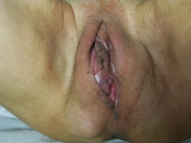
We report the case of 49-year-old unmarried patient complaining of perineal masses evolving for 10 years with no menstrual disorders. She also reported constipation. The rate of growth of theses masses was slow. Clinical examination shows two perineal masses lifting the skin of firm consistency on both sides of the vulva and near the anus. Theses masses had a diameter of 8 and 9 cm, respectively (Fig.1). At this stage, diagnostic difficulties were: the presumption of benignity or malignancy, and contiguity relations to the anus and vascular structures. Pelvic ultrasonography did not show any abnormality in uterus or adnexa. An exploration by MRI was performed and showed a non invasive muscular resonance process below the levator ani muscle. These perineal formations were presumed benign for: the slow clinical evolution and the absence of malignancy criteria on MRI.
Figure 1.

Bilateral perineal masses lifting the skin.
The patient was operated on the 24th February 2014: first, by laparoscopy: there was not any mass protruding into the pelvis. The exeresis was performed through bilateral incision in the crease of the thigh. Dissection was relatively easy (Figs.2 and 3). Two solid formations of 10 and 8 cm were removed with no evidence of locoregional invasion, including the anus. Some bleeding points in the cavities required ligature. After the resection the sphincter mechanism was noted to be intact. Histological examination confirmed the purely myomatous nature of these formations. The postoperative course was uneventful. The patient was seen a month later, she had a good perineal healing and no longer complains about constipation.
Figure 2.

Dissection of the right perineal myoma.
Figure 3.

Exeresis of the left perineal myoma.
Comment
Soft-tissue tumors of the perineum are rare and usually diagnosed in male patients. Liposarcoma and aggressive angiomyxoma are the most frequent histological subtypes 1. Few cases of perineal myomas were reported exclusively in male by London in 1953 and Galvan in 1972 2,3. Perineal leiomyomas are extremely rare with an incidence of 3.8% of all benign soft-tissue tumors 4.
Rare cases of unilateral perineal myoma in female patients were reported in the literature 4–8. This case report is historical: it's, in fact, the first case of bilateral perineal myoma in female reported in literature. The first case of perineal myoma was published by McCann in 1912 5. The patient was 54-year-old female with a large pendulous perineal tumor. The operation was carried out with two incisions: abdominal and perineal. After removing the myoma by supravaginal hysterectomy, a swelling was found occupying the left side of the pelvis anteriorly. As the enucleation proceeded the tumor was found to be continuous with the perineal growth and appeared to pass through a large foramen in the pelvic fascia. The tumor was gradually enucleated. He stated that the tumor had probably originated from the visceral layer of the pelvic fascia.
More recent publications suggest the diagnostic difficulties in managing perineal masses. For Von Waagner and al, a low-grade sarcoma should be considered as a differential diagnosis 6. For the same author, magnetic resonance imaging with intravenous contrast (MRI) is an excellent imaging tool for the characterization and diagnosis of perineal soft-tissue lesions. However, perineal leiomyomas can have variable and misleading MRI presentations 7.
Most perineal myomas reported in the literature were managed by perineal incisions. For Sistal and al an abdominoperineal surgical approach is recommended for such a lesion 8. We recommend performing a laparoscopy prior to perineal surgery.
In our case report, the patient had perineal formations evolving for 10 years. This case had presented to us diagnostic difficulties that have been partially resolved by imaging techniques (MRI). Diagnostic difficulties were the presumption of benignity or malignancy, and contiguity relations to the anus and vascular structures. In our opinion, achieving an MRI in perineal formations is necessary to predict the complete resectability of the tumor, rather than to presume of its nature.
Conflict of Interest
None declared.
References
- Behranwala KA, Clark MA. Thomas JM. Soft-tissue tumours of the perineum. Eur. J. Surg. Oncol. 2002;28:437–442. doi: 10.1053/ejso.2002.1267. [DOI] [PubMed] [Google Scholar]
- London MZ. Perineal myoma. Calif. Med. 1953;78:63–64. [PMC free article] [PubMed] [Google Scholar]
- Galvan ES. Giant fibroma of the perineum. Am. J. Proctol. 1972;23:68–71. [PubMed] [Google Scholar]
- Oliveira Brito LG, Falcão Motoki L, Magnani PS, Sabino-de-Freitas MM, Magnani Landell GA. Quintana SM. Giant perineal leiomyoma incidentally manifested at a recent episiotomy site: case report. J. Minim. Invasive Gynecol. 2011;18:267–269. doi: 10.1016/j.jmig.2010.12.010. [DOI] [PubMed] [Google Scholar]
- McCann FJ. Fibroma of the pelvic fascia forming a large perineal tumour. Proc. R. Soc. Med. 1912;5:38–41. doi: 10.1177/003591571200500903. [DOI] [PMC free article] [PubMed] [Google Scholar]
- Von-Waagner W, Liu H. Picon AI. Giant perineal leiomyoma: a case report and review of the literature. Case Rep. Surg. 2014;2014 doi: 10.1155/2014/629672. doi: 10.1155/2014/629672. [DOI] [PMC free article] [PubMed] [Google Scholar]
- Koc O, Sengul N. Gurel S. Perineal leiomyoma mimicking complex Bartholin mass. Int. Urogynecol. J. 2010;21:495–497. doi: 10.1007/s00192-009-0998-3. [DOI] [PubMed] [Google Scholar]
- Sistla SC, Reddy R, Sankar G. Elangovan S. Pelvic leiomyoma presenting as perineal hernia. Hernia. 2009;13:213–215. doi: 10.1007/s10029-008-0415-8. [DOI] [PubMed] [Google Scholar]


