

Abstract
This study investigated the mechanism underlying electroacupuncture therapy for vascular dementia through electroacupuncture at the acupoints of Baihui (DU20), Dazhui (DU14), and bilateral Shenshu (BL23) in a rat model of vascular dementia produced by bilateral middle cerebral artery occlusion. Morris water maze test showed that electroacupuncture improved the learning ability of vascular dementia rats. Western blot assay revealed that the expression of p70 ribosomal protein S6 kinase and ribosomal protein S6 in vascular dementia rats was significantly increased after electroacupuncture, compared with the model group that was not treated with acupuncture. The average escape latency was also shortened after electroacupuncture, and escape strategies in the spatial probe test improved from edge and random searches, to linear and trending swim pathways. The experimental findings indicate that electroacupuncture improves learning and memory ability by up-regulating expression of p70 ribosomal protein S6 kinase and ribosomal protein S6 in the hippocampus of vascular dementia rats.
Keywords: vascular dementia, electroacupuncture, hippocampus, p70 ribosomal protein S6 kinase, ribosomal protein S6, search strategy, neural regeneration
INTRODUCTION
Although definitive medical or surgical treatments for vascular dementia (VD) still remain limited[1], numerous clinical studies have revealed that electroacupuncture is an effective therapy for VD through the enhancement of cognitive capacity, improvement of dementia symptoms and electrical activity in the brain[2,3,4,5]. At the same time, strong evidence suggests that there are complex mechanisms underlying electroacupuncture, involving many signaling pathways. First, electroacupuncture rescues long-term potentiation impairment of VD rats, and N-methyl-D-aspartate receptor subtype 1 may be involved in this procedure[6,7]. Second, electroacupuncture exerts significant effects on VD model rat by antagonizing apoptosis signaling[8,9], which may be closely related with its role in attenuating the expression of P53, Noxa, bax, caspase-3[10,11], and by increasing the expression of bcl-2. Furthermore, electroacupuncture improves learning and memory of VD rats by limiting the formation of free radicals[12]. Together, these data suggest the mechanism associated with electroacupuncture therapy is through multiple pathways; however the molecular mechanisms remain to be studied.
p70 ribosomal protein S6 (p70S6) kinase/ribosomal protein S6 signaling pathway has been shown to promote neuronal growth and long-term potentiation[13,14,15,16,17,18]. We hypothesized that the p70S6 kinase/ribosomal protein S6 pathway may be related to VD. This study sought to analyze the expression of p70S6 kinase and ribosomal protein S6 in the hippocampus of VD rats by western blot assay, and further investigated the effects of electroacupuncture on the expression of p70S6 kinase and ribosomal protein S6 in VD rats.
RESULTS
Quantitative analysis of experimental animals
A total of 48 Sprague-Dawley rats were used in this study. They were randomly divided into three groups: sham-surgery (n = 10; only bilateral middle carotid artery was exposed), VD (n = 19; VD models were established by bilateral middle cerebral artery occlusion) and electroacupuncture (n = 19; VD models established were treated with electroacupuncture at the acupoints of Baihui (DU20), Dazhui (DU14), and bilateral Shenshu (BL23). During the operations, two rats in the sham-surgery group died within 2 days, nine rats in the VD group and seven rats in the electroacupuncture group died within 7 days. Finally, eight rats in the sham-surgery group, ten rats in the VD model group and twelve rats in the electroacupuncture group were included in the final analysis.
Learning and memory abilities of VD rats by electroacupuncture
The navigation test in the Morris water maze test was used to evaluate learning and memory ability of the rats. Results showed that the average escape latency in the VD model group was significantly longer than that in the sham-surgery group and electroacupuncture group (23.99 ± 15.82 s vs. 6.93 ± 2.34 s and 23.99 ± 15.82 s vs. 13.44 ± 9.64 s). The chi-square test showed that the data of escape strategies in the spatial probe test had significant differences among the three groups (P < 0.05), and partition of the chi-square test demonstrated significant differences of escape strategies between the VD model group and the electroacupuncture group (P < 0.05). Escape strategies of the VD rats improved from edge and random searches, to linear and trend swim pathways, after the electroacupuncture (Figure 1).
Figure 1.
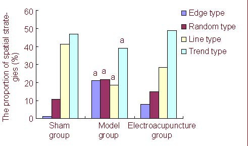
Search strategies of each group of rats in the spatial probe test (Morris water maze test). aP < 0.05, vs. electroacupuncture group. Partition of chi-square tests was used to analyze the difference between groups. The proportion of spatial strategies shows that electroacupuncture improved spatial strategies of vascular dementia rats. Data are expressed as mean. There were eight rats in the sham group, 10 in the model group, and 12 in the electroacupuncture group. Sham: sham-surgery.
Expression of p70S6 kinase protein and ribosomal protein S6 in VD rats by electroacupuncture
Western blot assay was used to detect the expression of p70S6 kinase protein and ribosomal protein S6 in the rat hippocampus (Figures 2, 3). Compared with the model group, the expression of p70S6 kinase protein and ribosomal protein S6 was significantly increased in the hippocampus of VD rats in the electroacupuncture group (P < 0.05).
Figure 2.
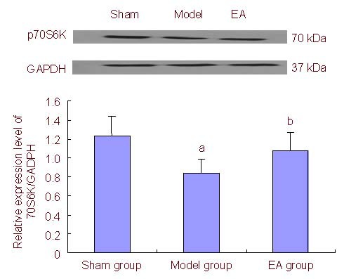
p70 ribosomal protein S6 kinase (p70S6K) expression in the hippocampus of vascular dementia rats by electroacupuncture (EA). Data are represented as the ratio of p70S6K/GAPDH. aP < 0.05, vs. sham group; bP < 0.05, vs. model group. Data are expressed as mean ± SD in six rats for each group (one-way analysis of variance and LSD-t). Sham: Sham-surgery; GAPDH: glyceraldehyde-3-phosphate dehydrogenase.
Figure 3.
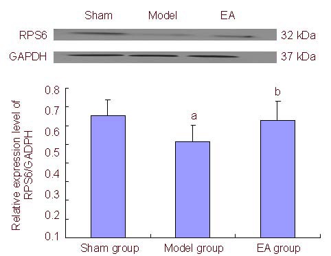
Ribosomal protein S6 (RPS6) expression in the hippocampus of vascular dementia rats by electroacupuncture (EA). Data are represented as the ratio of RPS6/GAPDH. aP < 0.05, vs. sham group; bP < 0.05, vs. model group. Data are expressed as mean ± SD in six rats for each group (one-way analysis of variance and LSD-t). Sham: Sham-surgery; GAPDH: glyceraldehyde-3-phosphate dehydrogenase.
DISCUSSION
p70S6 kinase is activated by kinases such as mammalian target of rapamycin and mitogen-activated protein kinase[19,20,21,22], which results in ribosomal protein S6 phosphorylation[23,24], that then promotes cell regeneration, protein synthesis and long-term potentiation[25,26]. Thus, dysregulation of p70S6 kinase and ribosomal protein S6 may be important in the pathogenesis of VD. The present study showed that the expression of p70S6 kinase and ribosomal protein S6 in the hippocampus of VD rats was less than that of normal rats. This is evidence that p70S6 kinase/ribosomal protein S6 pathway is involved in the pathogenesis of VD.
The theory of traditional Chinese medicine believes that deficiency of marrow-reservoir underlies the pathogenesis of VD, and the BL 23 point controls the spinal marrow[27]. BL23 is the back-shu-acupoint of the kidney, and DU 20 and DU14 belong to the Du meridian which is closely associated with the regulation of the brain's function, so we electroacupunctured at BL23, DU20, and DU14 to invigorate the kidney, replenish the essence and improve the brain's function. The present study demonstrated that electroacupuncture improved learning and spatial memory of VD rats. To further explore its mechanism, this study confirmed that electroacupuncture was able to increase the expression of p70S6 kinase and ribosomal protein S6 in the hippocampus of VD rats, which might protect neurons and facilitate long-term potentiation in the hippocampus.
In conclusion, data from this study demonstrated that the decreasing expression of p70S6 kinase and ribosomal protein S6 plays important roles in the pathogenesis of VD. Electroacupuncture improved the learning and spatial memory abilities of VD rats by up-regulating expression of p70S6 kinase and ribosomal protein S6.
MATERIALS AND METHODS
Design
A randomized, controlled animal study.
Time and setting
This study was performed at the Molecular Biology Laboratory of Fujian University of Traditional Chinese Medicine, China between January and July 2011.
Materials
A total of 48 healthy, female, Sprague-Dawley rats, aged 12 months, of specific pathogen-free grade, weighing 400 ± 30 g, were provided by the Research Center of Laboratory Animals, Academy of Military Medical Sciences, China (certificate No. SCXK (Jun) 2007-004). The experimental procedures were performed in accordance with the Guidance Suggestions for Care and Use of Laboratory Animals, issued by the Ministry of Science and Technology of China[28].
Methods
Establishment of VD model rats
VD models were established in rats through bilateral occlusion of common carotid arteries[29]. In brief, under anesthesia (3 mg/kg chloral hydrate), rats were fixed in a supine position, and a midline incision of the cervical skin was made to expose the bilateral carotid arteries, which were ligated with double silk suture, then the rats were fed and regularly observed.
Electroacupuncture interventions
One month after surgery, the rats in the electroacupuncture group were electroacupunctured with an electronic acupuncture treatment instrument (SDZ-II, Suzhou, China). The rats were fixed in a fitting web. No. 30, 1.3-cm filiform needles were inserted obliquely 1.3 cm at the DU20 point of the model rats, perpendicularly 1.3 cm at the DU14 and 2.5-cm filiform needles were inserted obliquely 0.6 cm at both sides of BL23. DU20 is located at the center of the parietal bone of rats. DU14 is located between the seventh cervical vertebrae and the first thoracic vertebrae. BL23 is beside the second lumbar vertebra[8,30] (Figure 4).
Figure 4.
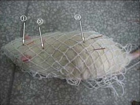
Schematic diagram of a vascular dementia rat electroacupunctured at Baihui (DU20, ①), Dazhui (DU14, ②) and Shenshu (BL23, ③).
The electronic acupuncture treatment instrument was connected to supply continuous waveform with a frequency of 4 Hz. The stimulation intensity was within rat resting tolerance (about 2.0 mA). This treatment was administered once a day for 30 days, and the needle was retained for 20 minutes during each treatment.
Behavioral test analyzed by Morris water maze test
Behavior was monitored using the Morris water maze test[29]. A circular tank (150 cm diameter, 50 cm deep) was filled 17 cm deep with water at 22–26°C. Four entering points were marked on the wall of the pool, and the pool was divided into four quadrants. The platform (12.5 cm diameter, 15 cm high) was placed in the third quadrant, and was submerged 2 cm below the water surface. The behavioral test began immediately after the treatment.
Learning and memory ability of rats analyzed by navigation test
The test lasted for 5 days with four trials per day. Each trial began by releasing the rat into the water at one of the four placement points with its face toward the pool wall. The time to find the platform within a 2-minute limit was recorded as the escape latency. If the rat failed to find the platform within 2 minutes, the experimenter moved the animal to the platform, where it remained for 10 seconds, and the escape latency was recorded as 2 minutes. The mean values of escape latency in four tests were calculated.
Spatial memory ability of rats analyzed by spatial probe test
The platform was removed from the pool, and rats were allowed to search for the platform for 120 seconds. Swim paths were recorded to determine search strategies, which included search along the edge (edge type), random swim path (random type), a trend towards a straight swim path (trend type) and a linear swim path (line type).
Expression of p70S6 kinase and ribosomal protein S6 in hippocampus of rats detected by western blot assay
Six rats per group (surplus rats were used for paraffin imbedding) were used for western blot analysis. Hippocampus tissues were homogenized and centrifuged, and protein level was determined by using a Micro BCA protein assay kit (Beyotime Institute of Biotechnology, Shanghai, China) with bovine serum albumin as the standard. A total of 100 μg protein preparations were used for the assay. Proteins were separated by sodium dodecyl sulfate-polyacrylamide gel electrophoresis in 8% gel, then transferred electrophoretically to polyvinylidene difluoride membrane (millipore, 0.45 mm). The membranes were blocked in phosphate buffered saline (PBS) containing 10% nonfat milk overnight at 4°C, then incubated with primary antibodies (1: 1 000) for 3 hours at room temperature. The following primary antibodies were used: rabbit anti-rat p70S6 kinase monoclonal antibody, rabbit anti-rat ribosomal protein S6 monoclonal antibody, and rabbit anti-rat glyceraldehyde-3-phosphate dehydrogenase monoclonal antibody (Cell Signal Biotechnology, Boston, MA, USA). The membranes were washed three times with 1% PBS Tween-20, then incubated with a peroxidase-conjugated goat anti-rabbit secondary antibody (1: 4 000; Santa Cruz Biotechnology, Santa Cruz, CA, USA) for 1 hour at room temperature. The membranes were washed three times with PBS Tween 20, and treated with enhanced chemiluminescence reagent (Santa Cruz Biotechnology), then exposed to X-ray film in a dark room. Results were scanned, and phoretiX 1D software (TotalLab Ltd.; Newcastle Upon Tyne, NE, UK) was used to calculate protein quantification. The result of p70S6 kinase and ribosomal protein S6 was corrected by GAPDH, and the relative expression level ratio of ribosomal protein S6/GAPDH and absorbance ratio of p70S6 kinase/GAPDH was respectively represented as the relative amount of ribosomal protein S6 and p70S6 kinase in rat hippocampus.
Statistical analysis
Data were statistically evaluated by the first author using SPSS 13.0 software (SPSS, Chicago, IL, USA). Data for escape latency and the results of the western blot analysis were analyzed with one-way analysis of variance. Post hoc test used the LSD-t test to identify the differences between means. Chi-square test and partition of chi-square tests were used for the spatial probe analysis. A P value less than 0.05 was considered to indicate statistical significance, and corrected P values less than 0.017 were used to indicate statistical significance in the partition of chi-square tests.
Acknowledgements:
We wish to thank Huasong Wu from the Animal Laboratory of Fujian Institute of Traditional Chinese Medicine, for raising the rats, and Zhihong Zheng from Department of Biochemistry, Fujian Medical University for instructing empirical methods.
Footnotes
Conflicts of interest: None declared.
Funding: This project was supported by the National Natural Science Foundation of China, No. 81001541.
Ethical approval: All procedures involved in animal experiments were in accordance with the Experimental Animals Administered Regulations of Fujian Province in China.
(Edited by Wei JZ, Song XG/Yang Y/Wang L)
REFERENCES
- [1].Erkinjuntti T, Román G, Gauthier S, et al. Emerging therapies for vascular dementia and vascular cognitive impairment. Stroke. 2004;35(4):1010–1017. doi: 10.1161/01.STR.0000120731.88236.33. [DOI] [PubMed] [Google Scholar]
- [2].Lai X, Mo F, Jiang G. Observation of clinical effect of acupuncture on vascular dementia and its influence on superoxide dismutase, lipid peroxide and nitric oxide. Zhongguo Zhongxiyi Jiehe Zazhi. 1998;18(11):648–651. [PubMed] [Google Scholar]
- [3].Zhao L, Zhang H, Zheng Z, et al. Electroacupuncture on the head points for improving gnosia in patients with vascular dementia. J Tradit Chin Med. 2009;29(1):29–34. doi: 10.1016/s0254-6272(09)60027-3. [DOI] [PubMed] [Google Scholar]
- [4].Zhang H, Zhao L, He CQ, et al. Clinically multi-central randomized controlled study on scalp electroacupuncture for treatment of vascular dementia. Zhongguo Zhen Jiu. 2008;28(11):783–787. [PubMed] [Google Scholar]
- [5].Han JS. Acupuncture analgesia: Areas of consensus and controversy. Pain. 2011;152(3S):S41–48. doi: 10.1016/j.pain.2011.03.021. [DOI] [PubMed] [Google Scholar]
- [6].Pan Y, Dai TL, Yang Q, et al. The effect of ear acupuncture and aniracetam to hippocampal dentate gyrus long-term potential in vascular dementia model rats. Linchuang Shenjing Dianshengli Xue Zazhi. 2009;18(3):131–134. [Google Scholar]
- [7].Lin YW, Hsieh CL. Electroacupuncture at Baihui acupoint (GV20) reverses behavior deficit and long-term potentiation through N-methyl-d-aspartate and transient receptor potential vanilloid subtype 1 receptors in middle cerebral artery occlusion rats. J Integr Neurosci. 2010;9(3):269–282. doi: 10.1142/s0219635210002433. [DOI] [PubMed] [Google Scholar]
- [8].Lai XS, Wang L. Effects of electroacupuncture on learning and memory abilities and the apoptosis of cerebral cells in rats with vascular dementia. Zhongguo Linchuang Kangfu. 2004;8(34):7861–7863. [Google Scholar]
- [9].Zhang HZ, Zhang LX, She YF, et al. Effect of electroacupuncture on apoptosis of hippocampus tissue in mice with vascular dementia. Zhen Ci Yan Jiu. 2008;33(6):377–381. [PubMed] [Google Scholar]
- [10].Zhu Y, Zeng Y. Electroacupuncture protected pyramidal cells in hippocampal CA1 region of vascular dementia rats by inhibiting the expression of P53 and Noxa. CNS Neurosci Ther. 2011;17(6):599–604. doi: 10.1111/j.1755-5949.2010.00192.x. [DOI] [PMC free article] [PubMed] [Google Scholar]
- [11].Han JS, Ho YS. Global trends and performances of acupuncture research. Neurosci Biobehav Rev. 2011;35(3):680–687. doi: 10.1016/j.neubiorev.2010.08.006. [DOI] [PubMed] [Google Scholar]
- [12].Wang L, Tang C, Lai X. Effects of electroacupuncture on learning, memory and formation system of free radicals in brain tissues of vascular dementia model rats. J Tradit Chin Med. 2004;24(2):140–143. [PubMed] [Google Scholar]
- [13].Steitz TA. A structural understanding of the dynamic ribosome machine. Nat Rev Mol Cell Biol. 2008;9(3):242–253. doi: 10.1038/nrm2352. [DOI] [PubMed] [Google Scholar]
- [14].Hutchinson JA, Shanware NP, Chang H, et al. Regulation of ribosomal protein S6 phosphorylation by casein kinase 1 and protein phosphatase 1. J Biol Chem. 2011;286(10):8688–8896. doi: 10.1074/jbc.M110.141754. [DOI] [PMC free article] [PubMed] [Google Scholar]
- [15].Raiker SJ, Lee H, Baldwin KT, et al. Oligodendrocyte-myelin glycoprotein and Nogo negatively regulate activity-dependent synaptic plasticity. J Neurosci. 2010;30(37):12432–12445. doi: 10.1523/JNEUROSCI.0895-10.2010. [DOI] [PMC free article] [PubMed] [Google Scholar]
- [16].Harada H, Andersen JS, Mann M, et al. P70S6 kinase signals cell survival as well as growth, inactivating the pro-apoptotic molecule BAD. Proc Natl Acad Sci U S A. 2001;98(17):9666–9670. doi: 10.1073/pnas.171301998. [DOI] [PMC free article] [PubMed] [Google Scholar]
- [17].Panja D, Dagyte G, Bidinosti M, et al. Novel translational control in Arc-dependent long term potentiation consolidation in vivo. J Biol Chem. 2009;284(46):31498–31511. doi: 10.1074/jbc.M109.056077. [DOI] [PMC free article] [PubMed] [Google Scholar]
- [18].Sui L, Wang J, Li BM. Role of the phosphoinositide 3-kinase-Akt-mammalian target of the rapamycin signaling pathway in long-term potentiation and trace fear conditioning memory in rat medial prefrontal cortex. Learn Mem. 2008;15(10):762–776. doi: 10.1101/lm.1067808. [DOI] [PubMed] [Google Scholar]
- [19].Sanghera KP, Mathalone N, Baigi R, et al. The PI3K / Akt / mTOR pathway mediates retinal progenitor cell survival under hypoxic and superoxide stress. Mol Cell Neurosci. 2011;47(2):145–153. doi: 10.1016/j.mcn.2011.03.010. [DOI] [PubMed] [Google Scholar]
- [20].Xu R, Nakand K, Iwasaka H, et al. Dual blockade of phosphatidylinositol 3’-kinase and mitogen-activated protein kinase pathways overcomes paclitaxel-resistance in colorectal cancer. Cancer Lett. 2011;306(2):151–160. doi: 10.1016/j.canlet.2011.02.042. [DOI] [PubMed] [Google Scholar]
- [21].Harhaji-Trajkovic L, Vilimanovich U, Kravic-Stevovic T, et al. AMPK-mediated autophagy inhibits apoptosis in cisplatin-treated tumor cells. J Cell Mol Med. 2009;13(9B):3644–3654. doi: 10.1111/j.1582-4934.2009.00663.x. [DOI] [PMC free article] [PubMed] [Google Scholar]
- [22].Pang X, Yi Z, Zhang J, et al. Celastrol suppresses angiogenesis-mediated tumor growth through inhibition of AKT/mammalian target of rapamycin pathway. Cancer Res. 2010;70(5):1951–1959. doi: 10.1158/0008-5472.CAN-09-3201. [DOI] [PMC free article] [PubMed] [Google Scholar]
- [23].Knaup KX, Jozefowski K, Schmidt R, et al. Mutual regulation of hypoxia-inducible factor and mammalian target of rapamycin as a function of oxygen availability. Mol Cancer Res. 2009;7(1):88–98. doi: 10.1158/1541-7786.MCR-08-0288. [DOI] [PubMed] [Google Scholar]
- [24].Leicht M, Simm A, Bertsch G, et al. Okadaic acid induces cellular hypertrophy in AKR-2B fibroblasts: involvement of the p70S6 kinase in the onset of protein and rRNA synthesis. Cell Growth Differ. 1996;7(9):1199–1209. [PubMed] [Google Scholar]
- [25].Guo R, Wang T, Shen H, et al. Involvement of mTOR and survivin inhibition in tamoxifen-induced apoptosis in human hepatoblastoma cell line HepG2. Biomed Pharmacother. 2010;64(4):249–253. doi: 10.1016/j.biopha.2009.06.007. [DOI] [PubMed] [Google Scholar]
- [26].Ma BB, Lui VW, Hui EP, et al. The activity of mTOR inhibitor RAD001 (everolimus) in nasopharyngeal carcinoma and cisplatin-resistant cell lines. Invest New Drugs. 2010;28(4):413–420. doi: 10.1007/s10637-009-9269-x. [DOI] [PubMed] [Google Scholar]
- [27].Tian JZ, Liu X. Explore the researching thoughts of senile dementia. Zhongguo Yiyao Xuebao. 2000;15(5):52–54. [Google Scholar]
- [28].The Ministry of Science and Technology of the People's Republic of China. Guidance Suggestions for the Care and Use of Laboratory Animals 2006-09-30 [Google Scholar]
- [29].Zhao XZ, Wang W, Kan ZH, et al. Structural changes in the hippocampal synapse of rats with vascular dementia. Zhongguo Linchuang Kangfu. 2003;7(31):4213–4215. [Google Scholar]
- [30].Ma RR, Li GQ, Wang JP, et al. Effect of electroacupuncture of different acupoints on GAP-43 expression after cerebral infarction in rats. Chongqing Yike Daxue Xuebao. 2011;36(1):38–41. [Google Scholar]