Abstract
Background
Femoroacetabular impingement is a recognized cause of chondrolabral injury. Although surgical treatment for impingement seeks to improve range of motion, there are very little normative data on dynamic impingement-free hip range of motion (ROM) in asymptomatic people. Hip ultrasound demonstrates labral anatomy and femoral morphology and, when used dynamically, can assist in measuring range of motion.
Questions/purposes
The purposes of this study were (1) to measure impingement-free hip ROM until labral deflection is observed; and (2) to measure the maximum degree of sagittal plane hip flexion when further flexion is limited by structural femoroacetabular abutment.
Methods
Forty asymptomatic adult male volunteers (80 hips) between the ages of 21 and 35 years underwent bilateral static and dynamic hip ultrasound examination. Femoral morphology was characterized and midsagittal flexion passive ROM was measured at two points: (1) at the initiation of labral deformation; and (2) at maximum flexion when the femur impinged on the acetabular rim. The mean age of the subjects was 28 ± 3 years and the mean body mass index was 25 ± 4 kg/m2.
Results
Mean impingement-free hip passive flexion measured from full extension to initial labral deflection was 68° ± 17° (95% confidence interval [CI], 65–72). Mean maximum midsagittal passive flexion, measured at the time of bony impingement, was 96° ± 6° (95% CI, 95–98).
Conclusions
Using dynamic ultrasound, we found that passive ROM in the asymptomatic hip was much less than the motion reported in previous studies. Measuring ROM using ultrasound is more accurate because it allows anatomic confirmation of terminal hip motion.
Clinical Significance
Surgical procedures used to treat femoroacetabular impingement are designed to restore or increase hip ROM and their results should be evaluated in light of precise normative data. This study suggests that normal passive impingement-free femoroacetabular flexion in the young adult male is approximately 95°.
Introduction
Femoroacetabular impingement (FAI) is an identified mechanical cause of hip pain and chondrolabral injury that may lead to end-stage hip arthrosis [2, 11]. Ganz et al. [8, 9, 15] have published various anatomic morphologic variations that are associated with pathologic mechanics and lead to restricted ROM, articular cartilage and labral injury, and hip pain. These morphologic alterations can be femoral-sided (cam impingement) or acetabular-sided (pincer impingement). Both open and arthroscopic surgical interventions are available to address cartilage damage and restricted ROM associated with FAI with an assumed goal of establishing impingement-free hip motion. Femoral head-neck junction osteochondroplasty for cam-type impingement increases femoral head-neck offset and resection of the acetabular rim or periacetabular osteotomy addresses acetabular overcoverage in pincer-type impingement, and these may be performed in combination [3, 10].
Although surgical interventions may increase range of movement, there are very few normative data on hip ROM, particularly the component of motion that is impingement-free. Previous studies have used physical examination and goniometric measurements to quantify hip ROM [1, 7, 13, 14, 16–18]. Although reproducible [12], this method is not capable of distinguishing soft tissue impingement from bony impingement or lumbosacral movement from hip movement and may overestimate total hip flexion. Ultrasonography of the hip permits characterization of labral anatomy and femoral morphology in multiple orientations [4, 6, 19]. It can also be used dynamically to monitor soft tissue changes of the hip during motion.
The purpose of this study conducted in asymptomatic young males is to answer two questions: (1) what is the impingement-free hip ROM until the point of labral deflection; and (2) what is the maximum degree of hip flexion when further flexion is limited by femoroacetabular abutment?
Materials and Methods
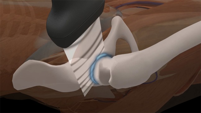
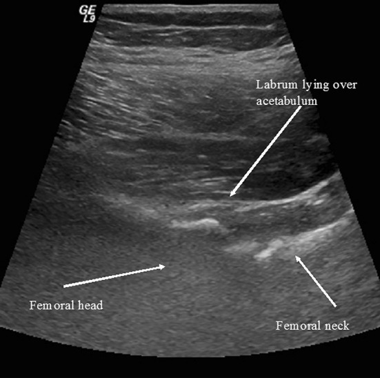
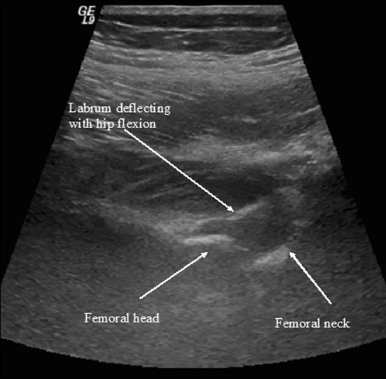
This study was approved by the institutional review board. Forty adult male volunteers (80 hips) between the ages of 21 and 35 years (mean age, 28 years) were recruited because males represent the majority of patients treated for symptomatic FAI. An a priori analysis was performed to determine an adequate sample size to detect a difference from values that have been previously reported in the literature. A sample size of 40 was found to be sufficient to detect differences within 5° based on a previously reported standard deviation of 8° [17]. Subjects were volunteers, primarily hospital residents, identified through verbal communications at the study institution. The mean age of the 40 subjects was 28 ± 3 years and the mean body mass index was 25 ± 4 kg/m2. As defined by US Census Bureau classification, 33 subjects were classified as white and seven subjects were classified as Asian. Volunteers were excluded if there was any history of groin and hip pain or injury. All ultrasounds were performed using a General Electric Logiq 9 (Wauwatosa, WI, USA) ultrasound system with an I739 linear array transducer. Imaging parameters were adjusted for best visualization of the anterior labrum of the hip. Each subject underwent bilateral static and dynamic hip ultrasound examinations. Two ultrasound-monitored measurements were performed for each hip. Patients were evaluated in the supine position with the contralateral hip and knee extended to control for lumbar position. The transducer was aligned with the inguinal crease and the transducer positioned parallel to the acetabulum (Fig. 1). During the static examination, the transducer was positioned approximately one-half centimeter distal to the anterior rim of the osseous acetabulum to image the anterior labrum. The static examination was performed to characterize the sphericity of the femoral head and the morphology of the head-neck region. Static assessment of the shape of the femoral head and the morphology of the femoral head-neck junction was performed for all hips. All femoral heads were found to be spherical. Thirteen subjects were identified with varying degrees of anterolateral head-neck junction cam morphology or limited offset that was bilateral in three and unilateral in 10 individuals. During passive hip flexion, the transducer was repositioned on the anterolateral proximal thigh so that its position would not interfere with hip movement. The transducer was parallel to the labrum so that the labrum appeared maximally hyperechoic and tissue anisotropy was minimized. The dynamic examination was performed as the hip was gradually passively flexed in the midsagittal plane until labral deflection was identified. Evaluation of labral deflection demonstrated deformation from a gently curved semicircular labrum in neutral position (Fig. 2) into a more boomerang-shaped labrum with hip flexion (Fig. 3). Impingement-free hip flexion was defined as the point at which labral morphology began to change. Maximal hip flexion was defined as the point when no further hip flexion was permitted as a result of bony abutment. Each point was monitored using the ultrasound image and the two points at which impingement occurred were measured using a large-sized goniometer that was adjusted with one limb in the midthigh and the second parallel to the examination table. A single observer (BL) performed all of the hip measurements with one of two experienced musculoskeletal ultrasonographers (MVH, PM) performing the examinations and determining the moment of initial labral deflection and terminal abutment.
Fig. 1.

Ultrasound probe position shown observing anterior labrum during hip flexion. Courtesy of M. van Holsbeeck MD.
Fig. 2.

Ultrasound view demonstrating labrum overlying the femoral neck.
Fig. 3.

Ultrasound image demonstrating initial labral deflection.
All analyses were performed in SPSS (Version 20; IBM Inc, Armonk, NY, USA). The intraclass correlation coefficient (ICC) was used to assess measurement reliability of the two measurements taken for impingement-free ROM. The ICC was found to be 0.946, which is considered to be excellent. Data were analyzed for normality using the Shapiro-Wilk test. A paired t-test was used to compare ROM between the right and left hips. To assess differences in ROM for normal versus cam morphology, an independent t-test was used for normal data and a Mann-Whitney rank sum test for nonnormal data. A Pearson’s correlation test (R) was used to analyze the correlation between impingement-free ROM and maximum flexion. In all tests, p < 0.05 was considered statistically significant.
Results
Soft tissue impingement was observed before bony abutment in all hips. The mean impingement-free hip flexion measured from neutral extension to initial labral deflection was 68° ± 17° on the right and 68° ± 16° on the left. The mean impingement-free hip flexion in all 80 hips was 68° ± 17 (95% confidence interval [CI], 65–72) (Table 1). In the cam-type hip, labral deflection occurred at 63° ± 20 (95% CI, 52–73) versus 70° ± 15 (95% CI, 66–74) in morphologically normal hips (Table 2). There was no significant difference detected between these values (p = 0.129).
Table 1.
Hip ROM measured with dynamic ultrasound
| ROM | Number | Mean | SD | Minimum | Maximum |
|---|---|---|---|---|---|
| Impingement-free passive flexion* | 80 | 68° | 17° | 30° | 111° |
| Maximum midsagittal passive flexion† | 80 | 96° | 6° | 84° | 112° |
* Impingement free-point of labral deflection; †maximum flexion-point of boney abutment; there was a mild but significant correlation between impingement-free ROM and maximum flexion, R = 0.398, p < 0.001.
Table 2.
Hip flexion in normal versus cam-type femoral head morphologies
| Femoral head morphology | Number | Impingement-free passive flexion | Maximum midsagittal passive flexion | ||||||
|---|---|---|---|---|---|---|---|---|---|
| Mean | SD | Minimum | Maximum | Mean | SD | Minimum | Maximum | ||
| Normal | 64 | 70° | 15° | 34° | 111° | 96° | 6° | 84° | 111° |
| Cam-type | 16 | 63° | 20° | 30° | 93° | 98° | 7° | 88° | 112° |
No significant difference was detected between normal versus cam-type morphologies for impingement-free passive flexion (p = 0.129) or maximum midsagittal passive flexion (p = 0.225).
The mean maximum midsagittal flexion measured at the time of bony abutment between the femoral head-neck junction and the acetabular rim was 97° ± 6° on the right and 96° ± 6° on the left. The mean maximum midsagittal flexion in all 80 hips was 96° ± 6° (95% CI, 95–98). There was a mild but significant correlation between impingement-free ROM and maximum flexion (Table 1). There was no difference with the numbers available when maximum terminal flexion was compared between cam-type hips (98° ± 7°; 95% CI, 95–102) and normal morphotype hips (96° ± 6°; 95% CI, 94–97; p = 0.225) (Table 2).
Discussion
FAI is associated with hip pain, chondral damage, and osteoarthrosis [2, 11]. Although treatment modalities continue to evolve, currently used open and arthroscopic techniques seek to repair or reconstruct chondrolabral injury and to relieve pathologic impingement. Whether the morphology of the head-neck junction or acetabular margin is altered in an effort to eliminate impingement, one surgical treatment goal is to increase impingement-free hip ROM. However, because the hip is a ball-in-socket articulation, terminal movement is limited by contact between the upper femur and acetabular rim. Currently, intraoperative surgical decision-making varies among surgeons and is often decided on a patient-by-patient basis with no normative data regarding hip ROM before bony abutment. We therefore sought to establish normative data for hip motion and to understand how the labrum moves with progressive flexion because these points are important for understanding both normal and abnormal hip kinematics and, ultimately, for surgeons treating patients with symptomatic FAI.
This study has certain limitations. First, the study was conducted only in young, primarily white males because athletic males are the most common patient treated for symptomatic FAI; therefore, the information is pertinent only to a narrow segment of the population. Second, because there is no accompanying plain radiographic imaging because our institutional review board determined that the risk from radiation exposure was not justifiable; therefore, this study is not able to correlate measured motions with specific skeletal morphology. Additionally, because cam-type morphology was only observed in 16 hips, the comparison between normal and cam-type hips was not highly powered, though differences may be detected with a larger population. However, it is interesting to observe that there was no major difference in flexion between normal and cam-type hips suggesting that there may be morphologic acetabular adaptations that permit better motion than what would be predicted by the presence of a head-neck junction deformity. Third, there were certain methodologic limitations all of which may affect the measurement precision that included using a goniometer for measurements, inability to stabilize the pelvis, and having a single observer perform all of the measurements, although similar limitations also affected previous reports [1, 5, 7, 12, 16, 17] thus justifying the contrasting ROM.
Our study found that impingement-free hip ROM in asymptomatic young adult males is approximately 65° and maximum hip flexion when further movement is prevented by bony abutment is approximately 95°, which is approximately 25% less than previously considered normal. There are multiple publications concerning normal hip ROM [1, 5, 7, 12, 16, 17] where maximum flexion was defined by maximum passive flexion that would have included lumbosacral motion (Table 3). Consequently, it is generally accepted that normal hip ROM is approximately 120°; however, all of these measurement observations were performed using goniometric techniques that were incapable of distinguishing lumbosacral movement from hip movement. Although our technique does not precisely segregate hip motion from lumbar motion, we feel that our observed flexion was lower than other studies because the measurements were performed in the supine position and based on observed intraarticular events; therefore, we feel that we have minimized the influence of lumbosacral movement on total observed hip motion. Furthermore, to our knowledge, no publication has defined impingement-free motion before the initiation of labral deformation.
Table 3.
Selected prior studies reporting normal hip ROM
| Study | Number of hips | ROM* |
|---|---|---|
| Boone and Azen [5] | 109 | 122° ± 6 |
| Roaas and Andersson [17] | 210 | 120° ± 8 |
| Roach and Miles [18] | 1892 | 121° (110–160) |
| Kubiak-Langer et al. [14] | 33 | 122° |
| Chevillotte et al. [7] | 21 | 104° ± 9 |
| Manning and Hudson [16] | 40 | 119° (115–121) |
* Mean ± SD with ranges in parentheses.
We believe these findings have several implications. First, they illustrate the importance of ascertaining normal data on which to base treatment goals. Second, they suggest that hips with substantial impingement may have less motion than previously considered, and this changes the expected normal ROM that may be established as a goal for treatment. Third, they suggest that because lumbosacral movement is a very important component of total hip motion, the loss of lumbosacral movement may independently adversely affect hip function in individuals with naturally low hip flexion. Fourth, they emphasize the need to obtain normative data on hip kinematics so that kinematics of painful hips can be studied. Future studies might involve methods to more accurately measure hip and lumbar motion using motion analysis techniques in a broader cross-section than was included in this study.
Acknowledgments
We thank Patrick Meyers MD, for assisting with the ultrasound examinations.
Footnotes
Each author certifies that he or she, or a member of his or her immediate family, has no funding or commercial associations (eg, consultancies, stock ownership, equity interest, patent/licensing arrangements, etc) that might pose a conflict of interest in connection with the submitted article.
All ICMJE Conflict of Interest Forms for authors and Clinical Orthopaedics and Related Research ® editors and board members are on file with the publication and can be viewed on request.
Clinical Orthopaedics and Related Research ® neither advocates nor endorses the use of any treatment, drug, or device. Readers are encouraged to always seek additional information, including FDA-approval status, of any drug or device prior to clinical use.
Each author certifies that his or her institution approved the human protocol for this investigation, that all investigations were conducted in conformity with ethical principles of research, and that informed consent for participation in the study was obtained.
This work was performed at Henry Ford Hospital, Detroit, MI, USA.
References
- 1.Ahlbaeck SO, Lindahl O. Sagittal mobility of the hip-joint. Acta Orthop Scand. 1964;34310–34322. [DOI] [PubMed]
- 2.Beck M, Kalhor M, Leunig M, Ganz R. Hip morphology influences the pattern of damage to the acetabular cartilage: femoroacetabular impingement as a cause of early osteoarthritis of the hip. J Bone Joint Surg Br. 2005;87:1012. doi: 10.1302/0301-620X.87B7.15203. [DOI] [PubMed] [Google Scholar]
- 3.Beck M, Leunig M, Parvizi J, Boutier V, Wyss D, Ganz R. Anterior femoroacetabular impingement: part II. Midterm results of surgical treatment. Clin Orthop Relat Res. 2004;418:67–73. doi: 10.1097/00003086-200401000-00012. [DOI] [PubMed] [Google Scholar]
- 4.Birn J, Pruente R, Avram R, Eyler W, Mahan M, van Holsbeeck M. Sonographic evaluation of hip joint effusion in osteoarthritis with correlation to radiographic findings. J Clin Ultrasound. 2014;42:205–211. doi: 10.1002/jcu.22112. [DOI] [PubMed] [Google Scholar]
- 5.Boone DC, Azen SP. Normal range of motion of joints in male subjects. J Bone Joint Surg Am. 1979;61:756–759. [PubMed] [Google Scholar]
- 6.Buck FM, Hodler J, Zanetti M, Dora C, Pfirrmann CW. Ultrasound for the evaluation of femoroacetabular impingement of the cam type. Diagnostic performance of qualitative criteria and alpha angle measurements. Eur Radiol. 2011;21:167–175. doi: 10.1007/s00330-010-1900-x. [DOI] [PubMed] [Google Scholar]
- 7.Chevillotte CJ, Ali MH, Trousdale RT, Pagnano MW. Variability in hip range of motion on clinical examination. J Arthroplasty. 2009;24:693–697. doi: 10.1016/j.arth.2008.04.027. [DOI] [PubMed] [Google Scholar]
- 8.Espinosa N, Beck M, Rothenfluh DA, Ganz R, Leunig M. Treatment of femoro-acetabular impingement: preliminary results of labral refixation. Surgical technique. J Bone Joint Surg Am. 2007;89(Suppl 2):136–153. doi: 10.2106/JBJS.F.01123. [DOI] [PubMed] [Google Scholar]
- 9.Espinosa N, Rothenfluh DA, Beck M, Ganz R, Leunig M. Treatment of femoro-acetabular impingement: preliminary results of labral refixation. J Bone Joint Surg Am. 2006;88:925–935. doi: 10.2106/JBJS.E.00290. [DOI] [PubMed] [Google Scholar]
- 10.Ganz R, Gill TJ, Gautier E, Ganz K, Krugel N, Berlemann U. Surgical dislocation of the adult hip: a technique with full access to the femoral head and acetabulum without the risk of avascular necrosis. J Bone Joint Surg Br. 2001;83:1119. doi: 10.1302/0301-620X.83B8.11964. [DOI] [PubMed] [Google Scholar]
- 11.Ganz R, Parvizi J, Beck M, Leunig M, Nötzli H, Siebenrock KA. Femoroacetabular impingement: a cause for osteoarthritis of the hip. Clin Orthop Relat Res. 2003;417:112–120. doi: 10.1097/01.blo.0000096804.78689.c2. [DOI] [PubMed] [Google Scholar]
- 12.Holm I, Bolstad B, Lütken T, Ervik A, Røkkum M, Steen H. Reliability of goniometric measurements and visual estimates of hip ROM in patients with osteoarthrosis. Physiother Res Int. 2000;5:241–248. doi: 10.1002/pri.204. [DOI] [PubMed] [Google Scholar]
- 13.Kettunen A. Factors associated with hip joint rotation in former elite athletes. Br J Sports Med. 2000;34:44–48. doi: 10.1136/bjsm.34.1.44. [DOI] [PMC free article] [PubMed] [Google Scholar]
- 14.Kubiak-Langer M, Tannast M, Murphy SB, Siebenrock KA, Langlotz F. Range of motion in anterior femoroacetabular impingement. Clin Orthop Relat Res. 2007;458:117–124. doi: 10.1097/BLO.0b013e318031c595. [DOI] [PubMed] [Google Scholar]
- 15.Leunig M, Beck M, Kalhor M, Kim YJ, Werlen S, Ganz R. Fibrocystic changes at anterosuperior femoral neck: prevalence in hips with femoroacetabular impingement. Radiology. 2005;236:237–246. doi: 10.1148/radiol.2361040140. [DOI] [PubMed] [Google Scholar]
- 16.Manning C, Hudson Z. Comparison of hip joint range of motion in professional youth and senior team footballers with age-matched controls: an indication of early degenerative change? Phys Ther Sport. 2009;10:25–29. doi: 10.1016/j.ptsp.2008.11.005. [DOI] [PubMed] [Google Scholar]
- 17.Roaas A, Andersson GB. Normal range of motion of the hip, knee and ankle joints in male subjects, 30–40 years of age. Acta Orthop Scand. 1982;53:205–208. doi: 10.3109/17453678208992202. [DOI] [PubMed] [Google Scholar]
- 18.Roach KE, Miles TP. Normal hip and knee active range of motion: the relationship to age. Phys Ther. 1991;71:656–665. doi: 10.1093/ptj/71.9.656. [DOI] [PubMed] [Google Scholar]
- 19.Vollman A, Hulen R, Dulchavsky S, Pinchcofsky H, Amponsah D, Jacobsen G, Dulchavsky A, van Holsbeek M. Educational benefits of fusing magnetic resonance imaging with sonograms. J Clin Ultrasound. 2014;42:257–263. doi: 10.1002/jcu.22136. [DOI] [PubMed] [Google Scholar]


