Abstract
Murine and human esophageal myofibroblasts are generated via enzymatic digestion. Neonate (8-12 day old) murine esophagus is harvested, minced, washed, and subjected to enzymatic digestion with collagenase and dispase for 25 min. Human esophageal resection specimens are stripped of muscularis propria and adventitia and the remaining mucosa is minced, and subjected to enzymatic digestion with collagenase and dispase for up to 6 hr. Cultured cells express α-SMA and vimentin and express desmin weakly or not at all. Culture conditions are not conducive to growth of epithelial, hematopoietic, or endothelial cells. Culture purity is further confirmed by flow cytometric evaluation of cell surface marker expression of potential contaminating hematopoietic and endothelial cells. The described technique is straightforward and results in consistent generation of non-hematopoieitc, non-endothelial stromal cells. Limitations of this technique are inherent to the use of primary cultures in molecular biology studies, i.e., the unavoidable variability encountered among cultures established across different mice or humans. Primary cultures however are a more representative reflection of the in vivo state compared to cell lines. These methods also provide investigators the ability to isolate and culture stromal cells from different clinical and experimental conditions, allowing comparisons between groups. Characterized esophageal stromal cells can also be used in functional studies investigating epithelial-stromal interactions in esophageal disorders.
Keywords: Cellular Biology, Issue 95, Cellular biology, mouse, human, esophagus, mesenchymal stromal cells, myofibroblasts, primary cells
Introduction
Epithelial-stromal interactions are involved in the regulation of a variety of gastrointestinal tract functions including mucosal regeneration, repair, fibrosis, and carcinogenesis1,2. These interactions have been best studied in the small intestine and colon and may similarly play a role in esophageal mucosal disorders3. A subpopulation of intestinal and colonic stromal cells termed myofibroblasts has been demonstrated to participate in mediating tissue injury, inflammation and repair4,5. In the distal GI tract, these spindle shaped cells are located adjacent to the basement membrane at the interface between the epithelium and lamina propria and are defined as α-SMA and vimentin positive, pan-cytokeratin negative, and weakly positive or desmin negative5.
The esophageal stroma has not been rigorously characterized at a cellular or molecular level. Our work in the murine esophagus has demonstrated α-SMA and vimentin cells in the esophageal stroma, occasionally subjacent to the squamous epithelium6. Epithelial-stromal interactions have been implicated in esophageal mucosal disorders such as gastro-esophageal mediated injury6 and eosinophilic esophagitis3. Fibrotic strictures are also a known complication of esophageal injury and stromal cells have been implicated in the pathogenesis of gastrointestinal fibrosis. Isolation of these cells will help accomplish the necessary studies to investigate deranged signaling pathways.
This submission provides the techniques necessary to establish primary cultures of α-SMA positive, vimentin positive myofibroblasts such that existing gaps in knowledge regarding signaling pathways mediating these interactions can be addressed. The technique described has been successfully used by the authors to establish primary murine colonic myofibroblasts7 and further adapted for establishment of murine6 and human myofibroblast-like esophageal stromal cells.
Herein we describe conditions needed to establish and characterize these cultures established from mouse or human esophagus prior to use in future functional studies. Cultures can be grown and utilized for up at 15 passages. Isolation and establishment of primary cultures via the methods outlined below generates stromal cells with a myofibroblast phenotype; α-SMA, vimentin positive, and weakly positive or negative for desmin, and cytokeratin negative. This phenotype is distinct from the phenotype of the esophageal fibroblast which is predominantly vimentin positive, α-SMA negative3 or the α-SMA positive, vimentin negative phenotype of the muscularis mucosae6.
Protocol
The protocol to perform animal experiments, the rationale and objectives of the research, were submitted and approved by the University of Southern California Institutional Animal Care and Use Committee.
The protocol for establishment of primary cultures from de-identified human esophagectomy specimens was approved by the University of Southern California Institutional Review Board.
1. Obtain Mouse or Human Esophagus
- Obtain Mouse Esophagus:
- Euthanize the 8-12 day old mouse with 2% isoflurane inhalant overdose followed by cervical dislocation.
- Pin the animal down with belly facing up and wet the ventral surface with 70% ethanol. With forceps, grab and lift up the skin anterior to the urethral opening with forceps. Use standard straight surgical scissors to cut along the ventral midline from the urethral opening to the chin. Be careful to cut only the skin.
- Make an incision from the urethral opening downward to the knee on both sides of the animal, forming an upside down “Y”. Make another incision on both sides of the animal along the rib cage.
- Carefully peel the skin on both sides, off of the underlying peritoneum and the rib cage, and lay to the sides. Examine the rib cage overlying the thoracic cavity and the peritoneum overlying the abdominal cavity.
- Lift up the peritoneum with forceps and cut through it up to the diaphragm and rib cage, pointing scissors upward to prevent damage to abdominal contents. Gently lift up the left lobe of the liver, expose the underlying stomach, the esophageal-gastric junction and the abdominal portion of the esophagus.
- Use autoclaved surgical scissors to cut through the sternum and ribcage along the midline up to the cervical girdle. Point scissors upward to prevent damage to the thoracic contents. Fully expose the contents of the thoracic cavity by pulling the rib cage to the sides.
- Gently remove the heart and both lobes of the lungs. Absorb excess blood with Q-tips. The thoracic esophagus is a narrow, flexible tube lying posterior to the trachea and anterior to the thoracic vertebra. NOTE: The esophagus can be difficult to identify in murine neonates. Localization of the esophageal-gastric junction makes esophageal identification straightforward.
- Carefully follow the esophagus up from the stomach, dissecting the surrounding vasculature, fat and mesentery all the way up to the esophageal origin in the cervical cavity. Surgical scissors with blunt tip work best for this purpose.
- Resect the entire length of the esophagus and place in Hanks’ balanced salt solution (HBSS) for further processing described below. Remove a portion of the stomach for orientation purposes if desired. NOTE: Due to small tissue size, separation of muscularis propria from the muscularis mucosa is not achievable in murine neonate esophagus and the entire esophagus is subjected to processing described below.
- Obtain Human Esophagus:
- Wash esophageal resection specimens (typically 5 cm or less) with HBSS and remove any attached connective tissue, fat, or vasculature.
- Resect a portion of the specimen and place in formalin for future histologic examination if desired.
- Separate the remaining mucosa from underlying muscularis propria by sharp dissection.
- Cut mucosa into sub centimeter fragments and subject to the protocol described below. NOTE: Human esophagus resections can be stored in PBS for up to 6 hr prior to processing described below. Source of esophagectomy specimens will dictate specimen size and will be laboratory dependent.
2. Isolate Murine and Human Esophageal Stromal Cells
- Isolate esophageal stromal cells from murine and human tissue by a combination of mechanical and enzymatic digestion.
- Mechanically digest by mincing the tissue with scissors and multiple washes with HBSS. Enzymatically digest by incubating the tissue with 300 U/ml collagenase XI and 0.1 mg/ml dispase for 25 min (murine tissue) or for up to 6 hr (human tissue). Ensure that instruments used for mincing have been autoclaved and sterilized. NOTE: Collagenases are enzymes that break down collagen, an extracellular matrix protein responsible for holding animal tissue together. Collagenase XI has high collagenase activity and has seen success in this isolation protocol. Dispase is a bacterial enzyme with mild proteolytic dissociation activity that preserves cell membrane activity and is used in combination with collagenase as a secondary enzyme.
- Place mucosal fragments obtained from mouse or human tissue in 1.7 ml micro-centrifuge or 5 ml tubes respectively, containing HBSS. Mince tissue further into 2-3 mm pieces using scissors with an open tip width that will fit into respective tubes. Allow enough time to allow for esophageal mucosal fragments to sediment to the bottom of the tube.
- Slowly decant HBSS, being careful not to inadvertently discard the tissue. Replace with fresh HBSS following by gentle shaking. Alternatively, remove HBSS with a 1 ml pipette.
- Wash tissue in this manner a total of 8 times with gentle shaking in between washes allowing time for esophageal fragments to sediment between each wash. NOTE: Human tissue will require more mincing that neonate mouse tissue.
- Incubate murine tissue with 300 U/ml collagenase XI and 0.1 mg/ml dispase for 25 min on a rocking shaker set at a slow speed at room temperature.
- Incubate human tissue with 300 U/ml XI collagenase and 0.1 mg/ml dispase for up to 6 hr, on a rocking shaker set at a slow speed at room temperature. NOTE: Availability of human esophagus is variable. Human esophageal mucosa incubated with enzymes for up to 6 hr results in successful generation of primary cultures.
After enzymatic digestion, mince tissue further with scissors and centrifuge at 200 x g for 10 min.
Discard the supernatant. Transfer the pellet, consisting of a mixed cell suspension to a 5 ml tube and wash 5 times in Dulbecco’s Modified Eagle Medium (DMEM) supplemented with 2% sorbitol to eliminate nonviable cells and debris.
Seed cells in 6-well plates and culture in DMEM with 10% fetal bovine serum (FBS), 10 mg/ml insulin, 10 µg/ml transferrin,10 µg/ml gentamicin, and 2 ng/ml epidermal growth factor (EGF). Vacuum filter (0.22 µm) all components except EGF which can be added after filtration. Distribute cells obtained from human resection specimens among multiple 6-well plates, depending on the resection size.
- Once wells are 80% confluent, passage adherent cells to T25 flasks using 0.05% trypsin/ethylenediaminetetraacetic acid (EDTA). Neutralize with media and spin at 400 x g.
- For a 6 well passage to a T25 flask, passage cells at a 1:1 ratio. Confluent T25 flasks can be passaged at a 1:2 or 1:3 ratio. To passage from a T25 flask to a T75 flask, use a 1:1 ratio. Change media every 2-3 days regardless of the vessel.
- Grow cells at 37 ºC in a humidified 5% CO2 incubator and culture in the myofibroblast media described above. NOTE: Epithelial cells do not survive under these culture or passage conditions. Cells used for characterization and in the studies described below are between passages 5 and 15.
- Cryo-preserve cells in liquid N2 by adding 10% dimethyl sulfoxide to the myofibroblast media.
3. Characterize Murine and Human Esophageal Stromal Cells
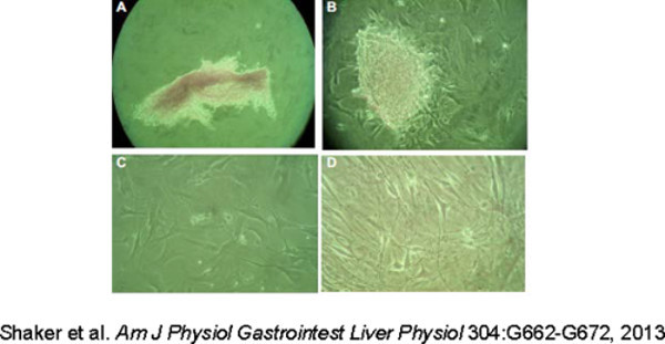
Examine the morphology of cells with an inverted microscope. Observe the spindle-shaped morphology of adherent cells (Figure 1).
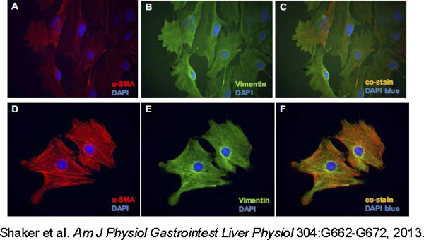
- Characterize cultured cells by evaluation of the following cytoskeletal markers: α-SMA, vimentin, desmin, and cytokeratin. Stromal cells with a myofibroblast-like phenotype express cytoskeletal myofibroblast markers α-SMA and vimentin (Figure 2).
- To further distinguish cultured stromal cells from muscle cells, immunostain for desmin. To perform immunostaining on cultured cells, plate 1.5 x 10⁴ cells in 4-well chamber slides and grow in myofibroblast media for 24 hr. Stromal cells with a myofibroblast-like phenotype will have weak or absent desmin expression. Stromal cells lack expression of the epithelial marker pan-cytokeratin (data not shown). NOTE: Assessment of cultures includes evaluation of cytoskeletal markers by immunocytochemistry and cell surface markers by flow cytometry. Immunocytochemistry is used as the primary method of characterization as the expression profile of cytoskeletal proteins is unique for the fibroblast, myofibroblast or muscle cell. Flow cytometry establishes culture purity by evaluating cell surface expression of epithelial, endothelial, and hematopoietic cells markers. CD90 is helpful as an adjunctive method of identifying human stromal cells as it is expressed on both human fibroblasts and myofibroblasts. CD90 is not helpful in characterization of murine stromal cells as it also expressed by murine T cells.
After cells reach 60% confluence, rinse slides with phosphate-buffered saline (PBS) and fix with methanol. Incubate cells in methanol for 10 min at -20 °C, then store in PBS at 4 °C until use.
Follow standard immunostaining protocol.
- Briefly, prior to applying the primary antibody, block the fixed cells with 5% goatserum in PBS. Dilute primary and secondary antibodies in 5% goat serum as well.
- For detection of α-SMA and vimentin, use the following antibodies and concentrations: Mouse mAB to α-SMA at 1:250 dilution and Rabbit pAB to vimentin at 1:500 dilution.
- Incubate with primary antibody at room temperature overnight at 4 °C, rinse 3 times with PBS and add secondary antibodies Rhodamine conjugated Goat anti mouse at a dilution of 1:200 and Cy2 conjugated Goat anti Rabbit at a dilution of 1:1,000 for 1 hr at room temperature.
- Counterstain nuclei with 4’, 6-Diamidino-2Phenylindole (DAPI) at a concentration of 1 µg/ml for 1 min and then wash with PBS. Analyze slides with an inverted microscope.
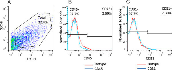
- Establish purity of cultures by examination of murine and human esophageal cells for hematopoietic (CD45) and endothelial (CD31) cell surface markers by flow cytometry (Figure 3). Characterize human cells further by expression of the stromal cell surface marker CD90. NOTE: CD90 cannot be used to identify murine stromal cells as murine T cells also express CD90.
- Detach cultured mouse esophageal stromal cells from the culture flasks by treatment with non-enzymatic Cell Dissociation Solution at 37 °C for 10 min, washed with FACS buffer twice, counted and stained with FITC-CD45 and APC-CD31 with relevant isotype controls, according to standard staining procedures.
- Collect cells using FACS CAlibur and analyze data with FlowJo software. Prior to staining cells, optimize antibodies and titrate concentrations using isotype controls, normal mouse spleen, and bone marrow cells.
Representative Results
Esophageal stromal cells isolated using mechanical and enzymatic digestion are initially plated and cultured in 6 well plates. Examination of cells with an inverted microscope within hours of plating demonstrates a mixed cell suspension loosely adherent to the plate bottom (Figure 1A). Over the next 24 hr, spindle-shaped cells firmly adherent to the plate bottom are observed shooting from the mixed cell suspension (Figure 1B).These sprouting cells cover the entire area of a well within 5 days and can be passaged successfully, retaining morphology for at least 15 passages. Spindle-shaped adherent cells are observed at low density (Figure 1C) and when near-confluent (Figure 1D).
Immunostaining of primary cultures of esophageal stromal cells grown in chamber slides demonstrates abundant expression of myofibroblast cytoskeletal markers α-SMA and vimentin (Figure 2), weak expression of desmin, and absent cytokeratin.
Primary cultures of esophageal myofibroblasts can be further examined for cell purity by examination of hematopoietic and endothelial cell surface markers. Forward and side scatter are established for primary murine esophageal stromal cells followed by gating and analysis of live cells for cell surface proteins (Figure 3A). Primary murine esophageal stromal cells lack expression of hematopoietic CD45 (Figure 3B) and endothelial cell CD31 (Figure 3C) cell surface markers.
 Figure 1: Primary cultures of murine esophageal stromal cells at different passages. Examination of murine stromal cells with an inverted microscope within hours of isolation and plating demonstrates a cluster of mixed cells loosely adherent cells to the plate bottom (A). After 24-48 hours in culture, spindle-shaped cells sprout from the now adherent cluster (B). Spindle-shaped morphology of adherent cells persists at low (C) and high (D) density. Please click here to view a larger version of this figure. (Figures used with permission from Shaker et al.6).
 Figure 2: Murine esophageal stromal cells grown in primary culture express myofibroblast markers α-SMA and vimentin. Primary murine esophageal stromal cells were grown on chamber slides and immunostained for α-SMA and vimentin at moderate (A-C) and low density (D-F). Esophageal stromal cells abundantly express α-SMA (A, D) and vimentin (B, E). Murine esophageal stromal cells co-express these myofibroblast markers (C, F). (Figures used with permission from Shaker et al.6). Please click here to view a larger version of this figure.
 Figure 3: Primary cultures of murine esophageal myofibroblasts cells lack hematopoietic and endothelial cell surface markers. The dot plot of murine esophageal stromal cells, demonstrates the FSC and SSC characteristics of this population of cells. A total of 32.4% of events were gated (A), Expression of hematopoietic (CD45) and endothelial (CD31) cell surface markers were evaluated by a single immunostain according to standard staining protocols. Nonspecific staining was determined for each antibody using isotype controls. Primary cultures of murine esophageal stromal cells do not express CD45 (B) or CD31 (C). FSC: forward scatter; SSC: side scatter. Please click here to view a larger version of this figure.
Discussion
The techniques for primary culture generation are generally similar when using murine or human tissue with mincing and digestion times adapted for tissue size. Our experience with murine tissue suggests that using the methods described above consistently result in successful establishment of primary cultures. Critical steps within the protocol include sterilization of surgical equipment and following standard sterile techniques of tissue culture. The age of the mice is also a critical step. 8 - 12 day old neonates consistently yield successful primary cultures. Stromal cells isolated from younger neonates have not been not been successfully generated. Attempts to establish myofibroblast-like stromal cells from older mice are ongoing. Others have reported successful generation of esophageal fibroblasts from four to eight week old mice8.
Unlike establishment of colonic myofibroblasts, contamination is an infrequent issue in establishment of cultures of murine myofibroblasts when antibiotics are included in the culture medium. Because cultures are established from neonate mice, a limiting factor is small tissue size and stripping of muscularis propria is not readily feasible. On the other hand, in the human esophagus, muscularis propria is easily distinguishable and separated from the muscularis mucosa. The enzymatic digestion and culture conditions are not conducive to growth of immune or epithelial cells in cultures established from mouse or human esophagus. Human resection specimens remain vulnerable to bacterial contamination despite the use antibiotics in the culture medium, perhaps because of the larger quantify of tissue undergoing processing. Following standard tissue culture techniques and rigorously sterilizing surgical instruments mitigates risk of contamination.
The advantage of primary culture is that these cells are relatively unmanipulated and may better reflect the in vivo environment compared to immortalized cell lines9.
The described technique is straight-forward and is within the means of most laboratories. The disadvantage of primary cultures versus cell lines is that cells beyond passage 15 are susceptible to genetic mutation and eventual senescence. In addition,primary cultures obtained from mice or humans have inherent variability not observed in cell lines. Alternative methods for esophageal fibroblast isolation have been described10. Characterization of these cells in culture however has been limited to vimentin expression or have demonstrated cells that express vimentin and are only weakly α-SMA positive10.
Once this technique has been mastered, primary cultures can be established from resection specimens of diseased human esophagus. These methods provide investigators the ability to isolate and culture stromal cells from different clinical and experimental conditions, allowing comparisons between groups. Cultured myofibroblast-like stromal cells can also be potentially adapted for use in organotypic or co-culture models with other cells such as eosinophils3 or in organotypic culture8.
Disclosures
This work is supported by NIH/NCI K08CA153036-01A1 (AS), AGA-General Mills Bell Institute of Health and Nutrition Research Scholar Award in Gut Physiology and Health (AS) and a Wright Foundation Pilot Award (AS).
Acknowledgments
Authors have nothing to disclose.
References
- Stappenbeck TS, Miyoshi H. The role of stromal stem cells in tissue regeneration and wound repair. Science. 2009;324(5935):1666–1669. doi: 10.1126/science.1172687. [DOI] [PubMed] [Google Scholar]
- Powell DW, Pinchuk IV, Saada JI, Chen X, Mifflin RC. Mesenchymal cells of the intestinal lamina propria. Annu Rev Physiol. 2011;73:213–237. doi: 10.1146/annurev.physiol.70.113006.100646. [DOI] [PMC free article] [PubMed] [Google Scholar]
- Rieder F, et al. T-Helper 2 Cytokines, Transforming Growth Factor beta1, and Eosinophil Products Induce Fibrogenesis and Alter Muscle Motility in Patients With Eosinophilic Esophagitis. Gastroenterology. 2014;146(5):1266–1277. doi: 10.1053/j.gastro.2014.01.051. [DOI] [PMC free article] [PubMed] [Google Scholar]
- Powell DW, et al. Paracrine cells important in health and disease. The American Journal of Physiology. 1999;277(1 Pt. 1):C1–9. doi: 10.1152/ajpcell.1999.277.1.C1. [DOI] [PubMed] [Google Scholar]
- Powell DW, et al. Intestinal subepithelial myofibroblasts. The American Journal of Physiology. 1999;277(2 Pt. 1):C183–201. doi: 10.1152/ajpcell.1999.277.2.C183. [DOI] [PubMed] [Google Scholar]
- Shaker A, et al. Stromal cells participate in the murine esophageal mucosal injury response. American Journal of Physiology Gastrointestinal and Liver Physiology. 2013;304(7):662–672. doi: 10.1152/ajpgi.00225.2012. [DOI] [PMC free article] [PubMed] [Google Scholar]
- Shaker A, et al. Epimorphin deletion protects mice from inflammation-induced colon carcinogenesis and alters stem cell niche myofibroblast secretion. The Journal of Clinical Investigation. 2010;120(6):2081–2093. doi: 10.1172/JCI40676. [DOI] [PMC free article] [PubMed] [Google Scholar]
- Kalabis J, et al. Isolation and characterization of mouse and human esophageal epithelial cells in 3D organotypic culture. Nature Protocols. 2012;7(2):235–246. doi: 10.1038/nprot.2011.437. [DOI] [PMC free article] [PubMed] [Google Scholar]
- Khalil H, Nie W, Edwards RA, Yoo J. Isolation of primary myofibroblasts from mouse and human colon tissue. Journal of Visualized Experiments. 2013. [DOI] [PMC free article] [PubMed]
- Rieder F, et al. Gastroesophageal reflux disease-associated esophagitis induces endogenous cytokine production leading to motor abnormalities. Gastroenterology. 2007;132(1):154–165. doi: 10.1053/j.gastro.2006.10.009. [DOI] [PubMed] [Google Scholar]


