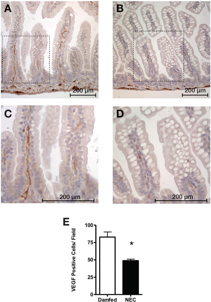
Figure 5. VEGF staining is decreased in intestinal tissues of mice submitted to a NEC protocol.
After immunostaining tissues for VEGF, we found significantly fewer VEGF-positive cells in the small intestinal lamina propria of mice exposed to our NEC protocol (B, enlarged in D) compared to dam-fed controls (A, enlarged in C) (E, *: p < 0.005).

