

Abstract
Background/Aim:
Galectins affect diverse physiological and pathophysiological processes such as development, inflammation, and tumor growth. We aimed to compare serum galectin-3 levels in three patient groups with chronic hepatitis B and C virus (HBV, HCV), cirrhosis secondary to HBV or HCV, and hepatocellular carcinoma (HCC) secondary to HBV or HCV and evaluate the role of galectin-3 during HCC progression.
Patients and Methods:
Nineteen patients with hepatocellular cancer, 22 patients with cirrhosis, and 24 patients with chronic hepatitis B and C were included in this study. Serum galectin-3 levels in different liver diseases were assessed by enzyme-linked immunosorbent assay.
Results:
The mean galectin-3 levels were 4.61 ng/mL (±2.32) in HCC patients, 5.68 ng/mL (±2,2) in cirrhotic patients, 1.98 ng/mL (±1.50) in chronic viral hepatitis group. There were no statistical differences between HCC and cirrhotic patients (P = 0.5), but lower in chronic hepatitis group statistically compared with cirrhosis and HCC (P < 0.001, P = 0.002, respectively). In case of cirrhotic patients, galectin-3 levels were significantly higher in patients with cirrhosis secondary to HCV compared with HBV (P = 0.03). When we evaluated galectin-3 levels in HCC patients, it was found to be 3.92 ng/mL in HCC secondary to hepatitis B and 5.37 ng/mL in HCC secondary to hepatitis C.
Conclusion:
Serum galectin-3 levels in patients with chronic HBV or HCV may guide us about progression to cirrhosis or HCC and prognosis of the disease. Especially, galectin-3 levels may be more pronounced in case of HCV.
Keywords: Chronic viral hepatitis, cirrhosis, galectin-3, HCC
Hepatocellular carcinoma (HCC) is the fifth most common solid tumor and the third most common cause of cancer-related mortality every year. Infections with hepatitis B virus (HBV) and hepatitis C virus (HCV) are some of the major risk factors for cirrhosis and HCC development.[1] Many etiologic factors and molecular biology of HCC were studied in the recent years.[2,3,4] Galectins are members of a newly defined and growing family of the animal lectins. They play an essential role in the function and development of multicellular organisms, including development, differentiation, cell-cell adhesion, cell-matrix interaction, growth regulation, apoptosis, RNA splicing, and tumor metastasis.[5] Galectin-3 is the most extensively studied. Oncogenic and viral stimulation can change galectin-3 expression and galectin-3 increases in several human tumors. Galectin-3 is a nonintegrin β-galactoside-binding lectin (also known as Mac-2, CBP-35, CBP-30, IgEBP, RL-29, L-29, L-31, L-34, and LBL) with an estimated molecular weight varying between 26,200 and 30,300 Da. These molecules are expressed by macrophages, neutrophils, mast cells, and Langerhans cells, and exhibit pleiotropic biological function, playing a key role in many physiological and pathological processes.[6] In many studies, galectin-3 overexpression was demonstrated in cancer tissues and use of it in case of differentiation between benign and malign tissue was emphasized in the study.[6,7,8,9,10]
Hsu et al.[11] demonstrated that normal hepatocytes do not express galectin-3, but this protein can be present in HCC. The investigation revealed that galectin-3 expression in HCC is independent of whether the patient had prior HBV infection. It was found that focal regenerating nodules of cirrhotic tissue also express galectin-3.[11] A study by Matsuda et al., showed that galectin-3 expression was involved in the tumor progression and related to the tumor prognosis of HCC.[12]
In this study, we aimed to compare serum galectin-3 levels in three patient groups with chronic hepatitis (HBV, HCV), hepatic cirrhosis secondary to HBV or HCV, and HCC secondary to HBV or HCV and evaluate the role of galectin-3 during HCC progression.
PATIENTS AND METHODS
In 2009–2011, 65 patients (HCC: 19 patients, cirrhosis: 22 patients, and chronic hepatitis B or C: 24 patients) were included in this study. Blood samples were taken after 10 h of fasting from antecubital vein into vacuum and gel seperator tubes and centrifuged 3000 ×g for 10 min. Subsequently, all serum samples were divided into aliquotes and frozen and preserved at −80°C until test time. At the same time when blood samples were obtained, whole blood count (hemoglobin (hb), white blood cell (WBC), platelet (plt)), liver function tests (aspartate aminotransferase, AST; alanine aminotransferase, ALT; gamma-glutamyl transferase, GGT; alkaline phosphatase, ALP), and prothrombin time (PT) were also studied. The study was approved by the local ethical commitee of the hospital.
Serum galectin-3 levels in different liver diseases (HCC, cirrhosis, and chronic hepatitis B and C) were measured with enzyme-linked immunosorbent assay (ELISA) according to manufacturer's instructions (eBioscience, Inc, CA, USA). All results are given as ng/mL.
All data were recorded in “Excel XP for windows.” Statistics were performed using SPSS 16.0 for Windows. For comparison of the means of two groups, Student's t-test was used and a P < 0.05 was considered as significant.
RESULTS
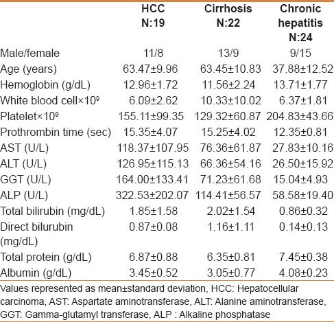
There were 65 patients (HCC: 19 patients, cirrhosis: 22 patients, and chronic hepatitis B or C: 24 patients) in this study. All patients with HCC did not have metastatic diseases, their diseases were limited to liver. Male/female ratios were 11/8, 13/9, and 9/15, respectively. The median age was 63.47 ± 9.96 in HCC and 63.45 ± 10.83 in the cirrhosis group. In the chronic hepatitis group, median age was 37.88 ± 12.52. The demographic features and laboratory values are given in Table 1. AST, ALT, GGT, and ALP levels were statistically different in each group. They were found to be highest in HCC compared with low levels in chronic hepatitis group.
Table 1.
The demographic features and laboratory levels of all patients
The mean galectin-3 levels were 4.61 ng/mL (±2.32) in HCC patients, 5.68 ng/mL (±2.2) in cirrhotic patients, 1.98 ng/mL (±1.50) in chronic viral hepatitis group. There was no statistical difference between HCC and cirrhotic patients (P = 0.5) in case of galectin-3 serum levels but chronic hepatitis group statistically compared with cirrhosis and HCC (P < 0.001, P = 0.002, respectively) [Table 2 and Figure 1].
Table 2.
Serum galectin-3 levels according to patient group
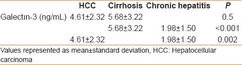
Figure 1.
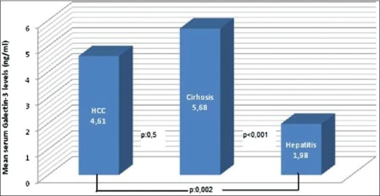
Serum galectin-3 levels according to diseases
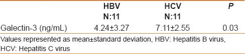
When we evaluated galectin-3 levels in HCC patients, it was found to be 3.92 ng/mL in HCC secondary to hepatitis B, 5.37 ng/mL in HCC secondary to hepatitis C. It seemed to be low in HCC secondary to HBV, but this difference was not statistically significant (P = 0.201, Table 3). In case of cirrhotic patients, galectin-3 levels were 4.27 and 7.11 ng/mL in patients with cirrhosis secondary to HBV and HCV, respectively. This difference was found to be statistically significant (P = 0.03, Table 4).
Table 3.
Galectin-3 levels in HCC patients according to viral etiology
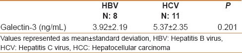
Table 4.
Galectin-3 levels in patients with cirrhosis according to viral etiology
DISCUSSION
Galectins are an ancient family of carbohydrate-binding proteins found in multicellular organisms from fungi to mammals. Galectin-3 is a nonintegrin β-galactoside-binding lectin that has a role in tissue homeostasis and cancer progression. This protein is expressed in a variety of tissues and cell types and mainly found in the cytoplasm. Galectin-3 exhibits pleiotropic function, playing a key role in many physiological and pathological processes.[13] Increased galectin-3 expression was found to be related with cellular motility and extracellular matrix invasion, thus related with tumor metastasis.[14,15] Matarrese et al.[16] showed that overexpression of galectin-3 can cause mitochondrial homeostasis and protect the cell from damage and apoptosis.
Although galectin-3 is minimally expressed in normal hepatocytes, it was found to be significantly highly expressed in the liver biopsies of patients with HCC or hepatic cirrhosis. Further investigation showed that galectin-3 expression in HCC is independent of whether the patient had prior HBV infection or not. In this study, 14 of 18 HCC cases from HBV-negative patients and 5 of 7 cases from HBV patients demonstrated positive galectin-3 immunohistochemistry. In addition, galectin-3 was abundantly expressed in cirrhotic liver in peripheral distribution within regenerating nodules, which may be a result of the high mitotic index.[11]
In another study, Matsuda et al.[12] studied galectin-3 expression in HCC by immunohistochemical analysis from specimen and also studied serum level of galectin-3 by ELISA. It was found to be statistically correlated with histological differentiation and vascular invasion. They concluded that galectin-3 expression was involved in the tumor progression and related to the prognosis of HCC.[12]
In our study, serum galectin-3 levels were found to be significantly higher in HCC and cirrhosis than in chronic viral hepatitis. Our results were correlated with the literature. We also observed that galectin-3 levels were higher in HCV-related cirrhosis and HCC compared with HBV, but this was only statistically significant in the HCV-related cirrhotic patient. This may be explained by low number of patients. This was the main limitation of our study. Another limiting aspect of this study was the age difference between the study groups. The chronic viral hepatitis group consisted of younger cases than the other two groups, respectively. This may be due to difficulty in finding hepatitis carrier patient in older age group. However whether galectin-3 improves with age or not, has not been shown in any study.
In this study, we preferred chronic hepatitis B and C patients as control group. Healthy individuals who were not exposed to hepatitis were not included. However, no studies have been conducted so far to measure changes in galectin-3 levels in case of hepatitis carriers in whom liver fibrosis or carcinogenesis has not begun, compared with healthy subjects.
In conclusion, serum galectin-3 levels in patients with chronic HBV or HCV may be guiding us about progression to cirrhosis or HCC and prognosis of the disease. Especially, galectin-3 levels may be more pronounced in case of HCV. In these patients, if galectin-3 levels were found to be high, serum alpha-feto protein level and ultrasonographic examination could be repeated at more frequent intervals. This may also guide us in terms of the treatment plan. However, whether serum galectin-3 level is a prognostic indicator or not, should be supported by further randomized trials.
Footnotes
Source of Support: Nil
Conflict of Interest: Mehmet Ulu, Ahmet Alacacioglu, Esma Yuksel, Baris Pamuk, Giray Bozkaya, Alpay Ari, Arif Yuksel, Gulten Sop and Inci Alacacioglu declare that they have no conflict of interest. Additional informed consent was obtained from all patients for whom identifying information is included in this article.
REFERENCES
- 1.Bacigalupo ML, Manzi M, Rabinovich GA, Troncoso MF. Hierarchical and selective roles of galectins in hepatocarcinogenesis, liver fibrosis and inflammation of hepatocellular carcinoma. World J Gastroenterol. 2013;19:8831–49. doi: 10.3748/wjg.v19.i47.8831. [DOI] [PMC free article] [PubMed] [Google Scholar]
- 2.Pang RW, Poon RT. From molecular biology to targeted therapies for hepatocellular carcinoma: The future is now. Oncology. 2007;72(Suppl 1):30–44. doi: 10.1159/000111705. [DOI] [PubMed] [Google Scholar]
- 3.Lovet JM, Bruix J. Molecular targeted therapies in hepatocellular carcinoma. Hepatology. 2008;48:1312–27. doi: 10.1002/hep.22506. [DOI] [PMC free article] [PubMed] [Google Scholar]
- 4.Shen YC, Hsu C, Cheng AL. Molecular targeted therapy for advanced hepatocellular carcinoma: Current status and future perspectives. J Gastroenterol. 2010;45:794–807. doi: 10.1007/s00535-010-0270-0. [DOI] [PubMed] [Google Scholar]
- 5.Danguy A, Camby I, Kiss R. Galectins and cancer. Biochim Biophys Acta. 2002;1572:285–93. doi: 10.1016/s0304-4165(02)00315-x. [DOI] [PubMed] [Google Scholar]
- 6.Coli A, Bigotti G, Zucchetti F, Negro F, Massi G. Galectin-3, a marker of well-differentiated thyroid carcinema, is expressed in thyroid nodules with cytological atypia. Histopathology. 2002;40:80–7. doi: 10.1046/j.1365-2559.2002.01304.x. [DOI] [PubMed] [Google Scholar]
- 7.Abd-El Raouf S, Ibrahim TR. Immunohistochemical expression of HBME-1 and galectin-3 in the differential diagnosis of follicular-derived thyroid nodules. Pathol Res Pract. 2014;210:971–8. doi: 10.1016/j.prp.2014.06.010. [DOI] [PubMed] [Google Scholar]
- 8.Nechifor-Boilã A, Cãtanã R, Loghin A, Radu TG, Borda A. Diagnostic value of HBME-1, CD56, Galectin-3 and Cytokeratin-19 in papillary thyroid carcinomas and thyroid tumors of uncertain malignant potential. Rom J Morphol Embryol. 2014;55:49–56. [PubMed] [Google Scholar]
- 9.Takenaka Y, Fukumori T, Raz A. Galectin-3 and metastasis. Glycoconj J. 2004;19:543–9. doi: 10.1023/B:GLYC.0000014084.01324.15. [DOI] [PubMed] [Google Scholar]
- 10.Hossaka TA, Ribeiro DA, Focchi G, André S, Fernandes M, Lopes Carapeto FC, et al. Expression of Galectins 1, 3 and 9 in normal oral epithelium, oral squamous papilloma, and oral squamous cell carcinoma. Dent Res J (Isfahan) 2014;11:508–12. [PMC free article] [PubMed] [Google Scholar]
- 11.Hsu DK, Dowling CA, Jeng KC, Chen JT, Yang RY, Liu FT. Galectin-3 expression is induced in cirrhotic liver and hepatocellular carcinoma. Int J Cancer. 1999;81:519–26. doi: 10.1002/(sici)1097-0215(19990517)81:4<519::aid-ijc3>3.0.co;2-0. [DOI] [PubMed] [Google Scholar]
- 12.Matsuda Y, Yamaqiwa Y, Fukushima K, Ueno Y, Shimoseqawa T. Expression of galectin-3 involved in prognosis of patients with hepatocellular carcinoma. Hepatol Res. 2008;38:1098–111. doi: 10.1111/j.1872-034X.2008.00387.x. [DOI] [PubMed] [Google Scholar]
- 13.Krzeslak A, Lipinska A. Galectin-3 as a multifunctional protein. Cell Mol Biol Lett. 2004;9:305–28. [PubMed] [Google Scholar]
- 14.Oestreicher-Kedem Y, Halpern M, Roizman P, Hardy B, Sulkes J, Feinmesser R, et al. Diagnostic value of galectin-3 as a marker for malignancy in follicular patterned thyroid lesions. Head Neck. 2004;26:960–6. doi: 10.1002/hed.20087. [DOI] [PubMed] [Google Scholar]
- 15.Fang QQ, Ni RZ, Xiao MB, Jiang F, Lu CH. Serum and tissue expressions of galectin-3 in hepatocellular carcinoma and the clinical significances. Zhonghua Gan Zang Bing Za Zhi. 2011;19:527–31. doi: 10.3760/cma.j.issn.1007-3418.2011.07.014. [DOI] [PubMed] [Google Scholar]
- 16.Matarrese P, Tinari N, Semeraro ML, Natoli C, Iacobelli S, Malorni W. Galectin-3 overexpression protects from cell damage and death by influencing mitochondrial homeostasis. FEBS Lett. 2000;473:311–5. doi: 10.1016/s0014-5793(00)01547-7. [DOI] [PubMed] [Google Scholar]