Abstract
Background
The duration of antibiotic treatment of exacerbations of COPD (ECOPD) is controversial. Serum procalcitonin (PCT) is a biomarker of bacterial infection used to identify the cause of ECOPD.
Methods and Findings
We investigated whether a PCT-guided plan would allow a shorter duration of antibiotic treatment in patients with severe ECOPD. For this multicenter, randomized, non-inferiority trial, we enrolled 184 patients hospitalized with ECOPD from 18 hospitals in Italy. Patients were assigned to receive antibiotics for 10 days (standard group) or for either 3 or 10 days (PCT group). The primary outcome was the rate of ECOPD at 6 months. Having planned to recruit 400 patients, we randomized only 183: 93 in the PCT group and 90 in the standard group. Thus, the completed study was underpowered. The ECOPD rate at 6 months between PCT-guided and standard antibiotic treatment was not significant (% difference, 4.04; 90% confidence interval [CI], −7.23 to 15.31), but the CI included the non-inferiority margin of 15. In the PCT-guided group, about 50% of patients were treated for 3 days, and there was no difference in primary or secondary outcomes compared to patients treated for 10 days.
Conclusions
Although the primary and secondary clinical outcomes were no different for patients treated for 3 or 10 days in the PCT group, the conclusion that antibiotics can be safely stopped after 3 days in patients with low serum PCT cannot be substantiated statistically. Thus, the results of this study are inconclusive regarding the noninferiority of the PCT-guided plan compared to the standard antibiotic treatment. The study was funded by Agenzia Italiana del Farmaco (AIFA-FARM58J2XH). Clinical trial registered with www.clinicaltrials.gov (NCT01125098).
Trial Registration
ClinicalTrials.gov NCT01125098
Introduction
Exacerbations of chronic obstructive pulmonary disease (ECOPD) constitute 2.4% of all acute hospital admissions in England [1] and are associated with a mortality rate as high as 40% in the first year following hospitalization in patients needing mechanical support for acute ECOPD [2].
The most common causes of ECOPD are viral and/or bacterial respiratory tract infections and air pollution. However, a precise cause cannot be identified in a large number of patients [1–4].
Long-term antibiotic treatment (e.g., 12 months of 250 mg azithromycin daily) prevents ECOPD and hospitalization in patients with severe COPD [5,6], possibly by reducing the bacterial load and/or bronchial inflammation in the airways. However, it may also increase bacterial resistance, cardiovascular mortality due to prolongation of the heart rate-corrected QT (QTc) interval [7], and hearing loss. Thus, long-term antibiotic treatment is not recommended by most recent international guidelines [8].
The current approach to treatment of ECOPD due to bacterial infections is antibiotic treatment, even though the precise role of bacterial infections and antibiotic treatment in individual episodes of severe ECOPD remains controversial [9,10]. Identification of the bacterial etiology of ECOPD is often unclear, and the decision to treat with antibiotics is usually empirical; antibiotics are, of course, useful only for treating bacterial infections. Taking into account the limited evidence available, i.e., that from only one properly designed randomized clinical trial [11], current international guidelines recommend antibiotics only in ECOPD with (i) three cardinal symptoms, i.e., increased dyspnea, sputum volume, and sputum purulence (type 1) [11] or (ii) two cardinal symptoms, with one of the two being sputum purulence (type 2), and/or (iii) respiratory failure [11] with an arbitrary duration of 5–10 days [8,12].
Previous studies investigated the role of biomarkers of bacterial infection [13–15], particularly procalcitonin (PCT), in guiding antibiotic treatment in respiratory infections, i.e., pneumonia and ECOPD [16–20]. PCT is a prototype of a “hormokine” mediator that is released in bacterial infections but not in viral infections or noninfectious stimuli [21]. Indeed, PCT-guided antibiotic treatment may reduce the use of antibiotics in patients hospitalized for ECOPD [17,19,20]. However, none of those studies consisted only of patients with Anthonisen’s type 1 ECOPD [11] or respiratory failure, for whom guidelines recommend antibiotic treatment [8,12], and none investigated the role of PCT in reducing the duration of antibiotic treatment.
Antibiotic treatment for up to 10 days is supported by a single clinical trial published 28 years ago_[11]; in fact, both the previous (3–10 days) [22] and current (5–10 days) recommendations [8,12] have been given only a D level of evidence, i.e., panel consensus judgment. Also, although GOLD guidelines only mention PCT as a biomarker of bacterial infection_ [8], a recent JAMA clinical evidence synopsis [23] does not recommend its use.
For all these reasons, we decided to investigate whether antibiotic treatment could be safely stopped according to a PCT-guided 3-day treatment plan in patients hospitalized with ECOPD.
Methods
Study Design and Participants
The protocol for this trial and supporting CONSORT checklist are available as supporting information; see S7 CONSORT Checklist and S8 Protocol.
In a randomized, multicenter, open, controlled, parallel-group, noninferiority trial involving 18 university/city hospital pulmonary departments, we compared patients hospitalized with severe ECOPD who were receiving the standard 10-day course of antibiotic therapy recommended by the 2005 GOLD guidelines [22] with those receiving a 3- or 10-day antibiotic course guided by a PCT plan. According to current recommendations [22, 24], because all patients recruited for the study required hospitalization, the exacerbations were considered severe. Apart from the usual clinical investigations, which included routine blood tests, arterial blood gases, ECG, and chest X-ray, no other specific clinical investigation was used to assess the severity of exacerbations. Patients were recruited and followed up between 26 January 2007 and 25 July 2011.
Study participants were male or female, ≥18 years of age, current or former smokers, and diagnosed with COPD stages I-IV as defined by GOLD guidelines available at the time the study was designed, with protocol deviation [22]. Patients were hospitalized for severe ECOPD requiring antibiotic treatment, i.e., type 1 exacerbation (increased dyspnea, sputum volume, and sputum purulence verified by the attending clinician) according to Anthonisen [11], and/or characterized by respiratory failure. ECOPD was defined as “an acute event characterized by a worsening of the patient’s respiratory symptoms that is beyond normal day-to-day variations and leads to a change in medication” [22,24].
Exclusion criteria included bronchial asthma, unstable concomitant disease (cardiovascular, renal, hepatic, gastrointestinal, neurological, metabolic, musculoskeletal, neoplastic, respiratory or other disease), pregnancy and breastfeeding, clinically significant laboratory abnormalities suggestive of unstable concomitant disease, survival for 1 year unlikely, and inability to give written consent. Antibiotic pretreatment before hospital admission and radiographic signs of pneumonia did not preclude eligibility for the study. All patients underwent chest X-ray at admission. Only one patient recruited in the standard group had community-acquired pneumonia, and remained in the study group.
Patients’ clinical data, comorbidities, and routine blood test results were recorded at the time of recruitment. According to the protocol, all patients had to have a record of lung function testing confirming COPD (see below) or spirometric tests in the hospital according to guidelines [25]. All patients included in the analysis had hospital spirometry. Patients were included in the study if they had a FEV1/FVC <0.7 and FEV1 <80% predicted [8,22]. In addition, all patients underwent arterial blood gas analysis at admission, on Day 1, and on Day 2. Respiratory failure was defined as PaO2 <60 mm Hg with or without PaCO2 >50 mm Hg when breathing room air [8,22].
The study (which conformed to the Declaration of Helsinki) was approved by the ethics committees of each of the 18 participating centers. Names of members of the ethics committee/institutional review board(s) that approved the study are reported in Supporting Information. All patients gave informed written consent. The trial was approved and funded by the Agenzia Italiana del Farmaco (AIFA), the Italian agency for drugs that is an official body of the Italian Ministry of Health. The title of the protocol, approval and funding were registered in the AIFA registry (http://www.agenziafarmaco.gov.it/, <http://www.agenziaitalianadelfarmaco.gov.it,farm58j2xh/> FARM58J2XH) and the protocol was posted by AIFA in the European clinical trials register (https://www.clinicaltrialsregister.eu/ctr-search/search, 2006-005354-68) on 2007/07/04. The trial was registered by us at http://www.clinicaltrial.gov/ (NCT01125098) in 2010. The reason for the late registration at http://www.clinicaltrial.gov was that we thought that registration in the https://www.clinicaltrialsregister.eu/ctr-search/search represented sufficient evidence of trial publication. We confirm that all ongoing and related trials for this drug/intervention are registered.
Randomization and Treatment
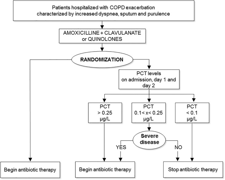
The independent Clinical Trials and Methodological Unit at the University of Modena carried out centralized randomization. Eligible patients were randomly assigned to receive standard antibiotic therapy (standard group) or PCT-guided antibiotic treatment (PCT group) according to a 1:1 permuted block computer-generated scheme, stratified according to hospital. The randomization was Web-based, and only statisticians and the website administrator knew the randomization sequence. On admission, all patients received a 3-day course of antibiotics (either amoxicillin plus clavulanate or quinolones) according to 2005 international guidelines [22]. PCT levels were measured on hospital admission, on Day 1, and on Day 2. On Day 2 each eligible patient was randomly assigned to one of the two treatment plans. Patients randomized to the standard group continued antibiotic therapy for 10 days, whereas patients randomized to the PCT group either continued treatment for 10 days or stopped on Day 3, depending on their PCT levels, according to previously recommended cut-off values [17,20]. Specifically, patients continued antibiotic treatment for 10 days if one or more of the PCT values on the first 3 days of hospitalization were ≥0.25 μg/L. When PCT values were <0.25 μg/L but ≥0.1 μg/L on any occasion, antibiotic treatment was continued for 10 days, but only if patients 0were clinically unstable or had acute respiratory failure; otherwise, treatment was stopped on Day 3. If all PCT values were consistently <0.1 μg/L, treatment was stopped on Day 3 (Fig. 1). Because the final decision about maximum duration of treatment in randomized patients was left to the referring investigator, who had the option of overruling the PCT-guided plan if he or she deemed it clinically inappropriate, 4 patients with low values of PCT received treatment between 4 and 10 days. All patients were also treated with (i) systemic corticosteroids for 14 days [22], plus (ii) regular inhaled short-acting or long-acting bronchodilators.
Fig 1. Trial protocol. Severe disease = respiratory failure or clinical instability.
Outcomes and Follow-up
We prospectively followed patients during hospitalization and after discharge. Patients were clinically assessed on admission, on Days 1 and 2 after hospitalization, and on Day 10 or at discharge. Blood samples were obtained for measurement of PCT and serologic testing (Mycoplasma pneumoniae, Chlamydia pneumoniae, and Legionella pneumophila [Virotech ELISA IgG and IgM; Vircell ELISA IgG and IgM; Virion Serion ELISA classic IgG and IgM]). Sputum was collected for Gram staining and culture. Microbiological analysis was carried out according to Isenberg [26], and all patients had a chest X-ray. Only one patient in the standard group had radiological evidence of pneumonia, and he was negative for serological detection of bacterial infection. In addition, each patient filled out quality-of-life questionnaires (Short Form 36, baseline [BDI] and transition [TDI] dyspnea indices, and CCIQ [Chronic Cough Impact Questionnaire]) on admission [27–30]. Follow-up visits were scheduled on Day 1, Day 3, and 6 months after discharge; telephone interviews were conducted at 2, 4, and 5 months after discharge.
The primary end point of the study was the percentage of patients with at least one exacerbation within 6 months after the index exacerbation. Secondary end points included hospital readmission, admission to the intensive care unit, change in lung function (ΔFEV1), length of hospital stay, and death from any cause.
The outcome of the index exacerbation was considered a clinical success when the signs and symptoms associated with the exacerbation were completely resolved or improved. Treatment failure was defined as a lack of resolution, lack of attenuation of signs and symptoms, worsening of signs and symptoms, or death.
The patient’s safety was monitored at each visit, including the assessment of adverse events related to the antibiotic treatment and of clinical events such as pneumonia, pulmonary embolism, acute pulmonary edema, and any cardiovascular accident.
Measurement of Procalcitonin
Quantitative measurements of PCT concentrations were performed in a centralized laboratory (Department of Laboratory Medicine, University of Padova, Italy), in duplicate in human serum samples (volume: 50 μL), using an automated immunofluorescent assay (Kryptor PCT; Brahms Diagnostica AG, Hennigsdorf/Berlin, Germany). The principle of the method is based on TRACE technology (time-resolved amplification of cryptate emission), which measures the signal emitted from an immunocomplex with time delay. The basis of TRACE technology is nonradiating energy transfer from a donor molecule (cryptate) to a fluorescent label (XL665) acceptor molecule; the long-life signal emitted is proportional to the concentration of the analyte to be measured (PCT) [31]. The characteristics of the test have been previously described [17,20]. Samples were couriered from each participating center to the Central Laboratory of Padova with the commitment to deliver the results of the 3-day samples within 24 hours of the previous sample.
Statistical Analysis
Sample size was determined according to the primary outcome of the study (cumulative ECOPD rate at 6 months), calculated by dividing the number of patients with at least one exacerbation during the 6-month follow-up by the total number of patients initially randomized. Data from previous studies [20], used to estimate the frequency of the primary outcome of the present study, suggested that exacerbations within 6 months occur in approximately 50% of patients. To define the noninferiority of the PCT-guided algorithm compared to the standard guidelines-recommended plan, we settled on a 15% difference in the percentage of patients with an exacerbation within the 6-month follow-up as a clinically tolerable upper limit. We hypothesized that exacerbation rates for both treatment plans would be equal (at 50%), and we considered a difference of ≤15% to be irrelevant; then, setting β = 0.20 and α = 0.05 (one-tail), the estimated sample size was 140 patients per arm. Taking into account an estimated 30% drop-out rate of patients at the 6-month follow-up, we increased the sample size to 200 patients per arm.
Data were summarized as mean ± standard deviation (SD), or as median and interquartile range (IQR) when appropriate, for continuous variables or as number and percentage for categorical variables.
To compare PCT-guided and standard groups in terms of primary outcome, we estimated the risk difference and its relative 90% confidence interval (CI). For secondary outcomes, risks and mean differences as well as their 95% CIs were calculated for comparing binary and continuous outcomes, respectively. Hazard ratios and their 95% CIs were estimated to compare time-to-event data. Categorical data were compared with the use of the chi-square test or the Fisher exact test, when appropriate. Continuous data were compared with the use of Student’s t test. Cox proportional hazards regression analysis was used to estimate hazard ratios and 95% CIs for time-to-event data (e.g., length of hospital stay).
Statistical analyses were performed by using STATA version 12 (StataCorp LP, College Station, TX).
Results
Study Population
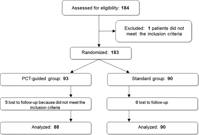
We planned to recruit 400 patients. However, because of slow recruitment by the centers and possibly because of the very strict inclusion criteria (Anthonisen type 1 and/or respiratory failure), we were able to screen only 184 patients and randomize 183: 93 into the PCT group, and 90 into the standard group (Fig. 2). Five patients in the PCT group were not included in the analyses because they were randomized by mistake; they actually did not meet the inclusion criteria. No patient in the standard group was lost to follow-up. According to the protocol, within the PCT group, 45 patients stopped antibiotics after a 3-day course, and 43 patients continued antibiotics for 10 days. Clinical characteristics of the study participants were similar in all groups (Table 1). Overall, 18.5% of all patients received antibiotics before admission, with no difference between groups (p = 0.481), and the duration of antibiotic treatment was similar even in the subgroups (p = 0.942). Use of other medications prescribed before admission was no different between groups (Table 1). FEV1 (% of predicted value) was similar in the two groups (41.3% in the PCT group and 42.7% in the standard group; p = 0.597). Overall, 80% of patients (78% in the standard group and 83% in the PCT group; p = 0.385) had relevant comorbidities.
Fig 2. Trial profile: screening, enrollment, randomization, and follow-up.
Table 1. Characteristics of Patients Randomized to PCT (Procalcitonin) Group (3 or 10 Days of Antibiotics) or Standard Group (10 Days of Antibiotics) at Recruitment.
| Characteristic | PCT | Standard | ||
|---|---|---|---|---|
| 3 days | 10 days | All PCT patients | 10 days | |
| (n = 45) | (n = 43) | (N = 88) | (N = 90) | |
| Sex | ||||
| Female | 5/45 (11.1) | 6/43 (14.0) | 11/88 | 13/90 (14.4) |
| Age, median (IQR) | 72 (69–78) | 74 (67–78) | 73.5 (68.5–78) | 72.5 (65–78) |
| Smoking history, pack-yr | 48.5 (27.5–66) | 45 (30–60) | 45 (30–60) | 45 (30–68) |
| Current smokers | 13/45 (28.9) | 10/43 (23.3) | 23/88 (26.14) | 22/88 (25.0) |
| Duration of ECOPD, days | 5 (2–7) | 3 (0–6) | 3.5 (2–7) | 5 (2–8) |
| Symptoms | ||||
| Cough | 42/44 (95.5) | 40/42 (95.2) | 82/86 (95.4) | 82/87 (94.3) |
| Increased sputum production | 36/43 (83.7) | 36/41 (87.8) | 72/84 (85.7) | 72/86 (83.7) |
| Sputum purulence | 29/43 (67.4) | 35/42 (83.3) | 64/85 (75.3) | 64/84 (76.2) |
| Dyspnea | 41/44 (93.2) | 41/42 (97.6) | 82/86 (95.4) | 87/87 (100) |
| Fever | 11/43 (25.6) | 15/42 (35.7) | 26/85 (30.6) | 25/86 (29.1) |
| Maintenance therapy at hospital admission | ||||
| Inhaled β2-bronchodilators | 8/45 (17.8) | 8/43 (18.6) | 16/88 (18.2) | 16/90 (17.8) |
| Inhaled anticholinergics | 22/45 (48.9) | 25/43 (58.1) | 47/88 (53.4) | 48/90 (53.3) |
| Inhaled corticosteroids | 4/45 (8.9) | 6/43 (14.0) | 10/88 (11.4) | 18/90 (20.0) |
| Oral corticosteroids | 0/45 (0) | 0/43 (0) | 0/88 (0) | 2/90 (2.2) |
| Combination inhaled β2-bronchodilators + corticosteroids | 18/45 (40.0) | 23/43 (53.5) | 41/88 (46.6) | 35/90 (38.9) |
| Combination inhaled β2-bronchodilators + anticholinergics | 3/45 (6.7) | 2/43 (4.7) | 5/88 (5.7) | 6/90 (6.7) |
| Theophylline | 9/45 (20.0) | 12/43 (27.9) | 21/88 (23.9) | 22/90 (24.4) |
| Long-term oxygen therapy | 1/45 (2.2) | 3/43 (7.0) | 4/88 (4.6) | 3/90 (3.3) |
| Previous therapy with antibiotics at hospital admission | 7/45 (15.6) | 9/43 (20.9) | 16/88 (18.2) | 17/90 (18.9) |
| Length of hospital stay, days | 6.2 ± 5.4 | 5.2 ± 3.0 | 5.7 ± 4.2 | 5.8 ± 3.7 |
| Home treatment for ECOPD | ||||
| Oral or parenteral corticosteroids | 7/45 (15.56) | 6/43 (13.95) | 13/88 (14.77) | 16/90 (17.78) |
| Inhaled steroids | 0/45 (0) | 2/43 (4.65) | 2/88 (2.27) | 5/90 (5.56) |
| β2-agonist and/or anticholinergic agents | 0/45 (0) | 2/43 (4.65) | 2/88 (2.27) | 9/90 (10.0) |
| Theophylline | 1/45 (2.22) | 2/43 (4.65) | 3/88 (3.41) | 2/90 (2.22) |
| Comorbidities | ||||
| Arterial hypertension | 24/45 (53.3) | 22/43 (51.2) | 46/88 (52.3) | 51/90 (56.7) |
| Chronic heart failure | 5/45 (11.1) | 5/43 (11.6) | 10/88 (11.4) | 7/90 (7.8) |
| Peripheral artery disease | 3/45 (6.7) | 4/43 (9.3) | 7/88 (8.0) | 4/90 (4.4) |
| Other cardiovascular disease | 11/45 (24.4) | 13/43 (30.2) | 24/88 (27.3) | 20/90 (22.2) |
| Hypercholesterolemia | 1/45 (2.2) | 5/43 (11.6) | 6/88 (6.8) | 5/90 (5.6) |
| Diabetes | 13/45 (28.9) | 13/43 (30.2) | 26/88 (29.6) | 19/90 (21.1) |
| Cerebrovascular disease | 2/45 (4.4) | 2/43 (4.7) | 4/88 (4.6) | 2/90 (2.2) |
| Osteoporosis | 0/45 (0) | 1/43 (2.3) | 1/88 (1.1) | 3/90 (3.3) |
| Cancer | 0/45 (0) | 3/43 (7.0) | 3/88 (3.4) | 4/90 (4.4) |
| Chronic renal failure | 0/45 (0) | 3/43 (7.0) | 3/88 (3.4) | 6/90 (6.7) |
| Anxiety/depression | 2/45 (4.4) | 5/43 (11.6) | 7/88 (8.0) | 6/90 (6.7) |
| Severity of COPD | ||||
| GOLD stage I | 1/43 (2.3) | 1/42 (2.4) | 2/85 (2.4) | 1/82 (1.2) |
| GOLD stage II | 12/43 (27.9) | 9/42 (21.4) | 21/85 (24.7) | 23/82 (28.1) |
| GOLD stage III | 22/43 (51.2) | 18/42 (42.9) | 40/85 (47.1) | 31/82 (37.8) |
| GOLD stage IV | 8/43 (18.6) | 14/42 (33.3) | 22/85 (25.9) | 27/82 (32.9) |
| Heart rate, beats/min | 81 ± 12 | 83 ± 15 | 82 ± 13 | 84 ± 14 |
| BP, mm Hg | ||||
| Systolic | 127 ± 14 | 133 ± 20 | 130 ± 17 | 133 ± 1 |
| Diastolic | 76 ± 9 | 76 ± 11 | 76 ± 10 | 78 ± 10 |
| SpO2 | 93 (91–95) | 91.5 (87–94) | 92.5 (90–95) | 93 (90–95) |
| pH | 7.41 (7.40–7.43) | 7.40 (7.36–7.42) | 7.41 (7.39–7.43) | 7.41 (7.38–7.44) |
| PaO2 | 66.0 (60.0–74.5) | 60.5 (52.0–67.0) | 63.0 (56.0–71.0) | 66.0 (58.0–73.0) |
| PaCO2 | 42.0 (38.5–49.5) | 44.0 (41.0–51.0) | 43.0 (39.0–51.0) | 42.0 (38.0–50.0) |
Values are given as no. (%), mean ± SD, or median (IQR).
The rate of positive cultures from sputum was similar in the standard group (16.7%) and the PCT group (17%) (p = 0.946). Gram-negative bacteria accounted for 73% of all microorganisms recovered, and gram-positive bacteria were found in 27% of samples. In both groups, the predominant bacteria detected were Pseudomonas spp. (4%), Haemophilus influenzae (3.37%), Streptococcus pneumoniae (1.68%), and Staphylococcus aureus (1.68%). Bacterial infections were serologically diagnosed in 30 patients (C. pneumoniae, n = 13; M. pneumoniae, n = 13; L. pneumophila, n = 4), even though only one patient in the standard group had radiological evidence of pneumonia; he was negative for the serological detection of bacterial infection.
On admission, the median PCT level (IQR) was 0.12 μg/L (25th-75th centile, 0.08–0.18) in the standard group, 0.21 μg/L (0.13–0.57) in the PCT subgroup who continued antibiotics for 10 days, and 0.08 μg/L (0.06–0.12) in the PCT subgroup who stopped antibiotics after 3 days. As expected, PCT levels decreased on Day 1 and Day 2 (Table 2).
Table 2. Blood Levels of PCT at Days 0, 1, and 2 by Randomization Arm.
| Characteristic | PCT | Standard | ||
|---|---|---|---|---|
| 3 days | 10 days | All PCT patients | 10 days | |
| (n = 45) | (n = 43) | (N = 88)* | (N = 90) | |
| PRT levels | ||||
| Day 0 (at admission) | ||||
| n | 43 | 43 | 86 | 86 |
| Mean | 0.09 | 0.46 | 0.27 | 0.55 |
| Median | 0.08 | 0.15 | 0.1 | 0.11 |
| SD | 0.04 | 1.43 | 1.02 | 3.67 |
| Min-max | 0.02–0.2 | 0.04–9.4 | 0.02–9.4 | 0.02–34.1 |
| No. (%) patients with PCT values <0.1 μg/L | 29 (64.44) | 10 (23.26) | 39 (44.32) | 41 (45.56) |
| No. (%) patients with PCT values 0.1–0.25 μg/L | 14 (31.11) | 21 (48.84) | 35 (39.77) | 34 (37.78) |
| No. (%) patients with PCT values >0.25 μg/L | 0 (0.00) | 12 (27.91) | 12 (13.64) | 11 (12.22) |
| Missing (%) | 2 (4.44) | 0 (0.00) | 2 (2.27) | 4 (4.44) |
| Day 1 | ||||
| n | 43 | 41 | 84 | 84 |
| Mean | 0.07 | 0.34 | 0.20 | 0.46 |
| Median | 0.07 | 0.13 | 0.09 | 0.09 |
| SD | 0.04 | 0.64 | 0.47 | 2.84 |
| Min-max | 0.01–0.18 | 0.05–4.00 | 0.01–4.00 | 0.03–26.1 |
| No.(%) patients with PCT values <0.1 μg/L | 35 (77.78) | 9 (20.93) | 44 (50.00) | 45 (50.00) |
| No.(%) patients with PCT values 0.1–0.25 μg/L | 8 (17.78) | 20 (46.51) | 28 (31.82) | 30 (33.33) |
| No.(%) patients with PCT values >0.25 μg/L | 0 (0.00) | 12 (27.91) | 12 (13.64) | 9 (10.00) |
| Missing (%) | 2 (4.44) | 2 (4.65) | 4 (4.55) | 6 (6.67) |
| Day 2 | ||||
| n | 43 | 40 | 83 | 88 |
| Mean | 0.08 | 0.28 | 0.17 | 0.27 |
| Median | 0.07 | 0.15 | 0.1 | 0.08 |
| SD | 0.04 | 0.34 | 0.26 | 1.47 |
| Min-max | 0.02–0.19 | 0.05–1.9 | 0.02–1.9 | 0.03–13.8 |
| No.(%) patients with PCT values <0.1 μg/L | 32 (71.11) | 9 (20.93) | 41 (46.59) | 49 (54.44) |
| No.(%) patients with PCT values 0.1–0.25 μg/L | 11 (24.44) | 19 (44.19) | 30 (34.09) | 33 (36.67) |
| No.(%) patients with PCT values > 0.25 μg/L | 0 (0.00) | 12 (27.91) | 12 (13.64) | 6 (6.67) |
| Missing (%) | 2 (4.44) | 3 (6.98) | 5 (5.68) | 2 (2.22) |
*Originally 91 patients in this group; three were lost to follow-up.
Sputum purulence was observed in 72% of patients (n = 128). Interestingly, PCT levels were no different between patients with sputum purulence and patients with respiratory failure without sputum purulence (8.4%, n = 15) (p = 0.894).
Primary and Secondary Outcomes
Fifty-three patients experienced at least one exacerbation in the 6 months after hospital discharge. The number of patients with at least one exacerbation within 6 months after hospital discharge was no different between the PCT (n = 28) and standard (n = 25) groups (Table 3) (risk difference [90% CI], 4.04 (−7.23;15.31), even when the upper limit of the 90% CI (15.31) went beyond the predefined noninferiority margin of 15. The number of exacerbations was also no different between groups (Table 4).
Table 3. Clinical Outcome Parameters at 6-Month Follow-up in Patients with ECOPD According to Treatment Plan.
| Procalcitonin | Standard | Measure of association(CI)* | |||
|---|---|---|---|---|---|
| Outcome | 3 days | 10 days | All PCT patients | 10 days | |
| (n = 45) | (n = 43) | (N = 88) | (N = 90) | ||
| Primary outcome, no. (%) of patients with at least 1 exacerbation | RD (90% CI) | ||||
| 12 (26.67) | 16 (37.21) | 28 (31.82) | 25 (27.78) | 4.04 (−7.23;15.31) | |
| Secondary outcomes | |||||
| Binary outcomes, no. (%) | RD (95% CI) | ||||
| Subsequent antibiotic use for the treatment of ECOPD within 6 months | 10 (22.22) | 8 (18.60) | 18 (20.45) | 12 (13.33) | 7.12 (−3.85;18.09) |
| Clinical success | 27 (60.00) | 31 (72.09) | 58 (65.91) | 51 (56.67) | 9.24 (−5.00;23.49) |
| Hospital readmission for ECOPD within 6 months | 4 (8.89) | 9 (20.93) | 13 (14.77) | 8 (8.89) | 5.88 (−3.58;15.35) |
| Hospital readmission for any cause | 5 (11.11) | 10 (23.26) | 15 (17.05) | 10 (11.11) | 5.93 (−4.26;16.17) |
| Need for ICU stay | 0 (0) | 1 (2.32) | 1 (1.14) | 0 (0) | 1.14 (−1.08;3.35) |
| Respiratory failure | 10 (22.22) | 20 (46.51) | 30 (34.09) | 27 (30.00) | 4.09 (−9.61;17.79) |
| Death from any cause within 6 mo. | 1 (2.22) | 2 (4.65) | 3 (3.41) | 2 (2.22) | 1.19 (−3.67;6.05) |
| Continuous outcomes, mean ± SD | MD (95% CI) | ||||
| Change in lung function (ΔFEV1, liters) | 0.04 ± 0.31 | 0.13 ± 0.29 | 0.08 ± 0.30 | 0.11 ± 0.24 | −0.03 (−0.11;0.05) |
| Change in lung function (ΔFEV1, % pred.) | 2.97 ± 13.12 | 5.5 ± 11.11 | 4.11 ± 12.20 | 4.10 ± 13.11 | 0.01 (−3.74;3.76) |
| Time-to-event outcome, median (IQR) | HR (95% CI) | ||||
| Length of hospital stay, days | 5 (2–6) | 5.5 (4–7) | 5 (4–7) | 5 (4–7) | 1.08 (0.74;1.60) |
* Association measures and CIs were calculated by comparing the standard group and the PCT (“All PCT patients”) groups.
IQR = interquartile range; RD = risk difference; MD = mean difference; HR = hazard ratio; CI = confidence interval.
Table 4. Number of Exacerbations During the 6-Month Follow-up.
| PCT 3 days n = 45 | PCT 10 days n = 43 | All PCT N = 88 | Standard N = 90 | p-value | |
|---|---|---|---|---|---|
| No (%) of patients with 1 exacerbation | 8 (17.78) | 12 (27.91) | 20 (22.73) | 17 (18.89) | 0.798 |
| No (%) of patients with 2 exacerbations | 4 (8.89) | 3 (6.98) | 7 (7.95) | 7 (7.78) | |
| No (%) of patients with 3 exacerbations | 0 (0) | 1 (2.33) | 1 (1.14) | 1 (1.11) | |
| Total no. of exacerbations | 16 | 21 | 37 | 34 |
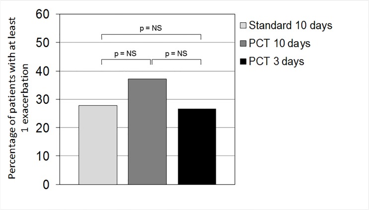
Antibiotic treatment during the 6 months of follow-up was no different between groups (PCT, 20.45% vs. standard, 13.33%; risk difference [95% CI], 7.12 (−3.85;18.09]) (Table 3 and Fig. 3). Time to the next exacerbation in patients treated with antibiotics in the PCT and standard groups was also no different (hazard ratio [95% CI], 1.31[0.69;2.48]). All other clinical measures of outcome and hospitalization were similar between the two groups at the 6-month follow-up (Table 2). Similarly, the scores on quality-of-life questionnaires were no different between groups. The incidence of subsequent pneumonia was also no different between groups (p = 0.365). Few adverse events were reported: 3 deaths in the PCT group and 2 deaths in the standard group (Table 5).
Fig 3. Percentage of patients with at least one exacerbation during the 6 months of follow-up in the three groups: patients who received antibiotics for 3 days (PCT 3 days) or 10 days (PCT 10 days) according to a PCT-guided plan, or for 10 days according to the standard plan. P = not significant.
Table 5. Adverse Events among Study Participants.
| Characteristic | PCT | Standard | ||
|---|---|---|---|---|
| 3 days | 10 days | All PCT patients | 10 days | |
| (n = 45) | (n = 43) | (N = 88) | (N = 90) | |
| No. of patients with at least one serious adverse event (SAE) | 1 | 2 | 3 | 3 |
| No. of patients with at least one non-SAE | - | 1 | 1 | 2 |
| Reported SAE | ||||
| Death | 1 | 2 | 3 | 2 |
| Cause of death* | SCLC | MI,SD | AHF,ARF | |
| Pneumonia | - | - | - | 1 |
| Reported non-SAE | ||||
| Cough | - | - | - | 1 |
| High blood pressure level | - | - | - | 1 |
| Gastric pain | - | 1** | 1** | - |
| Fever | - | - | - | 1 |
| Vomitus | - | 1** | 1** | - |
| Dyspnea | - | - | - | 1 |
| Facial paralysis | - | - | - | 1 |
*SCLC = small cell lung cancer; MI = myocardial infarction; SD = sudden death; AHF = acute heart failure; ARF = acute respiratory failure.
**Probably related to the study medication.
Discussion
Antibiotic treatment of ECOPD patients is widespread, even though evidence for both the bacterial etiology and the efficacy of antibiotic treatment in ECOPD is still controversial. The recognition of clinical/biochemical characteristics that would (i) identify patients with ECOPD due to bacterial infection, (ii) select patients who could most benefit from antibiotic treatment, and (iii) determine the appropriate duration of antibiotic treatment would improve ECOPD management and reduce antibiotic use.
The results of our study failed to demonstrate the noninferiority of PCT-guided antibiotic treatment compared to standard treatment, even though the difference in the upper limit of the 90% CI (15.31) went just slightly beyond the predefined non-inferiority margin (15.31 versus 15). However, we found no significant difference in primary or secondary outcomes between the PCT and standard groups and, more important, between the patients who received antibiotics for 3 days and those who received antibiotics for 10 days. Thus, the results of this study suggest that, in a subject with low levels of PCT, the duration of antibiotic treatment might safely be reduced to 3 days.
Even if a properly designed and powered study is required for corroboration before a specific recommendation can be made, our results are consistent with the efficacy and safety of the PCT-guided antibiotic treatment plan recently suggested in two important articles, i.e., a Cochrane review [32] and an authoritative synopsis of clinical evidence [23].
Our study has several limitations. First, it was underpowered, because we planned to enroll 400 patients and eventually randomized only 183. Thus, the conclusion that antibiotics can be safely stopped after 3 days in patients with low serum PCT cannot be substantiated statistically. Second, we arbitrarily decided on a 10-day duration for the standard group even though the 2005 guidelines recommended 3–10 days [22]. This decision was made because of the severity of ECOPD, which in our study required hospitalization. Third, the calculation of the sample was based on a 50% re-exacerbation risk at 6 months according to a previous similar study [20], whereas the incidence we observed in the standard group was 27.78%. We can only speculate on this discrepancy, but we believe that the lower incidence at 6 months might have been due to two major differences in our study compared to the previous study: (1) We included only patients with either all three criteria of Anthonisen (increased dyspnea, sputum volume, and sputum purulence) or respiratory failure; and (2) we treated all patients for 10 days with full-dose inhaled bronchodilator/combination therapy and a course of oral steroids. Fourth, because we anticipated that the primary outcome (exacerbations of COPD) would be strong and easy to identify, and thus unlikely to be biased by investigator influence, we did not adopt any procedure to reduce bias during the follow-up part of the trial.
To the best of our knowledge, this study is the first randomized multicenter clinical trial testing a PCT-guided antibiotic treatment plan to include only hospitalized patients with severe ECOPD with clinical characteristics requiring antibiotic treatment according to guidelines [8,22]. Previous studies have tested the PCT-guided antibiotic treatment plan in patients hospitalized because of lower respiratory infections and/or ECOPD, but none has specifically included only patients with type 1 Anthonisen’s ECOPD and/or respiratory failure requiring antibiotic treatment [16,17,19,20]. In addition, all previous studies were designed and powered to assess the effect of a PCT plan on the prescription/exposure of patients to antibiotics, and were not designed and powered on clinical outcomes. Also, none of the previous studies investigated the role of a PCT-guided plan for deciding the duration of treatment with antibiotics. Therefore, previous studies could not provide evidence to support or modify the current guidelines regarding duration of antibiotic treatment [8].
In contrast with previous studies, our study was conducted only in patients with clinical characteristics requiring antibiotics, and thus could provide novel evidence about the role of a PCT-guided plan to decide the duration of antibiotic treatment, even though—because of lack of statistical significance regarding noninferiority—our study cannot be considered conclusive. Another strength of our study that was inconsistently present in previous studies is that, according to guidelines, all patients were given oral or intravenous corticosteroids and inhaled bronchodilators for at least 3 days. This allowed us to properly investigate the effects of antibiotics in addition to bronchodilator and steroid treatment. Surprisingly, considering the entry criteria, the prevalence of positive cultures from sputum was low (17%) compared to previously reported data (32%) [33]. We do not have a clear explanation for this discrepancy.
In conclusion, because of the low statistical power of our study, the results leave us with uncertainty about the role of PCT-guided antibiotic treatment in deciding the duration of antibiotic treatment for patients hospitalized with severe ECOPD. PCT may help to identify patients in whom a shorter duration of antibiotic treatment could be safely prescribed, but adequately powered studies are needed to affirm this possibility.
Supporting Information
(DOC)
(PDF)
(PDF)
(PDF)
(PDF)
(DOCX)
(DOCX)
(PDF)
Acknowledgments
We thank the patients who participated in this study. We thank Mary McKenney (professional editor, Michigan, USA) for editing the manuscript, and Elisa Veratelli (University of Ferrara, Ferrara, Italy) for assistance with the preparation of the manuscript.
FARM58J2XH Study Group
Leonardo M. Fabbri (Modena, Coordinating Center, leonardo.fabbri@unimore.it), Bianca Beghé (Modena), Alessia Verduri (Modena), Francesca Franco (Modena), Maria Rosaria De Carlo (Modena), Valentina Ruggieri (Modena), Marco Confalonieri (Trieste), Giuseppe Milani (Trieste), Ernesto Pozzi (Pavia), Maurizio Luisetti (Pavia), Isa Cerveri (Pavia), Rossana Niniano (Pavia), Serafino Marsico (Napoli), Cecilia Calabrese (Napoli), Dario Olivieri (Parma), Panagiota Tzani (Parma), Olga Torre (Parma), Giorgio W. Canonica (Genova), Fulvio Braido (Genova), Antonio Spanevello (Cassano delle Murge, BA), Francesco Salerno (Cassano delle Murge, BA), Mauro Carone (Cassano delle Murge, BA), Luigi Zucchi (Reggio Emilia), Francesco Menzella (Reggio Emilia), Claudia Castagnetti (Reggio Emilia), Pier Luigi Paggiaro (Pisa), Barbara Vagaggini (Pisa), Francesco Costa (Pisa), Alberto Papi (Ferrara), Marco Contoli (Ferrara), Brunilda Marku (Ferrara), Maria Pia Foschino Barbaro (Foggia), Maria Grazia Cagnazzo (Foggia), Nunzio Crimi (Catania), Claudio Mastruzzo (Catania), Michele Ciccarelli (Milano), Stefano Calabro (Bassano del Grappa, VI), Elisabetta Balestro (Bassano del Grappa, VI), Andrea Rossi (Bergamo), Giulio Donazzan (Bolzano), Lucio Bonazza (Bolzano), Renzo Zuin (Padova), Massimo Pistolesi (Firenze), Francesca Bigazzi (Firenze).
Notation of prior abstract publication/presentation
Verduri A, D’Amico R, Ruggieri V, Vicini R, Liverani A, Plebani M, Papi A, Fabbri LM, Beghé B. Pro-Calcitonin guided antibiotic treatment of acute exacerbations of COPD. Am J Respir Crit Care Med 185;2012:A3026. ATS annual meeting; May 18–23, 2012 • San Francisco, California
Data Availability
All relevant data are within the paper and its Supporting Information files. The protocol is available at the following website: http://servertrial.unimore.it/proct/.
Funding Statement
This research was supported by Agenzia Italiana del Farmaco (AIFA, Roma, Italy) (AIFA-FARM58J2XH); Consorzio Ferrara Ricerche (CFR, Ferrara, Italy); and Fondazione Chiesi, Parma, Italy. The funders had no role in study design, data collection and analysis, decision to publish, or preparation of the manuscript.
References
- 1. Donaldson GC, Wedzicha JA (2006) COPD exacerbations. 1: Epidemiology. Thorax 61: 164–168. [DOI] [PMC free article] [PubMed] [Google Scholar]
- 2. Hurst JR (2011) Exacerbation phenotyping in chronic obstructive pulmonary disease. Am J Respir Crit Care Med 184: 625–626. 10.1164/rccm.201106-1136ED [DOI] [PubMed] [Google Scholar]
- 3. White AJ, Gompertz S, Stockley RA (2003) Chronic obstructive pulmonary disease. 6: The aetiology of exacerbations of chronic obstructive pulmonary disease. Thorax 58: 73–80. [DOI] [PMC free article] [PubMed] [Google Scholar]
- 4. Miravitlles M, Anzueto A (2013) Antibiotics for acute and chronic respiratory infection in patients with chronic obstructive pulmonary disease. Am J Respir Crit Care Med 188: 1052–1057. 10.1164/rccm.201302-0289PP [DOI] [PubMed] [Google Scholar]
- 5. Albert R, Connett J, Bailey W, Casaburi R, Cooper JJ, et al. (2011) Azithromycin for prevention of exacerbations of COPD. N Engl J Med 365: 689–698. 10.1056/NEJMoa1104623 [DOI] [PMC free article] [PubMed] [Google Scholar]
- 6. Han M, Tayob N, Murray S, Dransfield M, Washko G, et al. (2014) Predictors of chronic obstructive pulmonary disease exacerbation reduction in. Am J Respir Crit Care Med 189: 1503–1508. 10.1164/rccm.201402-0207OC [DOI] [PMC free article] [PubMed] [Google Scholar]
- 7. Ray W, Murray K, Hall K, Arbogast P, Stein C (2012) Azithromycin and the risk of cardiovascular death. N Engl J Med 366: 1881–1890. 10.1056/NEJMoa1003833 [DOI] [PMC free article] [PubMed] [Google Scholar]
- 8.Global Strategy for the Diagnosis, Management and Prevention of COPD, Global Initiative for Chronic Obstructive Lung Disease (GOLD) (2014) Available: http://www.goldcopd.org/. Last accessed: 2014 April 9.
- 9. Vollenweider DJ, Jarrett H, Steurer-Stey CA, Garcia-Aymerich J, Puhan MA (2012) Antibiotics for exacerbations of chronic obstructive pulmonary disease. Cochrane Database Syst Rev 12: CD010257 10.1002/14651858.CD010257 [DOI] [PubMed] [Google Scholar]
- 10. Daniels JM, Snijders D, de Graaff CS, Vlaspolder F, Jansen HM, et al. (2010) Antibiotics in addition to systemic corticosteroids for acute exacerbations of chronic obstructive pulmonary disease. Am J Respir Crit Care Med 181: 150–157. 10.1164/rccm.200906-0837OC [DOI] [PubMed] [Google Scholar]
- 11. Anthonisen NR, Manfreda J, Warren CP, Hershfield ES, Harding GK, et al. (1987) Antibiotic therapy in exacerbations of chronic obstructive pulmonary disease. Ann Intern Med 106: 196–204. [DOI] [PubMed] [Google Scholar]
- 12. Woodhead M (2011) New guidelines for the management of adult lower respiratory tract infections. Eur Respir J 38: 1250–1251. 10.1183/09031936.00105211 [DOI] [PubMed] [Google Scholar]
- 13. Blasi F, Bocchino M, Di Marco F, Richeldi L, Aliberti S (2012) The role of biomarkers in low respiratory tract infections. Eur J Intern Med 23: 429–435. 10.1016/j.ejim.2012.05.002 [DOI] [PubMed] [Google Scholar]
- 14. Koutsokera A, Stolz D, Loukides S, Kostikas K (2012) Systemic biomarkers in exacerbations of COPD: the evolving clinical challenge. Chest 141: 396–405. 10.1378/chest.11-0495 [DOI] [PubMed] [Google Scholar]
- 15. Soler N, Esperatti M, Ewig S, Huerta A, Agusti C, et al. (2012) Sputum purulence-guided antibiotic use in hospitalised patients with exacerbations of COPD. Eur Respir J 40: 1344–1353. 10.1183/09031936.00150211 [DOI] [PubMed] [Google Scholar]
- 16. Bafadhel M, Clark TW, Reid C, Medina MJ, Batham S, et al. (2011) Procalcitonin and C-reactive protein in hospitalized adult patients with community-acquired pneumonia or exacerbation of asthma or COPD. Chest 139: 1410–1418. 10.1378/chest.10-1747 [DOI] [PMC free article] [PubMed] [Google Scholar]
- 17. Christ-Crain M, Jaccard-Stolz D, Bingisser R, Gencay MM, Huber PR, et al. (2004) Effect of procalcitonin-guided treatment on antibiotic use and outcome in lower respiratory tract infections: cluster-randomised, single-blinded intervention trial. Lancet 363: 600–607. [DOI] [PubMed] [Google Scholar]
- 18. Christ-Crain M, Stolz D, Bingisser R, Muller C, Miedinger D, et al. (2006) Procalcitonin guidance of antibiotic therapy in community-acquired pneumonia: a randomized trial. Am J Respir Crit Care Med 174: 84–93. [DOI] [PubMed] [Google Scholar]
- 19. Schuetz P, Christ-Crain M, Thomann R, Falconnier C, Wolbers M, et al. (2009) Effect of procalcitonin-based guidelines vs standard guidelines on antibiotic use in lower respiratory tract infections: the ProHOSP randomized controlled trial. JAMA 302: 1059–1066. 10.1001/jama.2009.1297 [DOI] [PubMed] [Google Scholar]
- 20. Stolz D, Christ-Crain M, Bingisser R, Leuppi J, Miedinger D, et al. (2007) Antibiotic treatment of exacerbations of COPD: a randomized, controlled trial comparing procalcitonin-guidance with standard therapy. Chest 131: 9–19. [DOI] [PubMed] [Google Scholar]
- 21. Christ-Crain M, Muller B (2005) Procalcitonin in bacterial infections—hype, hope, more or less? Swiss Med Wkly 135: 451–460. [DOI] [PubMed] [Google Scholar]
- 22.Global Strategy for the Diagnosis, Management and Prevention of COPD, Global Initiative for Chronic Obstructive Lung Disease (GOLD) 2005. Available: http://www.goldcopd.org/. Last accessed: 2014 April 9.
- 23. Schuetz P, Briel M, Mueller B (2013) Clinical outcomes associated with procalcitonin algorithms to guide antibiotic therapy in respiratory tract infections. JAMA 309: 717–718. 10.1001/jama.2013.697 [DOI] [PubMed] [Google Scholar]
- 24. Rodriguez-Roisin R (2000) Toward a consensus definition for COPD exacerbations. Chest 117: 398S–401S. [DOI] [PubMed] [Google Scholar]
- 25. Miller MR, Hankinson J, Brusasco V, Burgos F, ATS/ERS Task Force, et al. (2005) Standardization of spirometry. Eur Respir J. Aug;26(2):319–38 [DOI] [PubMed] [Google Scholar]
- 26. Isenberg H (1992) Clinical microbiology procedures handbook Washington, DC; Blackwell. [Google Scholar]
- 27. Baiardini I, Braido F, Fassio O, Tarantini F, Pasquali M, et al. (2005) A new tool to assess and monitor the burden of chronic cough on quality of life: Chronic Cough Impact Questionnaire. Allergy 60: 482–488. [DOI] [PubMed] [Google Scholar]
- 28. Brazier JE, Harper R, Jones NM, O'Cathain A, Thomas KJ, et al. (1992) Validating the SF-36 health survey questionnaire: new outcome measure for primary care. BMJ 305: 160–164. [DOI] [PMC free article] [PubMed] [Google Scholar]
- 29. Mahler DA, Tomlinson D, Olmstead EM, Tosteson AN, O'Connor GT (1995) Changes in dyspnea, health status, and lung function in chronic airway disease. Am J Respir Crit Care Med 151: 61–65. [DOI] [PubMed] [Google Scholar]
- 30. Mahler DA, Weinberg DH, Wells CK, Feinstein AR (1984) The measurement of dyspnea. Contents, interobserver agreement, and physiologic correlates of two new clinical indexes. Chest 85: 751–758. [DOI] [PubMed] [Google Scholar]
- 31. Mathis G (1993) Rare earth cryptates and homogeneous fluoroimmunoassays with human sera. Clin Chem 39: 1953–1959. [PubMed] [Google Scholar]
- 32. Schuetz P, Muller B, Christ-Crain M, Stolz D, Tamm M, et al. (2012)—Procalcitonin to initiate or discontinue antibiotics in acute respiratory tract.—Cochrane Database Syst Rev 2012 Sep 12;9:CD007498 doi:: T—epublish. 10.1002/14651858.CD007498.pub2 [DOI] [PMC free article] [PubMed] [Google Scholar]
- 33. Sethi S, Evans N, Grant BJ, Murphy TF (2002) New strains of bacteria and exacerbations of chronic obstructive pulmonary. N Engl J Med 347: 465–471. [DOI] [PubMed] [Google Scholar]
Associated Data
This section collects any data citations, data availability statements, or supplementary materials included in this article.
Supplementary Materials
(DOC)
(PDF)
(PDF)
(PDF)
(PDF)
(DOCX)
(DOCX)
(PDF)
Data Availability Statement
All relevant data are within the paper and its Supporting Information files. The protocol is available at the following website: http://servertrial.unimore.it/proct/.


