Abstract
Background
Chronic thromboembolic pulmonary hypertension (CTEPH) results from inadequate recanalization of the pulmonary circulation after pulmonary thromboembo-lism. Its 2-year prevalence is 1–4%. If untreated, patients with CTEPH have a mean life expectancy of less than three years. Fortunately, a number of effective treatments are now available.
Methods
This review is based on a selective search of PubMed for pertinent articles published from 1980 to 2014.
Results
The gold-standard test for the exclusion of CTEPH is perfusion scintigraphy: the predictive value of a negative test is nearly 100%. On the other hand, confirmation of a positive diagnosis for treatment planning requires right-heart catheterization and pulmonary angiography. The treatment of first choice for CTEPH is surgical pulmo-nary endarterectomy (PEA), with which about 70% of patients can be cured. The perioperative mortality of this procedure in experienced centers is now 2–4%. Thirty to 50% of all patients with CTEPH are considered inoperable; for these patients, and for patients with persistent pulmonary hypertension after PEA, the drug riociguat was introduced in Germany in 2014 (the first drug specifically introduced for the treatment of CTEPH). There is also a new interventional treatment option for inoperable patients—pulmonary balloon angioplasty, which is currently being performed in a small number of centers.
Conclusion
The timely diagnosis of CTEPH, followed by referral to a specialized center, is now more important than ever, because treatment options are now available for nearly all of the forms in which this disease can manifest itself.
Chronic thromboembolic pulmonary hypertension (CTEPH) is the name given to an increase in mean resting pulmonary arterial tension (≤25 mmHg) with underlying persistent obstruction of the airways after pulmonary thromboembolism, that persists de-spite at least 3 months of appropriate anticoagulation treatment (1). Left untreated, it has a poor prognosis. As recently as the 1980s, the 3-year mortality for advanced CTEPH was over 50% (2). Since then, many therapeutic options have become available, and the disease is now treatable in almost all cases, and in many can even be cured.
The present review is based on a selective search of PubMed that included works published on this topic in the period from 1980 to June 2014. The review concentrates on current methods of diagnosis and treatment of CTEPH.
Epidemiology and pathogenesis
Data on the prevalence of CTEPH in patients 2 to 4 years after experiencing pulmonary embolism range from 0.8% to 3.8% (3, 4). However, around 25% of all patients with CTEPH have no history of clinically manifest pulmonary embolism (5), and for this reason, CTEPH must form part of the differential diagnosis in every patient with unexplained pulmonary hypertension. Ultimately, we have no reliable data about the incidence or prevalence of this disease. Men and women are affected in equal numbers, and the disease may occur at any age, but peak prevalence is in the 60- to 70-year-old age group (5).
Pathogenetically, the primary cause of CTEPH is single or recurrent pulmonary emboli that fail to dissolve, or do so incompletely. In most cases, what causes the endogenous fibrinolysis to be ineffective is unknown. Classical risk factors for the development of CTEPH are thrombophilia, a history of splenectomy, ventriculoatrial shunts, intracardiac pacemaker probes, myelodysplastic syndrome, and chronic inflammatory bowel disease (5, 6). It is possible that the chronic inflammation and recurrent bacteremia often associated with these diseases have a central role in its development (6).
If the emboli do not dissolve completely, they turn into fibrotic scar tissue (7) and no longer regress under the action of anticoagulants. The raised pulmonary vascular resistance results in pulmonary hypertension, which in turn leads in time to microvascular lesions and vascular remodeling in the nonoccluded segments of the airways of the lung (7– 9). This makes CTEPH a progressive disease even in the absence of further thromboembolic events (7, 9). In late-stage CTEPH, right heart failure develops that if left untreated is usually fatal (9).
Diagnosis
The main symptom of CTEPH, like that of all other forms of pulmonary hypertension, is progressive exertional dyspnea, accompanied by fatigue, easy tiring, and in some cases syncope. Signs of manifest right heart failure do not appear until the later stages of the disease. Because the symptoms are nonspecific, especially those in the early disease stages, CTEPH is often diagnosed late or not at all (10). This is unfortunate, since echocardiography, a widely available and noninvasive diagnostic technique, will in most cases answer the question of whether further investigation is required (11). Screening for CTEPH after pulmonary embolism is not recommended (12), but patients with persistent or recurrent exertional dyspnea should be examined by echocardiography (13, 14). Electrocardiography and biomarkers also appear to enable CTEPH to be ruled out with reasonable certainty: a Dutch study found a negative predictive value of 0.99 when electrocardiographic indicators of right heart strain were absent and the values of N-terminal fragments of the pro brain natriuretic peptide (NT-proBNP) were not raised after pulmonary embolism (15). In addition to that, spiroergometry can also deliver important diagnostic clues (16, 17).
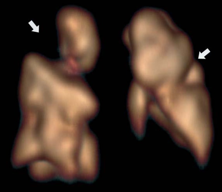
Demonstrating the presence or absence of CTEPH is an essential part of the diagnostic investigation of any patient with pulmonary hypertension of unknown etiology. A history of isolated or recurrent venous thromboembolism makes the diagnosis likely, but even in patients without a suggestive history, it is mandatory to investigate CTEPH thoroughly (5). The most important diagnostic procedure is ventilation–perfusion scintigraphy or (in patients with normal X-ray findings) perfusion scintigraphy alone (Figure 1), which, even in the era of high-resolution computed tomography (CT), still has the highest sensitivity for this disease (18). Its negative predictive value is close to 100%; i.e., a normal perfusion distribution rules out CTEPH with a probability that is close to certainty. On the other hand, the finding of a perfusion defect is not specific for CTEPH, since similar findings are occasionally observed in other forms of pulmonary hypertension or in vasculitis or malignancies of the pulmonary vessels, such as sarcoma (19– 21).
Figure 1.
Perfusion scintigraphy (3-D single photon emission computed tomography, SPECT) in a woman with chronic thromboembolic pulmonary hypertension (CTEPH), showing wedge-shaped perfusion defects (arrows)
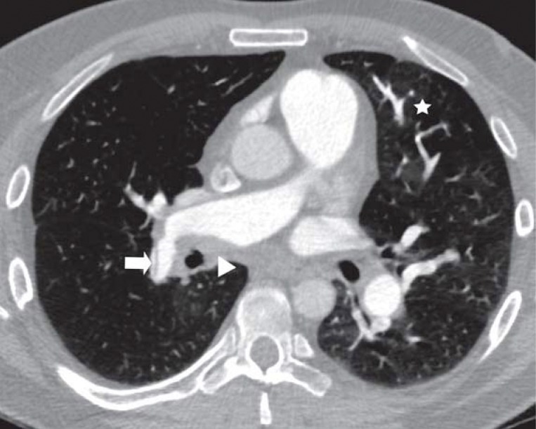
CT angiography can also provide important indicators of the presence of CTEPH. In addition to direct evidence of suggestive lesions in the pulmonary arteries and of proximal wall changes, these include indirect signs. Foremost among these is large bronchial arteries and so-called mosaic perfusion, the name given to the appearance of sharply demarcated adjacent areas of hyper- and hypoperfusion (Figure 2). However, these are all outweighed by the consideration that a normal-appearing angio-CT of the thorax does not rule out the presence of CTEPH with certainty (10, 12, 18).
Figure 2.
Multidetector computed tomography (MDCT) of the central pulmonary arteries in a 46-year-old man with chronic thromboembolic pulmonary hypertension (CTEPH), showing peripheral thrombi in the right main pulmonary artery (arrowhead) and a band-like stenosis in the right lower lobe artery (arrow). In the lung parenchyma, a hint of mosaic perfusion is seen, made up of sharply outlined, field-like hyperdense and hypodense areas—an indirect sign of CTEPH (*)
Modern CT and magnetic resonance techniques may make scintigraphy superfluous in future, if, in addition to the pulmonary vessels, regional lung perfusion can also be routinely imaged (22, 23).
If the history or scintigraphy are suggestive of CTEPH, right heart catheterization and invasive imaging in the form of pulmonary angiography are necessary for treatment planning. These investigations can be combined. Until recently, conventional pulmo-nary angiography was the method of choice because, if correctly carried out, it is generally well suited to answering the question of whether a patient is a candidate for surgery. However, now that interventional techniques have become available as additional treatment methods, there is an increasing need for high-resolution imaging of the smaller pulmonary arteries. There are many new developments in this field, e.g., C-arm CT (also known as cone beam CT or rotational angiography) (24), which combines classical angiography and 3-D CT, allowing segmental and subsegmental pulmonary arteries to be imaged in much greater detail.
Invasive diagnostic imaging for CTEPH should be carried out at centers where operative or interventional treatment can also be performed if required. This can save the patient multiple invasive investigations and exposures to radiation and contrast agents, and is a precondition for optimal treatment outcome. Today it is standard in all German expert centers for treatment to be planned jointly in conference between a multidisciplinary team of experts consisting of internal medical specialists, radiologists, and surgeons, once all the diagnostic findings have been received.
Treatment
Anticoagulation
Lifelong anticoagulation therapy is regarded as a matter of course in patients with CTEPH, although there are no controlled studies on this subject (13). Normally, vitamin K antagonists are given. The new (direct) oral anticoagulants have not yet been studied systematically in patients with CTEPH.
Supportive therapy
Oxygen therapy or diuretics are adjusted to the patient’s individual needs to support the treatment (12). No special studies in patients with CTEPH are available on this. As in other forms of pulmonary hypertension, physical overload should be avoided, since this can lead to syncope or right heart decompensation. Moderate exercise, on the other hand, is worthwhile and can be supported through targeted rehabilitation measures (25).
Vena cava filter
Given the pathogenesis of CTEPH, some experts consider that the use of a vena cava filter is indicated as a matter of principle (26). However, here again no controlled studies have been performed. In European registry data, the postoperative 1-year survival was independent of placement of a vena cava filter (27). In most German centers, these filters are used only on an individual basis, i.e., in patients who continue to suffer thromboembolic events despite appropriate anticoagulation, or in cases where adequate anticoagulation is not possible (12).
Pulmonary endarterectomy
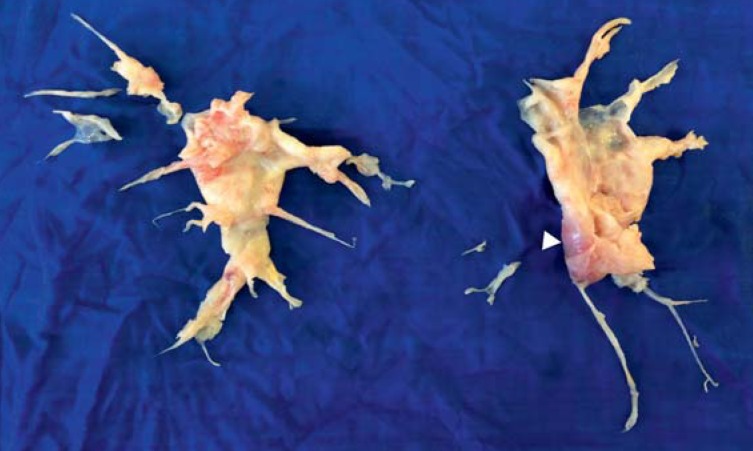
Surgical pulmonary endarterectomy (PEA) is the only potentially curative therapy for CTEPH. Although no controlled study exists, it is regarded as the standard treatment (13, 26). This operation should not be confused with pulmonary embolectomy (Trendelenburg operation) in patients with massive pulmonary embo-lism. PEA is a “true” endarterectomy, in which the intraluminal scar tissue is “peeled away” from the vascular walls (Figure 3). This operation requires the use of the heart–lung machine and intermittent periods of cardiovascular arrest in deep hypothermia. The pulmonary arteries are opened intrapericardially via a median sternotomy. From there, the obstructive material is dissected away as far peripherally as possible (28). The surgery is almost always performed bilaterally.
Figure 3.
Surgical specimens after pulmonary endarterectomy. This postthrombotic scar tissue was “peeled away” from the central pulmonary artery and is laid out here to correspond with the anatomy of the pulmonary arteries (right/left, cranial/caudal), appearing as a kind of cast specimen of the pulmonary arterial tree. Note the reddish thrombus at the bottom of the left-sided specimen (arrowhead)
Perioperative mortality is presently 2% to 4% in experienced centers (27, 29). Cognitive deficits have not been observed despite the intermittent cardiovascular arrest (30). Almost all patients show markedly improved hemodynamics postoperatively. About 70% of those affected show normal or nearly normal pressure values (mean resting pulmonary arterial pressure <25 mmHg), whereas residual pulmonary hypertension can be shown in up to 30% of patients (31, 32). The 10-year survival rate is around 75% (29).
Targeted medical therapy
Although PEA is the preferred treatment for CTEPH, 30% to 50% of patients are not candidates for surgery (5), whether because they have a predominantly peripheral form of the disease, or because concomitant conditions make the operative risk appear too high, or because they refuse surgery. In the past, these patients were mainly treated with drugs that were licensed for the treatment of pulmonary arterial hypertension (PAH) but not for CTEPH: endothelin receptor antagonists, phosphodiesterase-5 (PDE-5) inhibitors, or prostacy-clin analogs. This approach was supported by numerous case series and by open studies. Current guidelines with a low level of evidence recommend targeted medical therapy (12, 13), but so far it has not been confirmed by controlled studies (33, 34).
Until the beginning of 2014, inoperable CTEPH was a disease for which there was no licensed medical treatment. This situation has now changed, since riociguat was licensed on the basis of a positive phase III trial—the first and, so far, the only substance licensed for treatment of inoperable or postoperatively persis-tent CTEPH. Riociguat is a stimulator of soluble guanylate cyclase, which stimulates the target cells, which include the smooth muscle cells of the pulmo-nary arteries, to form guanosine monophosphate (cGMP). In this way a vasodilator effect develops.
The safety and efficacy of riociguat in patients with CTEPH was investigated in the CHEST-1 study, a randomized, placebo-controlled double-blind study. The study enrolled 261 patients with inoperable CTEPH or postoperatively persistent pulmonary hypertension (35). The primary endpoint of the study, a change in 6-minute walking distance after 16 weeks, was reached: in the drug arm, the 6-minute walking distance increased by 39 m, whereas in the placebo arm it dropped by 6 m. The mean placebo-corrected increase in 6-minute walking distance with riociguat treatment was 46 m (95% confidence interval [95% CI]: 25–67 m; p<0.0001). Other, secondary endpoints, including functional class, hemodynamic parameters, and plasma NT-proBNP concentration, were significantly improved in the riociguat group. The most frequent unwanted effects were:
Headache (25% with riociguat versus 14% with placebo)
Dyspepsia (18% versus 8%)
Dizziness (23% versus 12%)
Diarrhea (10% versus 5%)
Hypotension (9% versus 3%).
Hemoptysis was observed in 2% of the patients treated with riociguat; it is not yet clear whether there was a causal connection with the study drug. Long-term data on the course of the disease with riociguat treatment versus no treatment have not yet been published.
Riociguat is not a selective pulmonary vasodilator, but a potent peripheral vasodilator and can therefore cause symptomatic falls in blood pressure. This drug must not be combined with nitrates or PDE-5 inhibitors, and must be given in carefully adjusted doses in accordance with the study protocol (35).
Balloon pulmonary angioplasty
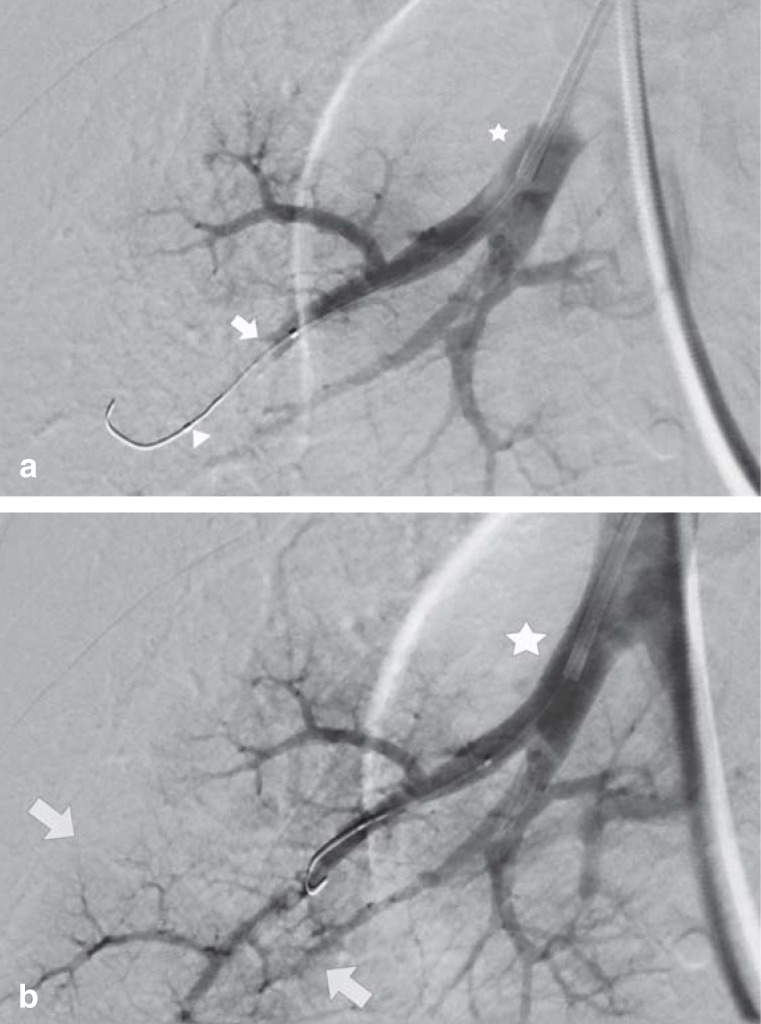
For patients with inoperable CTEPH, balloon pulmo-nary angioplasty (BPA) is another new treatment option, although one that has not yet been evaluated in controlled studies. This procedure was first described in 2001 by colleagues in the United States (36). It was not widely adopted at first because of the occurrence of serious, potentially fatal complications, in particular reperfusion edema and pulmonary hemorrhage. However, since then the technique has been refined, especially at Japanese centers (37, 38). The goal of the intervention is to eliminate the vascular obstruction. The obstruction is caused by the intraluminal postembolic scar-tissue strictures in the form of bands or webs, which are torn and pressed against the vascular walls by the BPA (Figure 4a, b). All the information so far suggests that the vessels remain open after the intervention, and that no restenosis occurs. Therefore, stents are apparently not needed (37– 39). Central stenoses and long vascular occlusions remain the domain of surgery. BPA, on the other hand, is targeted primarily at subsegmental levels that are often inaccessible to surgery. Combined therapeutic approaches may be possible in some cases, but as a matter of principle surgery should come first (12, 26).
Figure 4.
Pulmonary balloon angioplasty of the A8 segment of the right lower lobe.
a) Selective imaging of the right A8 segmental artery, in which the contrast is visibly interrupted at the right cardiac margin (arrow). The stenosis has already been successfully passed with a wire (arrowhead).
b) Selective DSA series (DSA, digital subtraction angiography) to follow up treatment outcome after dilatation of web stenosis with a suitable balloon catheter. There is clear contrast of the peripheral branches of the A8 segmental artery, with visualization of even the smallest subbranches of the pulmonary artery (arrows). The location of the guide catheter is the same in both series (*)
The interventions are carried out in several sessions, each restricted to one pulmonary lobe at most, to minimize the risk of serious bleeding or reperfusion edema, and the exposure to contrast agents and radiation (40). To avoid rupturing the thin-walled pulmonary arteries, balloons are used that have a smaller diameter than the internal diameter of the target vessels (37). This necessitates high-resolution imaging of the small pulmonary arteries, enabling positive identification of the target vessels and hence correct calibration of the balloons (24). In this way, it is now possible to recanalize even vessels that are completely occluded for a short stretch (38). Four to six sessions are required for treatment to be completed. The risk of serious or fatal complications has now been brought down to less than 1%. Published data now also show that, in addition to hemodynamics, right heart function significantly improves, and excercise capacity increases markedly (37, 38, 40). Nevertheless, BPA cannot yet be regarded as a standard treatment because no robust long-term data exist. Moreover, there is still no adequate definition of which patients are the most likely to benefit from this treatment technique.
Lung transplantation
Because of the treatment options outlined above, it has become very rare for CTEPH patients to require a lung transplant. Nevertheless, it remains an option in patients who cannot be adequately treated in any other way (26).
Summary
CTEPH is a rare but serious complication of pulmonary thromboembolism that must be included in the differential diagnosis of any case of pulmonary hypertension of unknown origin—the more so because not all patients with this condition report a history of venous thromboembolism. Perfusion scintigraphy is an essential part of the diagnostic investigations, because it is the only noninvasive imaging technique that allows CTEPH to be ruled out with almost 100% certainty.
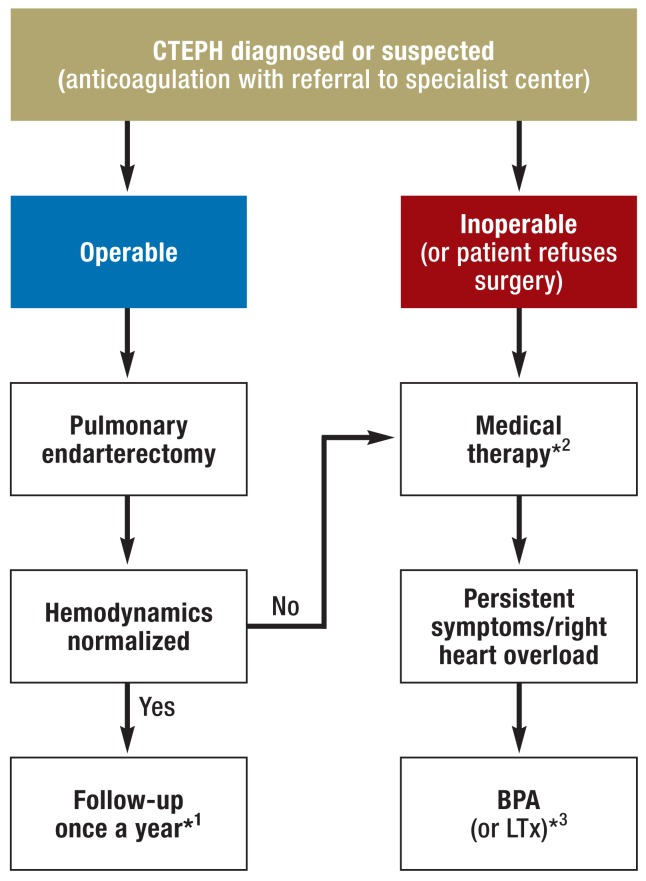
The final diagnostic investigations and treatment planning require a right heart catheter and high-resolu-tion pulmonary angiography, and should be carried out at the same center where treatment will be given. The treatment options for CTEPH have improved greatly in recent years, such that today almost every patient can be offered some form of effective treatment (Figure 5). The treatment of first choice is and remains PEA, as this is the only treatment that can offer the patient the prospect of cure. Both medical treatment with riociguat and BPA should be restricted to patients for whom surgery is not an option and those with persistent pulmonary hypertension after PEA, because at present there are no robust long-term data for these therapies.
Figure 5.
Treatment algorithm for chronic thromboembolic pulmonary hypertension (CTEPH) (adapted from [1]).
BPA, balloon pulmonary angioplasty; LTx, lung transplantation
*1 Clinical examination and echocardiography usually sufficient.
*2 At present, only riociguat is licensed for medical treatment of inoperable CTEPH.
*3 Lung transplantation is now only rarely required for CTEPH
In the near future, the focus of research will certainly be on the development of further medical treatments for patients with inoperable CTEPH. For this, in particular, long-term data are needed. In addition, it needs to be more precisely defined which patients are most likely to benefit from BPA, and whether in some cases hybrid procedures consisting of surgery and BPA might be valuable.
Key Messages.
Chronic thromboembolic pulmonary hypertension (CTEPH) is a life-threatening disease that occurs in about 1% to 4% of patients who have suffered a pulmonary thromboembolism.
The most important diagnostic investigation to rule out CTEPH is perfusion scintigraphy, which should be carried out in all patients with pulmonary hypertension of unknown origin—even if there is no indication of pulmonary embolism in the history.
The most effective treatment for CTEPH is surgical pulmonary endarterectomy (PEA), which results in marked improvement of hemodynamic and clinical parameters in almost all patients. In most cases, it is curative.
For patients in whom surgery is not possible, and those in whom pulmonary hypertension persists postoperatively, a medical therapy became available when riociguat was licensed in Germany at the beginning of 2014.
Pulmonary balloon angioplasty is another new therapeutic option for patients with inoperable CTEPH. This procedure is currently under evaluation at various centers.
Acknowledgments
Translated from the original German by Kersti Wagstaff, MA.
Footnotes
Conflict of interest statement
Dr. Olsson has received reimbursement of conference fees and travel expenses from Bayer Healthcare. She has received lecture fees from Actelion, Pfizer, GSK, and Bayer.
Dr. Meyer has received lecture fees and reimbursement of conference fees and travel expenses from Bayer.
Professor Hoeper has received study funding (third-party) and consultancy and lecture fees from Actelion, Bayer, GSK, and Pfizer.
Dr. Cebotari has received consultancy fees and reimbursement of conference fees and travel expenses from Bayer.
Drs. Hinrichs and Vogel-Claussen declare that no conflict of interest exists.
References
- 1.Hoeper MM, Madani MM, Nakanishi N, Meyer B, Cebotari S, Rubin LJ. Chronic thromboembolic pulmonary hypertension. Lancet Respir Medicine. 2014;2:573–582. doi: 10.1016/S2213-2600(14)70089-X. [DOI] [PubMed] [Google Scholar]
- 2.Riedel M, Stanek V, Widimsky J, Prerovsky I. Longterm follow-up of patients with pulmonary thromboembolism. Late prognosis and evolution of hemodynamic and respiratory data. Chest. 1982;81:151–158. doi: 10.1378/chest.81.2.151. [DOI] [PubMed] [Google Scholar]
- 3.Becattini C, Agnelli G, Pesavento R, et al. Incidence of chronic thromboembolic pulmonary hypertension after a first episode of pulmonary embolism. Chest. 2006;130:172–175. doi: 10.1378/chest.130.1.172. [DOI] [PubMed] [Google Scholar]
- 4.Pengo V, Lensing AW, Prins MH, et al. Incidence of chronic thromboembolic pulmonary hypertension after pulmonary embolism. N Engl J Med. 2004;350:2257–2264. doi: 10.1056/NEJMoa032274. [DOI] [PubMed] [Google Scholar]
- 5.Pepke-Zaba J, Delcroix M, Lang I, et al. Chronic thromboembolic pulmonary hypertension (CTEPH): results from an international prospective registry. Circulation. 2011;124:1973–1981. doi: 10.1161/CIRCULATIONAHA.110.015008. [DOI] [PubMed] [Google Scholar]
- 6.Bonderman D, Wilkens H, Wakounig S, et al. Risk factors for chronic thromboembolic pulmonary hypertension. Eur Respir J. 2009;33:325–331. doi: 10.1183/09031936.00087608. [DOI] [PubMed] [Google Scholar]
- 7.Lang IM, Pesavento R, Bonderman D, Yuan JX. Risk factors and basic mechanisms of chronic thromboembolic pulmonary hypertension: a current understanding. Eur Respir J. 2013;41:462–468. doi: 10.1183/09031936.00049312. [DOI] [PubMed] [Google Scholar]
- 8.Moser KM, Bloor CM. Pulmonary vascular lesions occurring in patients with chronic major vessel thromboembolic pulmonary hypertension. Chest. 1993;103:685–692. doi: 10.1378/chest.103.3.685. [DOI] [PubMed] [Google Scholar]
- 9.Delcroix M, Vonk Noordegraaf A, Fadel E, Lang I, Simonneau G, Naeije R. Vascular and right ventricular remodelling in chronic thromboembolic pulmonary hypertension. Eur Respir J. 2013;41:224–232. doi: 10.1183/09031936.00047712. [DOI] [PubMed] [Google Scholar]
- 10.Lang IM, Madani M. Update on chronic thromboembolic pulmonary hypertension. Circulation. 2014;130:508–518. doi: 10.1161/CIRCULATIONAHA.114.009309. [DOI] [PubMed] [Google Scholar]
- 11.Otero R, Oribe M, Ballaz A, et al. Echocardiographic assessment of pulmonary arterial pressure in the follow-up of patients with pulmonary embolism. Thromb Res. 2011;127:303–308. doi: 10.1016/j.thromres.2010.12.010. [DOI] [PubMed] [Google Scholar]
- 12.Konstantinides S, Torbicki A, Agnelli G, et al. 2014 ESC Guidelines on the diagnosis and management of acute pulmonary embolism. The task force for the diagnosis and management of acute pulmonary embolism of the European Society of Cardiology (ESC) endorsed by the European Respiratory Society (ERS) Eur Heart J. 2014 pii ehu283 [Epub ahead of print] [Google Scholar]
- 13.Galie N, Hoeper MM, Humbert M, et al. Guidelines for the diagnosis and treatment of pulmonary hypertension. The task force for the diagnosis and treatment of pulmonary hypertension of the European Society of Cardiology (ESC) and the European Respiratory Society (ERS) endorsed by the International Society of Heart and Lung Transplantation (ISHLT) Eur Respir J. 2009;34:1219–1263. doi: 10.1183/09031936.00139009. [DOI] [PubMed] [Google Scholar]
- 14.Surie S, Gibson NS, Gerdes VE, et al. Active search for chronic thromboembolic pulmonary hypertension does not appear indicated after acute pulmonary embolism. Thromb Res. 2010;125:e202–e205. doi: 10.1016/j.thromres.2009.12.016. [DOI] [PubMed] [Google Scholar]
- 15.Klok FA, Surie S, Kempf T, et al. A simple non-invasive diagnostic algorithm for ruling out chronic thromboembolic pulmonary hypertension in patients after acute pulmonary embolism. Thromb Res. 2011;128:21–26. doi: 10.1016/j.thromres.2011.03.004. [DOI] [PubMed] [Google Scholar]
- 16.Scheidl SJ, Englisch C, Kovacs G, et al. Diagnosis of CTEPH versus IPAH using capillary to end-tidal carbon dioxide gradients. Eur Respir J. 2012;39:119–124. doi: 10.1183/09031936.00109710. [DOI] [PubMed] [Google Scholar]
- 17.Held M, Grun M, Holl R, et al. Cardiopulmonary exercise testing to detect chronic thromboembolic pulmonary hypertension in patients with normal echocardiography. Respiration. 2014;87:379–387. doi: 10.1159/000358565. [DOI] [PubMed] [Google Scholar]
- 18.Tunariu N, Gibbs SJ, Win Z, et al. Ventilation-perfusion scintigraphy is more sensitive than multidetector CTPA in detecting chronic thromboembolic pulmonary disease as a treatable cause of pulmonary hypertension. J Nucl Med. 2007;48:680–684. doi: 10.2967/jnumed.106.039438. [DOI] [PubMed] [Google Scholar]
- 19.Kerr KM, Auger WR, Fedullo PF, Channick RH, Yi ES, Moser KM. Large vessel pulmonary arteritis mimicking chronic thromboembolic disease. Am J Respir Crit Care Med. 1995;152:367–373. doi: 10.1164/ajrccm.152.1.7599847. [DOI] [PubMed] [Google Scholar]
- 20.Bailey CL, Channick RN, Auger WR, et al. „High probability“ perfusion lung scans in pulmonary venoocclusive disease. Am J Respir Crit Care Med. 2000;162:1974–1978. doi: 10.1164/ajrccm.162.5.2003045. [DOI] [PubMed] [Google Scholar]
- 21.Kauczor HU, Schwickert HC, Mayer E, Kersjes W, Moll R, Schweden F. Pulmonary artery sarcoma mimicking chronic thromboembolic disease: computed tomography and magnetic resonance imaging findings. Cardiovasc Intervent Radiol. 1994;17:185–189. doi: 10.1007/BF00571532. [DOI] [PubMed] [Google Scholar]
- 22.Rajaram S, Swift AJ, Telfer A, et al. 3D contrast-enhanced lung perfusion MRI is an effective screening tool for chronic thromboembolic pulmonary hypertension: results from the ASPIRE Registry. Thorax. 2013;68:677–678. doi: 10.1136/thoraxjnl-2012-203020. [DOI] [PubMed] [Google Scholar]
- 23.Hong YJ, Kim JY, Choe KO, et al. Different perfusion pattern between acute and chronic pulmonary thromboembolism: evaluation with two-phase dual-energy perfusion CT. AJR Am J Roentgenol. 2013;200:812–817. doi: 10.2214/AJR.12.8697. [DOI] [PubMed] [Google Scholar]
- 24.Sugiyama M, Fukuda T, Sanda Y, et al. Organized thrombus in pulmonary arteries in patients with chronic thromboembolic pulmonary hypertension; imaging with cone beam computed tomography. Jpn J Radiol. 2014;32:375–382. doi: 10.1007/s11604-014-0319-8. [DOI] [PubMed] [Google Scholar]
- 25.Nagel C, Prange F, Guth S, et al. Exercise training improves exercise capacity and quality of life in patients with inoperable or residual chronic thromboembolic pulmonary hypertension. PloS one. 2012;7 doi: 10.1371/journal.pone.0041603. [DOI] [PMC free article] [PubMed] [Google Scholar]
- 26.Kim NH, Delcroix M, Jenkins DP, et al. Chronic thromboembolic pulmonary hypertension. J Am Coll Cardiol. 2013;62:D92–D99. doi: 10.1016/j.jacc.2013.10.024. [DOI] [PubMed] [Google Scholar]
- 27.Mayer E, Jenkins D, Lindner J, et al. Surgical management and outcome of patients with chronic thromboembolic pulmonary hypertension: results from an international prospective registry. J Thorac Cardiovasc Surg. 2011;141:702–710. doi: 10.1016/j.jtcvs.2010.11.024. [DOI] [PubMed] [Google Scholar]
- 28.Jenkins DP, Madani M, Mayer E, et al. Surgical treatment of chronic thromboembolic pulmonary hypertension. Eur Respir J. 2013;41:735–742. doi: 10.1183/09031936.00058112. [DOI] [PubMed] [Google Scholar]
- 29.Madani MM, Auger WR, Pretorius V, et al. Pulmonary endarter-ectomy: recent changes in a single institution’s experience of more than 2,700 patients. Ann Thorac Surg. 2012;94:97–103. doi: 10.1016/j.athoracsur.2012.04.004. discussion 103. [DOI] [PubMed] [Google Scholar]
- 30.Vuylsteke A, Sharples L, Charman G, et al. Circulatory arrest versus cerebral perfusion during pulmonary endarterectomy surgery (PEACOG): a randomised controlled trial. Lancet. 2011;378:1379–1387. doi: 10.1016/S0140-6736(11)61144-6. [DOI] [PubMed] [Google Scholar]
- 31.Skoro-Sajer N, Marta G, Gerges C, et al. Surgical specimens, haemodynamics and long-term outcomes after pulmonary endarterectomy. Thorax. 2014;69:116–122. doi: 10.1136/thoraxjnl-2013-203746. [DOI] [PMC free article] [PubMed] [Google Scholar]
- 32.Freed DH, Thomson BM, Berman M, et al. Survival after pulmonary thromboendarterectomy: effect of residual pulmonary hypertension. J Thorac Cardiovasc Surg. 2011;141:383–387. doi: 10.1016/j.jtcvs.2009.12.056. [DOI] [PubMed] [Google Scholar]
- 33.Jais X, D’Armini AM, Jansa P, et al. Bosentan for treatment of inoperable chronic thromboembolic pulmonary hypertension BENEFiT (Bosentan Effects in iNopErable Forms of chronic Thromboembolic pulmonary hypertension), a randomized, placebo-controlled trial. J Am Coll Cardiol. 2008;52:2127–2134. doi: 10.1016/j.jacc.2008.08.059. [DOI] [PubMed] [Google Scholar]
- 34.Suntharalingam J, Treacy CM, Doughty NJ, et al. Long-term use of sildenafil in inoperable chronic thromboembolic pulmonary hypertension. Chest. 2008;134:229–236. doi: 10.1378/chest.07-2681. [DOI] [PubMed] [Google Scholar]
- 35.Ghofrani HA, D’Armini AM, Grimminger F, et al. Riociguat for the treatment of chronic thromboembolic pulmonary hypertension. N Engl J Med. 2013;369:319–329. doi: 10.1056/NEJMoa1209657. [DOI] [PubMed] [Google Scholar]
- 36.Feinstein JA, Goldhaber SZ, Lock JE, Ferndandes SM, Landzberg MJ. Balloon pulmonary angioplasty for treatment of chronic thromboembolic pulmonary hypertension. Circulation. 2001;103:10–13. doi: 10.1161/01.cir.103.1.10. [DOI] [PubMed] [Google Scholar]
- 37.Kataoka M, Inami T, Hayashida K, et al. Percutaneous transluminal pulmonary angioplasty for the treatment of chronic thromboembolic pulmonary hypertension. Circulation Cardiovascular interventions. 2012;5:756–762. doi: 10.1161/CIRCINTERVENTIONS.112.971390. [DOI] [PubMed] [Google Scholar]
- 38.Mizoguchi H, Ogawa A, Munemasa M, Mikouchi H, Ito H, Matsubara H. Refined balloon pulmonary angioplasty for inoperable patients with chronic thromboembolic pulmonary hypertension. Circulation Cardiovascular Interventions. 2012;5:748–755. doi: 10.1161/CIRCINTERVENTIONS.112.971077. [DOI] [PubMed] [Google Scholar]
- 39.Sugimura K, Fukumoto Y, Satoh K, et al. Percutaneous transluminal pulmonary angioplasty markedly improves pulmonary hemodynamics and long-term prognosis in patients with chronic thromboembolic pulmonary hypertension. Circ J. 2012;76:485–488. doi: 10.1253/circj.cj-11-1217. [DOI] [PubMed] [Google Scholar]
- 40.Fukui S, Ogo T, Morita Y, et al. Right ventricular reverse remodelling after balloon pulmonary angioplasty. Eur Respir J. 2014;43:1394–1402. doi: 10.1183/09031936.00012914. [DOI] [PubMed] [Google Scholar]


