Abstract
Preliminary animal experiments have confirmed that sensory nerve fibers promote osteoblast differentiation, but motor nerve fibers have no promotion effect. Whether sensory neurons promote the proliferation and osteogenic differentiation of bone marrow mesenchymal stem cells remains unclear. No results at the cellular level have been reported. In this study, dorsal root ganglion neurons (sensory neurons) from Sprague-Dawley fetal rats were co-cultured with bone marrow mesenchymal stem cells transfected with green fluorescent protein 3 weeks after osteogenic differentiation in vitro, while osteoblasts derived from bone marrow mesenchymal stem cells served as the control group. The rat dorsal root ganglion neurons promoted the proliferation of bone marrow mesenchymal stem cell-derived osteoblasts at 3 and 5 days of co-culture, as observed by fluorescence microscopy. The levels of mRNAs for osteogenic differentiation-related factors (including alkaline phosphatase, osteocalcin, osteopontin and bone morphogenetic protein 2) in the co-culture group were higher than those in the control group, as detected by real-time quantitative PCR. Our findings indicate that dorsal root ganglion neurons promote the proliferation and osteogenic differentiation of bone marrow mesenchymal stem cells, which provides a theoretical basis for in vitro experiments aimed at constructing tissue-engineered bone.
Keywords: nerve regeneration, bone marrow mesenchymal stem cells, bone, osteoblasts, ganglion, spine, neurons, co-culture techniques, proliferation, differentiation, real-time quantitative PCR, NSFC grants, neural regeneration
Introduction
Although our understanding of the mechanisms underlying bone regeneration method has greatly improved, the clinical treatment of large bone defects remains difficult. Autogenous bone graft is regarded as the gold standard surgical treatment, but this approach induces some complications such as bone nonunion and additional damage (Mahendra and Maclean, 2007).
Construction of tissue-engineered bone tissue is a potential way to treat bone defects. Bone is not only a kind of hard tissue, but also a complex body containing abundant nerves, blood vessels and fascia (Togari et al., 2005). Nerves play an extensive and crucial role in the development, formation and metabolism of bone tissue (Imai et al., 1997). Nerve cells function to regulate bone formation and resorption. Growing evidence from animal experiments has shown that sensory nerves promote bone formation, but this effect was not observed with motor nerves (Dai et al., 2008). Therefore, both bone tissue and surrounding tissue (such as blood vessels and nerve tissue) require surgical repair prior to reconstruction (Haastert et al., 2006).
In the present study, we aimed to verify whether dorsal root ganglion (DRG) neurons promote the proliferation and differentiation of bone marrow mesenchymal stem cell (BMSC)-derived osteoblasts at the cellular level. We co-cultured DRG neurons with the osteoblasts in a broader attempt to explore the correlation between the two and to lay the foundation for further in vivo experiments.
Materials and Methods
Experimental animals
Female Sprague-Dawley rats at pregnant 15 days and healthy male 2-week-old Sprague-Dawley rats, weighing 80 g, were obtained from the Experimental Animal Center of Binzhou Medical University (license No. SYXK (Lu) 20130020) in China. All investigations conformed to the Guide for the Care and Use of Laboratory Animals published by the Ministry of Science and Technology of China in 2006.
Isolation, culture, identification and osteogenic differentiation of rat BMSCs
Healthy male 2-week-old Sprague-Dawley rats were killed by decapitation under anesthesia and soaked in 75% ethanol for 15 minutes. The rat femur and tibia were then harvested aseptically, and soft tissue attached to the skeleton was removed. After the medullary cavity was rinsed twice with sterile normal saline, bone marrow was directly collected and placed in a tube, and centrifuged at 300 × g for 10 minutes. The supernatant was discarded and the BMSCs were resuspended in standard culture medium (Cyagen Biosciences Inc., Sunnyvale, CA, USA) and counted. Cells were then cultured in 25 cm2 culture flasks at 37°C, in a 5% CO2 incubator. The culture medium was replenished 24 hours later and then changed every 2–3 days. When the cells reached 80–90% confluence (at 3–5 days of primary culture), cells were digested and subcultured using 0.25% trypsin. Passage 3 BMSCs were observed under an inverted phase contrast microscope and identified by flow cytometry analysis. After identification, passage 3 BMSCs were induced for 14 days in an osteogenic induction liquid, which consisted of high-glucose DMEM (HyClone, Logan, UT, USA), 10% fetal bovine serum (HyClone), 1 × 10⁻⁸ M dexamethasone, 10 mM β-glycerophosphate and 50 μg/mL vitamin C (Sigma, St. Louis, MO, USA). The induced cells were then detected using flow cytometry and alkaline phosphatase staining (Nanjing Jiancheng Bioengineering Co., Ltd., Nanjing, Jiangsu Province, China). The morphology of BMSCs was observed under an inverted phase contrast microscope (Nikon, Tokyo, Japan).
Isolation, culture and identification of rat DRG neurons
Female Sprague-Dawley rats at pregnant 15 days were anesthetized and sacrificed by cervical dislocation. After abdominal skin was disinfected with 75% ethanol, an axial incision was made, the uterus was harvested and placed in a dish containing D-Hank's solution. Fetal rats were removed and fetal spine was isolated, followed by removal of the DRG attached to the spine. DRG samples were rinsed with D-Hank's solution, separated under a microscope, placed in a 15 mL centrifuge tube, digested with 3–4 mL of 0.25% trypsin and 3–4 mL of 0.02% EDTA, and incubated at 37°C for 50 minutes. After digestion was terminated, cells were triturated and a cell suspension was collected. The obtained suspension was incubated with Neurabasal + B27 medium on cover slips. Half of the culture medium was changed every 24–48 hours and 1 × 10⁻⁵ nM cytarabine was added to the culture medium; cytarabine treatment was terminated 24–48 hours later. DRG neurons were identified using microtubule-associated protein 2 (MAP2) and 4′,6-diamidino-2-phenylindole (DAPI) double staining, and observed under a fluorescence microscope (Nikon, Tokyo, Japan).
Proliferation-promoting effect in a 24-well co-culture test
Osteoblasts differentiated from rat BMSCs (Cyagen) that were previously transfected with green fluorescent protein (GFP) were used in this study. After selection, the transfection efficiency was up to 90%. The 24-well culture plate was divided into nine regions at the bottom and cells were counted under an inverted phase contrast microscope (Nikon).
Osteoblasts were divided into two groups as follows. (1) DRG neurons group: DRGs previously cultured on cover slips were transferred to 24-well culture plates, and GFP-transfected osteoblasts were prepared into cell suspensions at a density of 1 × 10⁴/mL. Then, 80 μL of the cell suspension was placed in the 24-well plates and cells were cultured with high-glucose DMEM culture medium. (2) Blank group: GFP-transfected rat BMSCs were prepared into cell suspensions at a density of 1 × 10⁴/mL. Then, 80 μL of the cell suspension was placed in the 24-well plates and cells were cultured with high-glucose DMEM culture medium. Each well contained nine regions. At 1, 3, and 5 days of co-culture, cells were counted under a fluorescence microscope (Nikon, Tokyo, Japan). The experiments were repeated three times.
Real-time fluorescent quantitative PCR detection
Co-culture of DRG neurons and BMSC-derived osteoblasts: The following three groups were used: co-culture 3 days, co-culture 5 days, and control groups. In the co-culture groups, DRG neurons were cultured on cover slips and placed in 6-well plates. The osteoblasts were prepared into cell suspensions at 1 × 10⁴/mL, and 100 μL of the suspension was added to the 6-well plates; the cells were then co-cultured for 3 or 5 days. In the control group, osteoblasts were prepared into cell suspensions at 1 × 10⁴/mL and 100 μL of the suspension was added to the 6-well plates. There were three wells for each group. All cells were cultured in high-glucose DMEM culture medium to induce osteogenic differentiation. The experiment was repeated three times.
The levels of mRNAs for osteocalcin, osteopontin, alkaline phosphatase and bone morphogenetic protein 2 were detected using real-time fluorescent quantitative PCR (Bio-Rad, Hercules, CA, USA) (Table 1). Total RNA was extracted according to the instructions of the Trizol Isolation Reagent kit. During the experiment, all containers and pipettes were treated by diethylpyrocarbonate under high pressure. The optical density at 260 and 280 nm was read on a DU800 UV spectrophotometer, to determine total RNA purity and concentration. The 2 × AllinOne™ Q-PCR Mix was thawed at room temperature, and the PCR reaction mix was prepared on ice with the addition of the following: 2 × AllinOne™ Q-PCR Mix, 10 μL; AllinOne™ Q-PCR Primer at a final concentration of 2 μM, 2 μL; cDNA (1:5), 2 μL; ddH2O. The PCR reaction mix was placed into 96-well plates and centrifuged. A standard three-step method was adopted for the PCR procedure: denaturation at 95°C for 10 minutes; 40 cycles of denaturation at 95°C for 10 seconds, annealing at 60°C for 20 seconds, and extension at 72°C for 15 seconds. Real-time quantitative PCR data were analyzed using the 2−ΔΔCt method (Livak and Schmittgen, 2001).
Table 1.
Real-time fluorescent quantitative PCR primer sequences

Statistical analysis
Measurement data are expressed as the mean ± SD and were statistically analyzed using SPSS 16.0 software (SPSS, Chicago, IL, USA). Differences among groups were compared using one-way analysis of variance and intergroup comparisons were performed using the Student-Newman-Keuls method. Data from the co-culture test were analyzed using repeated measures analysis of variance, and intragroup pairwise comparisons were performed using the Bonferroni test. A P level < 0.05 was considered to indicate a significant difference.
Results
Identification of rat DRG neurons cultured in vitro
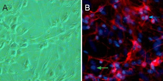
The results of MAP2 and DAPI double staining are shown in Figure 1. After 60 days in culture, rat DRG neurons positive for both MAP2 and DAPI were visible under a fluorescence microscope.
Figure 1.

Primary cultured rat dorsal root ganglion neurons, identified by MAP2 and DAPI double staining (fluorescence microscopy, × 200).
(A) Morphology of dorsal root ganglion neurons cultured for 60 hours. (B) Dorsal root ganglion neurons cultured for 60 days were detected using MAP2 and DAPI staining, MAP2-labeled neurons emitted red fluorescence in the cell bodies and neurites; DAPI-labeled neurons emitted blue fluorescence in the nuclei. Double-labeled cells are neurons; the blue arrow shows MAP2 and DAPI double-positive neurons, while the green arrow shows non-neuronal nuclei. MAP2: Microtubule-associated protein 2; DAPI: 4′,6-diamidino-2-phenylindole.
Morphology and osteogenic identification of rat BMSCs
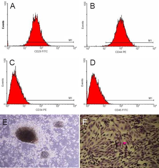
At 2 weeks in culture, passage 3 BMSCs were long and fusiform and had a clear boundary under an inverted fluorescence microscope. Alkaline phosphatase staining and flow cytometry results showed that the rat BMSCs were successfully differentiated (Figure 2A–F).
Figure 2.

Morphology, identification (flow cytometry) and osteogenic differentiation of rat bone marrow mesenchymal stem cells after 2 weeks of culture.
(A, B) Rat bone marrow mesenchymal stem cells were positive for CD29 and CD44. (C, D) Rat bone marrow mesenchymal stem cells were negative for CD34 and CD45. (E) Morphology of primary cultured rat bone marrow mesenchymal stem cells (inverted phase contrast microscope, × 40). (F) Morphology of rat bone marrow mesenchymal stem cells after alkaline phosphatase staining (inverted phase contrast microscope, × 40). The red arrow shows alkaline phosphatase-positive cells.
Effect of rat DRG neurons on the proliferation of BMSC-derived osteoblasts
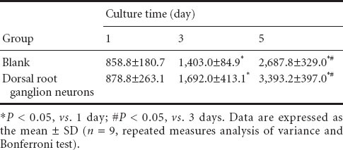
The results of the proliferation promotion test of DRG neurons are shown in Table 2. DRG neurons showed no obvious effect in terms of promoting the proliferation of BMSC-derived osteoblasts at 1 and 3 days after co-culture (P > 0.05); DRG neurons obviously promoted the proliferation of BMSC-derived osteoblasts at 5 days after co-culture (P < 0.05).
Table 2.
Effect of rat dorsal root ganglion neurons on the proliferation of osteoblasts derived from bone marrow mesenchymal stem cells

Osteocalcin, osteopontin, alkaline phosphatase and bone morphogenetic protein 2 mRNA expression after co-culture of rat DRG neurons and BMSC-derived osteoblasts
Real-time fluorescent quantitative PCR analysis showed that DRG neurons promoted the osteogenic differentiation of BMSC-derived osteoblasts after 3 and 5 days in co-culture. The co-culture groups showed higher expression levels of osteocalcin, osteopontin, alkaline phosphatase and bone morphogenetic protein 2 mRNA than did the control group (P < 0.05; Table 3).
Table 3.
Osteocalcin, osteopontin, alkaline phosphatase and bone morphogenetic protein 2 mRNA expression (2–ΔΔ Ct) after co-culture of rat dorsal root ganglion neurons with bone marrow mesenchymal stem cell-derived osteoblasts

Discussion
Bone contains abundant nerves, blood vessels and fascia (Takeda et al., 2002). Bone and nerve tissue are adjacent and closely linked to each other. In the past decade, several research groups have confirmed the presence of a variety of neurotransmitter receptors and peptidergic nerve fibers in bone cells (Goto and Tanaka, 2002; Togari, 2002, 2005), indicating that nerve cells regulate bone formation and resorption. The ultimate goal of ongoing research in the field is to construct a skeleton of normal physiological state using tissue engineering techniques; that is, tissue-engineered bone. For this purpose, we need to explore the relationship between nerve cells and osteoblasts at the cellular level. Sensory nerves have been shown to promote osteogenesis, while no significant effect of motor nerves was observed. The effect of sensory nerve bundles on promoting bone formation was similar to that of single vascular transplantation: both sensory nerve fibers and vascular bundles promoted the regeneration and restoration of tissue-engineered bone (Chen et al., 2010). In the process of constructing tissue-engineered bone, calcitonin gene-related peptide (CGRP), substance P (SP) and neuropeptide Y were dynamically monitored, and it was found that the levels of these neuropeptides increase during bone formation and modeling (Goto et al., 2007). This highlights the contribution of nerve tissue to bone growth. However, these findings were obtained in animal experiments, and verification at the cellular level is rare. Whether sensory neurons promote the proliferation and osteogenic differentiation of BMSCs remains an unanswered question. In this study, we verified this hypothesis through the co-culture of two kinds of cells.
In this study, the proliferation of BMSC-derived osteoblasts co-cultured with DRG neurons was more pronounced than that in the control group. This indicated that DRG neurons promoted the proliferation of BMSCs. How to effectively co-culture DRG neurons with BMSC-differentiated osteoblasts has been a difficult issue in previous studies, and no relevant experimental support was obtained. The culture medium for DRG neurons and BMSC induction are different, and DRG neurons do not survive well; if osteoblast culture medium is applied in the co-culture process, the survival of DRG neurons is sharply decreased, and the co-culture is prone to failure after 7 days. In addition, osteoblasts proliferate rapidly and are easy to count. After 3 and 5 days in co-culture, DRG neurons promoted the proliferation of BMSC-derived osteoblasts. This effect was mediated by the secretion of CGRP and SP by DRG neurons. CGRP and SP are the two main neuropeptides secreted by sensory nerve fibers (Quartu et al., 2014) and play a significant role in promoting bone formation. Among them, CGRP is the neuropeptide that provides the strongest dilation of blood vessels, and CGRP-positive nerve fibers are abundant at the growth plate and epiphyseal connection site, where bone metabolism is active (Bjurholm et al., 1988; Hill and Elde, 1991; Li et al., 2007). These nerve fibers also promote bone proliferation and reconstruction (Tuo et al., 2013) and have potential promotion effects in the construction of tissue-engineered bone. This finding is consistent with preliminary animal experiments by our research group, aiming to construct tissue-engineered bone in rabbits and showing the contribution of sensory nerves to bone formation. The majority of studies to date have demonstrated that DRG neurons promote the osteoblast proliferation through releasing neuropeptides. However, the mechanism of action and signaling pathways by which DRG-secreted neuropeptides promote the proliferation of BMSC-derived osteoblasts remain unclear and deserve further exploration.
To verify the effect of DRG neurons on the osteogenic differentiation of BMSCs, we co-cultured DRG neurons and BMSCs on 6-well plates and measured the expression levels of various factors by real-time quantitative PCR. The expression levels of osteocalcin, osteopontin and bone morphogenetic protein-2 mRNA were significantly higher in the co-culture group than in the control group. This finding suggests that DRG neurons promote the differentiation of BMSC-derived osteoblasts via the bone morphogenetic protein 2 pathway. However, this study only examined whether the co-cultured DRG neurons promote the proliferation and osteogenic differentiation of BMSCs at the cellular level, and there is no clear illustration of the underlying mechanism. DRG neurons can secrete a variety of neurotrophic factors, and these factors exert different effects on osteoblasts. The functions of DRG neurons at different stages of BMSC proliferation and differentiation are still unknown. In-depth studies with large sample sizes are needed. As part of our research topic (Construction Vascularized Blood Vessels Through In Vivo Experiments), the present study was designed to verify whether DRG neurons promote the proliferation and differentiation of BMSC-derived osteoblasts in the simulated in vivo environment at the cellular level, in an attempt to reveal the theoretical basis for the construction of tissue-engineered bone. Studies are needed to further explore the mechanism underlying the promotion of osteoblast proliferation and differentiation, and to examine the roles of vascular factors and neural factors in this effect on osteoblasts.
Footnotes
Funding: This study was supported by grants from the National Program on Key Basic Research Project of China (973 Program), No. 2014CB542200; the National Natural Science Foundation of China, No. 31271284, 81301570; Program for New Century Excellent Talents in University of Ministry of Education of China, No. BMU20110270; the Natural Science Foundation of Shandong Province of China, No. Y2008C18; Yantai Science and Technology Development Program of China, No. 2011207, 2011209.
Conflicts of interest: None declared.
Copyedited by McGowan D, Norman C, Wang J, Yang Y, Li CH, Song LP, Zhao M
References
- Bjurholm A, Kreicbergs A, Brodin E, Schultzberg M. Substance P- and CGRP-immunoreactive nerves in bone. Peptides. 1988;9:165–171. doi: 10.1016/0196-9781(88)90023-x. [DOI] [PubMed] [Google Scholar]
- Chen SY, Qin JJ, Wang L, Mu TW, Jin D, Jiang S, Zhao PR, Pei GX. Different effects of implanting vascular bundles and sensory nerve tracts on the expression of neuropeptide receptors in tissue-engineered bone in vivo. Biomed Mater. 2010;5:055002. doi: 10.1088/1748-6041/5/5/055002. [DOI] [PubMed] [Google Scholar]
- Dai J, GuoXian P, Yong L. Effects of nerve implantation on osteogenesis of large tissue-engineered bone: one-year observation. Zhonghua Chuangshang Guke Zazhi. 2008;10:354–358. [Google Scholar]
- Goto T, Tanaka T. Tachykinins and tachykinin receptors in bone. Microsc Res Tech. 2002;58:91–97. doi: 10.1002/jemt.10123. [DOI] [PubMed] [Google Scholar]
- Goto T, Nakao K, Gunjigake KK, Kido MA, Kobayashi S, Tanaka T. Substance P stimulates late-stage rat osteoblastic bone formation through neurokinin-1 receptors. Neuropeptides. 2007;41:25–31. doi: 10.1016/j.npep.2006.11.002. [DOI] [PubMed] [Google Scholar]
- Haastert K, Semmler N, Wesemann M, Rucker M, Gellrich NC, Grothe C. Establishment of cocultures of osteoblasts, Schwann cells, and neurons towards a tissue-engineered approach for orofacial reconstruction. Cell Transplant. 2006;15:733–744. doi: 10.3727/000000006783981512. [DOI] [PubMed] [Google Scholar]
- Hill EL, Elde R. Distribution of CGRP-, VIP-, D beta H-, SP-, and NPY-immunoreactive nerves in the periosteum of the rat. Cell Tissue Res. 1991;264:469–480. doi: 10.1007/BF00319037. [DOI] [PubMed] [Google Scholar]
- Imai S, Tokunaga Y, Maeda T, Kikkawa M, Hukuda S. Calcitonin gene-related peptide, substance P and tyrosine hydroxylase-immunoreactive innervation of rat bone marrows: an immunohistochemical and ultrastructural investigation on possible efferent and afferent mechanisms. J Orthop Res. 1997;15:133–140. doi: 10.1002/jor.1100150120. [DOI] [PubMed] [Google Scholar]
- Li J, Kreicbergs A, Bergstrom J, Stark A, Ahmed M. Site-specific CGRP innervation coincides with bone formation during fracture healing and modeling: A study in rat angulated tibia. J Orthop Res. 2007;25:1204–1212. doi: 10.1002/jor.20406. [DOI] [PubMed] [Google Scholar]
- Livak KJ, Schmittgen TD. Analysis of relative gene expression data using real-time quantitative PCR and the 2−ΔΔCt method. Methods. 2001;25:402–408. doi: 10.1006/meth.2001.1262. [DOI] [PubMed] [Google Scholar]
- Mahendra A, Maclean AD. Available biological treatments for complex non-unions. Injury. 2007;38(Suppl 4):S7–12. doi: 10.1016/s0020-1383(08)70004-4. [DOI] [PubMed] [Google Scholar]
- Quartu M, Carozzi VA, Dorsey SG, Serra MP, Poddighe L, Picci C, Boi M, Melis T, Del Fiacco M, Meregalli C, Chiorazzi A, Renn CL, Cavaletti G, Marmiroli P. Bortezomib treatment produces nocifensive behavior and changes in the expression of TRPV1, CGRP, and substance P in the rat DRG, spinal cord and sciatic nerve. Biomed Res Int 2014. 2014 doi: 10.1155/2014/180428. 180428. [DOI] [PMC free article] [PubMed] [Google Scholar]
- Takeda S, Elefteriou F, Levasseur R, Liu X, Zhao L, Parker KL, Armstrong D, Ducy P, Karsenty G. Leptin regulates bone formation via the sympathetic nervous system. Cell. 2002;111:305–317. doi: 10.1016/s0092-8674(02)01049-8. [DOI] [PubMed] [Google Scholar]
- Togari A. Adrenergic regulation of bone metabolism: possible involvement of sympathetic innervation of osteoblastic and osteoclastic cells. Microsc Res Tech. 2002;58:77–84. doi: 10.1002/jemt.10121. [DOI] [PubMed] [Google Scholar]
- Togari A, Arai M, Kondo A. The role of the sympathetic nervous system in controlling bone metabolism. Expert Opin Ther Targets. 2005;9:931–940. doi: 10.1517/14728222.9.5.931. [DOI] [PubMed] [Google Scholar]
- Tuo Y, Guo X, Zhang X, Wang Z, Zhou J, Xia L, Zhang Y, Wen J, Jin D. The biological effects and mechanisms of calcitonin gene-related peptide on human endothelial cell. J Recept Signal Transduct Res. 2013;33:114–123. doi: 10.3109/10799893.2013.770528. [DOI] [PubMed] [Google Scholar]


