Abstract
Background
Although Alkhumra haemorrhagic fever virus (AHFV) has been isolated from ticks, epidemiological data suggest that it is transmitted from livestock to humans by direct contact with animals or by mosquito bites, but not by ticks. This study was carried out to assess the ability of the virus to replicate in tick cells in vitro.
Methods
AHFV was inoculated into cell lines derived from the hard ticks Hyalomma anatolicum (HAE/CTVM9) and Rhipicephalus appendiculatus (RAE/CTVM1) and the soft tick Ornithodoros moubata (OME/CTVM24). Inoculated cells were directly examined every week for 4 weeks by real-time reverse transcription PCR and by IFAT using polyclonal antibodies.
Results
AHFV RNA was detected in all three inoculated tick cell lines throughout the 4-week observation period at levels up to almost twice that of the inoculum, but none of them exhibited a cytopathic effect. AHFV antigen could be detected in all three cell lines by IFAT. Titration of tick cell culture suspension in LLC-MK2 cells yielded AHFV titres of 106.6 50% tissue culture infective dose (TCID50)/ml for OME/CTVM24 and 105.5 TCID50/ml for RAE/CTVM1 cells after 4 weeks of culturing; no viable virus was detected in HAE/CTVM9 cells.
Conclusion
This is the first description of propagation of AHFV in tick cells.
Keywords: Alkhumra haemorrhagic fever virus, Propagation, Tick cell lines, Real time RT-PCR, Indirect fluorescent antibody test, IFAT
Introduction
Alkhumra haemorrhagic fever virus (AHFV) is a newly described haemorrhagic fever virus that was first identified in Saudi Arabia. It is a member of the tick-borne encephalitis group in the genus Flavivirus of the family Flaviviridae. It was first isolated in 1995 from six patients living in Alkhumra district in Jeddah, the main seaport in western Saudi Arabia.1 In 2001–2003, Madani described 20 confirmed cases in the holy city of Makkah, 75 km from Alkhumra district, and proposed the name ‘Alkhumra’ be given to the virus after the geographic location from which it was originally isolated.2,3 From 2003–2007, eight confirmed cases of AHFV infection were sporadically reported from Najran in southern Saudi Arabia.4 Subsequently, an outbreak of AHFV infection occurred in Najran in 2008–2009 with 70 confirmed cases reported. AHFV was reported only from Saudi Arabia until 2010 when two travellers returning to Italy from southern Egypt were confirmed to be infected with AHFV.5
Because of the close phylogenetic similarity between AHFV and the tick-borne Kyasanur Forest disease virus, ticks are believed by some to play an important role in the transmission cycle of AHFV.6,7 This is further supported by the PCR-based detection of a virus closely related to the human AHFV from an Ornithodoros tick in Jeddah, and from O. savignyi and Hyalomma dromedarii ticks in Najran, Saudi Arabia.8,9 Although clinico-epidemiological studies indicated that mosquito bites, but not tick bites, were a risk factor for AHFV infection, the role of ticks as reservoirs of the virus in its ecological niche and as vectors transmitting the virus between animals and perhaps also from animals to humans remains a possibility.2,3 Current epidemiological data suggest a clear association of human infection with livestock, particularly sheep, goats and camels, despite the absence of any manifestations of illness in such animals.3 Direct contact with these animals or handling of their fresh raw meat is strongly suspected as a primary mode of transmission. In addition, based on patient responses, mosquitoes may also be important vectors in the transmission of the virus from animals to humans.2,3
The susceptibility of a cell line derived from a particular arthropod vector to infection by a specific infectious agent (e.g. a virus) reflects the natural vector–virus relationship and can provide information about the determinants of virus transmission and viral persistence in the natural environment.10 Therefore, the recent report of successful propagation of AHFV in mosquito cells lends further support to the speculated mosquito-borne mode of transmission.11 The objective of this study was to examine whether tick cell lines can also support the growth of AHFV in an attempt to provide further insight into the potential vectors and/or reservoirs of this virus.
Materials and methods
Virus
The AHFV used in this study, designated AHFV/997/NJ/09/SA, was originally isolated from a patient's blood by inoculation into baby rat brains during the outbreak of the disease that occurred in Najran City, Southern Saudi Arabia, in 2008–2009.3 The virus was adapted and titrated in rhesus monkey kidney cell culture (LLC-MK2) monolayers as described previously.11 Its titre was 107.2 50% tissue culture infective dose (TCID50)/ml. The RNA control used in the real-time PCR was extracted, as described below, from AHFV grown in LLC-MK2 cell culture to a titre of 108.2 TCID50/ml.
Tick cell lines
The cell lines HAE/CTVM9 derived from the hard tick H. anatolicum, OME/CTVM24 derived from the soft tick O. moubata and RAE/CTVM1 derived from the hard tick Rhipicephalus appendiculatus (generated by the co-author Lesley Bell-Sakyi at the Roslin Wellcome Trust Tick Cell Biobank, Edinburgh, UK) were grown in L-15 (Leibovitz)-based medium supplemented with 10% tryptose phosphate broth, 20% fetal bovine serum, 2 mM l-glutamine, 100 U/ml penicillin and 100 μg/ml streptomycin (Gibco, Grand Island, NY, USA) at 28°C as previously described.12–14 The tick cells were maintained in 2.2 ml of medium in flat-sided culture tubes (Nunc, Rochester, NY, USA) or 6 ml of medium in 25 cm2 cell culture flasks (Corning Inc., Corning, NY, USA); medium was changed weekly by removal and replacement of two-thirds volume and cells were subcultured 1:1 as required.
Infection of tick cell lines
Four tubes of each tick cell line (HAE/CTVM9, OME/CTVM24 and RAE/CTVM1) were each inoculated with 0.1 ml of the virus suspension in the form of LLC-MK2 cell culture supernate, without removal of medium or any other disturbance. As it was not possible to subsequently wash the OME/CTVM24 cells to remove excess virus in the supernate owing to their extreme fragility,13 none of the tick cells were washed in order to treat all three lines identically. The tubes were incubated at 28°C and 140 μl of medium containing re-suspended cells was removed weekly (for 4 weeks) from each tube, pooled for each cell line, frozen and thawed, centrifuged at 492×g and the supernatant was tested in the real-time reverse transcription PCR (RT-PCR) for the presence of AHFV RNA as described below. Also from each tube of each cell line, 0.1 ml of cell suspension was gently collected each week, pooled and used to detect AHFV antigen by immunofluorescence as described below. A fifth, uninfected tube of each cell line was sampled as above to provide negative controls for real-time RT-PCR and immunofluorescence.
Real-time reverse transcription PCR
Tick cell culture suspensions harvested from infected and uninfected control tubes were clarified by centrifugation in a bench-top centrifuge (Eppendorf, Hamburg, Germany) at 12 298×g for 10 min. Viral RNA was extracted from 140 μl of the cell-free fluid using a QIAamp Viral RNA Kit (QIAGEN, Hilden, Germany) without modification. RNA was eluted in 50 μl of AVE elution buffer.
The One-Step Real-Time RT-PCR system combining SuperScript Reverse Transcriptase with Platinum Taq Polymerase (Life Technologies, Karlsruhe, Germany) was used in the 5′ nuclease assay. The reaction mix contained 10 μl of Master Mix provided with the kit (including the basic level of MgSO4), 40 ng of bovine serum albumin (BSA) (Sigma, Munich, Germany) per microlitre, and 2 μl of RNA. The 20 μl assay of the LightCycler reaction capillary for AHFV RT-PCR with 5′ nuclease probe detection involved reverse transcription at 50°C for 30 min, initial denaturation at 95°C for 15 min, and 45 cycles at 95°C for 1 s and then at 57°C for 30 s.15,16 Fluorescence was read at each of the combined annealing–extension steps at 57°C. A standard AHFV RNA preparation was used as a positive control in each real-time RT-PCR run. Ct values were used to calculate theoretical relative RNA copy numbers, which were then normalised against the values obtained for the positive control RNA on each sample day and were used to calculate percentage increase in RNA compared with the amount of RNA present in each culture immediately following inoculation.
IFAT
Tick cells infected with AHFV were examined by IFAT using a method described previously.11 AHFV-infected cells and uninfected control cells were deposited on Teflon-coated 8-well slides. The slides were air-dried inside a biosafety cabinet and were fixed in chilled acetone/methanol (1:1) for 20 min. The wells were overlaid with 20 μl of a 1:200 dilution in PBS of hyperimmune mouse ascitic fluid containing polyclonal antibodies against AHFV prepared as previously described.17 The slides were incubated in a moist chamber at 37°C for 60 min and were subsequently washed three times in PBS. The bound antibody was detected with fluorescein-isothiocyanate (FITC)-conjugated goat anti-mouse IgG (Sigma) in 0.2% Evans blue (Sigma) with 2% BSA. The slides were washed, mounted with Fluoprep (bioMérieux, Marcy L'Étoile, France) and were finally examined under a Leitz fluorescence microscope (Leitz, Barcelona, Spain) with appropriate excitation and barrier filters for FITC. Images were captured with an Olympus camera model BX51/BX52 # DP-72 (Olympus, Tokyo, Japan).
Virus titration in LLC-MK2 cell culture
The four tubes of each tick cell line were pooled 4 weeks after inoculation with AHFV, frozen and thawed, vortexed and centrifuged at 492×g. The supernatant was then collected and used for titration of the virus in LLC-MK2 cells as described previously.11 The endpoint titre was calculated as TCID50/ml.18 Uninfected control cultures of each tick cell line were similarly assayed.
Results
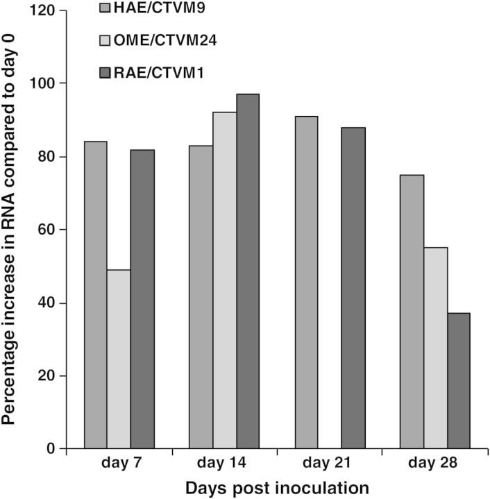
Following inoculation with AHFV, none of the tick cell lines showed any cytopathic effect over the subsequent 4-week test period. AHFV RNA was detected in the three inoculated tick cell lines on all sample days, with the real-time RT-PCR demonstrating a clear increase in RNA copy numbers in the 7 days following inoculation compared with the initial inoculum (Figure 1). By Day 14 post inoculation, RNA copy numbers had almost doubled in all three cell lines, followed by a gradual decrease to Day 28 when there was still more virus RNA present than on Day 0. Uninfected control cultures remained negative (no AHFV RNA amplification) throughout the test period.
Figure 1.
Real-time reverse transcription PCR for detection of Alkhumra haemorrhagic fever virus (AHFV) RNA in tick cell lines HAE/CTVM9 derived from the hard tick Hyalomma anatolicum, OME/CTVM24 derived from the soft tick Ornithodoros moubata and RAE/CTVM1 derived from the hard tick Rhipicephalus appendiculatus. The graph shows the amount of AHFV RNA detected in each cell line on Days 7, 14, 21 and 28 post inoculation, calculated as the percentage increase compared with the amount present in each culture immediately following inoculation on Day 0. OME/CTVM24 was not tested on Day 21 post inoculation.
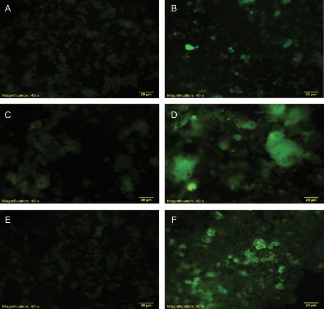
All of the inoculated tick cells showed specific fluorescence (Figure 2), with a progressively higher proportion of cells fluorescing each week until the end of the experiment (Table 1). Uninfected control cells did not show any specific fluorescence. The autofluorescence that is usually encountered with the OME/CTVM24 cells (authors' unpublished observations) was minimised by incorporation of BSA in the fluorescent conjugate.
Figure 2.
IFAT for detection of Alkhumra hemorrhagic fever virus (AHFV) in tick cell lines HAE/CTVM9 (A,B) derived from the hard tick Hyalomma anatolicum, OME/CTVM24 (C,D) derived from the soft tick Ornithodoros moubata and RAE/CTVM1 (E,F) derived from the hard tick Rhipicephalus appendiculatus. A,C,E: uninfected control cells; B,D,F: cells 7 days after inoculation with AHFV.
Table 1.
Results of IFAT and virus isolation for detection of Alkhumra haemorrhagic fever virus (AHFV) activity in infected and uninfected tick cell cultures following 4 weeks of incubation
| Tick cell linea and treatment | IFAT | Virus titre in LLC-MK2 cells (TCID50/ml) |
|---|---|---|
| AHFV-infected HAE/CTVM9 | + | NVID |
| Uninfected HAE/CTVM9 | – | NVID |
| AHFV-infected OME/CTVM24 | + | 106.6 |
| Uninfected OME/CTVM24 | – | NVID |
| AHFV-infected RAE/CTVM1 | + | 105.5 |
| Uninfected RAE/CTVM1 | – | NVID |
TCID50: 50% tissue culture infective dose; NVID: no virus infectivity detected.
a HAE/CTVM9 derived from the hard tick Hyalomma anatolicum; OME/CTVM24 derived from the soft tick Ornithodoros moubata; and RAE/CTVM1 derived from the hard tick Rhipicephalus appendiculatus.
As shown in Table 1, AHFV recovered from OME/CTVM24 and RAE/CTVM1 cultures at the end of the study (4 weeks) was infective for LLC-MK2 cells, yielding titres of 106.6 TCID50/ml and 105.5 TCID50/ml, respectively. No viable virus was recovered in LLC-MK2 cells from the inoculated HAE/CTVM9 or from the uninfected controls.
Discussion
Findings in the present study constitute the first record of growth of AHFV in tick cell lines. The results indicated that AHFV was able to multiply and produce viable virus both in the OME/CTVM24 and RAE/CTVM1 tick cell lines as confirmed by multiplication of AHFV in LLC-MK2 indicator cells to titres of 106.6 TCID50/ml and 105.5 TCID50/ml, respectively. As the titre of the original inoculum was 107.2 TCID50/ml, each tube of tick cells received 106.2 TCID50 and the nominal titre after inoculation would be 105.6 TCID50/ml. Therefore, it is clear that the AHFV multiplied by ≥10-fold in the OME/CTVM24 cells, whilst in RAE/CTVM1 cells the amount of virus recovered after 4 weeks in vitro was about the same as that inoculated on Day 0. However, any virus left in the supernate following inoculation would have been diluted at least 20-fold during the three medium changes on Days 7, 14 and 21, so it appears likely that some replication took place in both cell lines.
AHFV RNA was detected by real-time RT-PCR and viral antigen was detected by IFAT both in the OME/CTVM24 and RAE/CTVM1 tick cell lines. AHFV activity was also evident in the inoculated HAE/CTVM9 cultures as confirmed by real-time RT-PCR for AHFV RNA and by IFAT for viral antigen, but viable AHFV was not detected in the inoculated indicator LLC-MK2 cells, suggesting that the HAE/CTVM9 cell line did not support production of virus particles infective for mammalian cells. This could be due to failure of viruses to assemble properly, inability of mammalian-infective viruses to leave the tick cells, or inability to enter the mammalian cells. Increasing numbers of infected HAE/CTVM9 cells detected by IFAT indicate that the virus was able to leave and re-enter tick cells, suggesting that the mechanism used by the virus to enter arthropod cells may differ from that used to enter mammalian cells. Moreover, absence of infective AHFV in the HAE/CTVM9 cultures indicated that no virus from the original inoculum had survived the 4-week test period, thereby confirming that the AHFV detected in LLC-MK2 from OME/CTVM24 and RAE/CTVM1 cultures was most likely the result of replication and production of infective virus in these two tick cell lines.
Another factor that could affect the production of mammal-infective virus in a tick cell line is whether or not the line contains cells that are suitable for virus replication. Tick cell lines are both phenotypically and genotypically diverse19 and very little is known about the range of cell phenotypes within a particular line, apart from easily recognisable cell types such as hemocytes. It is not known which cells play an important role in AHFV replication in any arthropod. Certainly, one would expect that midgut and salivary gland cells would be important as the likely entry and exit points, respectively, for the virus, but other tissues may also be involved in virus persistence throughout the life cycle of the tick, for example during moult and during oogenesis if the virus is transovarially transmitted. Midgut cells and even salivary gland cells could be present in any of the tick cell lines used, as they were derived from whole tick embryos. Both RAE/CTVM1 and OME/CTVM24 are very heterogeneous and contain cells that resemble midgut cells by light microscopy (Lesley Bell-Sakyi, unpublished observations). HAE/CTVM9 is less heterogeneous and the cells are predominantly hemocyte-like.12 Although it is not possible to draw firm conclusions about the potential ability for a tick to transmit a virus in vivo from the phenotypic diversity of a cell line derived from that tick species which supports virus replication in vitro, the observation that the virus is capable of such replication in tick cells provides evidence in support of a possible role for the tick somewhere in the epidemiology of the virus.
We have recently confirmed that AHFV can propagate in the C6/36 cell line derived from the mosquito Aedes albopictus.11 AHFV can thus grow both in tick and mosquito cell lines. Such a phenomenon is not unique to AHFV as similar behaviour was previously reported for Langat virus, West Nile virus (WNV), Venezuelan equine encephalitis virus, Ganjam virus,20,21 and many other mosquito-borne arboviruses as reviewed recently.19
Early attempts to propagate arboviruses in arthropod cell lines were predominantly carried out in mosquito-derived cell lines.22 With the introduction of tick-derived cell lines, a range of arboviruses were found to replicate in these cell lines.19,20 Tick cell lines were subsequently employed to investigate different aspects of the relationship between tick-borne encephalitis virus and ticks at the cellular and molecular levels.19,23
Growth of arboviruses in vector and non-vector tick cell lines produced interesting findings. In a study by Růzek et al., it was found that there was a clear difference between tick-borne encephalitis virus growth in vector and non-vector tick cell lines.24 For example, cell lines derived from the vector Ixodes ricinus produced 100–1000-fold higher virus yield than non-vector cell lines.24 Similarly, Crimean–Congo haemorrhagic fever virus replication as measured by real-time RT-PCR was 10–100-fold higher in cell lines derived from the vector tick species H. anatolicum than in non-vector cell lines.19 In the present study, yield of viable AHFV from the O. moubata cell line (OME/CTVM24) was ≥10-fold higher than that from the R. appendiculatus cell line (RAE/CTVM1), whilst the H. anatolicum cell line (HAE/CTVM9) appeared to be incapable of supporting production of virus capable of infecting and causing a cytopathic effect in LLC-MK2 cells. It is unclear whether this in vitro pattern reflects real differences in natural vectorial or reservoir host capacity of these tick genera in vivo. AHFV has been isolated in Saudi Arabia from ticks of the genera Ornithodoros and Hyalomma,8 whilst there are no reports of Rhipicephalus spp. ticks being found positive for this virus.
A study examining the susceptibility of some tick and mosquito cell lines to 13 arthropod-borne flaviviruses and an alphavirus showed that the mosquito cell line was permissive to all of the tested mosquito-borne flaviviruses but not to the tick-borne viruses except Langat virus.20 On the other hand, the tick cell lines were susceptible to all the tested tick-borne viruses as well as to two mosquito-borne viruses, namely WNV and Venezuelan equine encephalitis virus, but not to other mosquito-borne viruses. The authors concluded that although a tick- or mosquito-derived cell line can become infected by a particular arbovirus, this does not always reflect vector association. Although naturally transmitted by mosquitoes, WNV has been isolated from ticks and can be experimentally transmitted by O. moubata.25 To the best of the authors' knowledge, no Flavivirus has been confirmed to be naturally transmitted both by ticks and mosquitoes.
In conclusion, although the vector(s) and reservoir(s) of AHFV have yet to be identified, the findings of this study and of a previous study11 indicate that both mosquitoes and ticks may play a role in the epidemiology of AHFV. Since epidemiological observations during AHFV outbreaks in Saudi Arabia suggested that mosquitoes were the likely vectors transmitting the virus from animals to humans, ticks may play a role as a reservoir of the virus in its ecological niche and/or as vectors transmitting the virus between animals as previously proposed.2,3,11 More studies are needed to define the exact roles of ticks and mosquitoes in the epidemiology and transmission of AHFV in nature.
Acknowledgments
Authors' contributions: TAM, EMEA, LBS, EIA and HMSA conceived and designed the study; LBS provided the tick cell lines; LBS, AMH, BEM and EMEA maintained the tick cell lines; TGK provided hyperimmune mouse ascitic fluid containing polyclonal antibodies against Alkhumra haemorrhagic fever virus; EMEA, AMH and BEM performed the viral infection of tick cell lines and the IFAT; EIA, AMH and BEM performed the real-time RT-PCR; all of the authors analysed and interpreted the data; TAM and EMEA wrote the manuscript; LBS, EIA, HMSA, HA, AMH, BEM and TGK critically revised the manuscript. All authors read and approved the final version. TAM is guarantor of the paper.
Acknowledgements: The authors thank Sheikh Mohammad Hussein Al-Amoudi for funding this research and the Scientific Chair for Viral Hemorrhagic Fever at King Abdulaziz University (Jeddah, Saudi Arabia). The authors acknowledge the Roslin Wellcome Trust Tick Cell Biobank (Edinburgh, UK) for kindly providing the tick cell lines. They also thank the technologist Mrs Noora A. Al-Kaiedi for excellent technical assistance in performing the real-time RT-PCR.
Funding: This study is one of the research products of the Scientific Chair of Sheikh Mohammad Hussein Al-Amoudi for Viral Hemorrhagic Fever, King Abdulaziz University (Jeddah, Saudi Arabia). The sponsor, Sheikh Mohammad Hussein Al-Amoudi, had no involvement in the study design, in the collection, analysis and interpretation of data, in the writing of the manuscript, or in the decision to submit the manuscript for publication.
Competing interests: None declared.
Ethical approval: Ethical approval was obtained from the Research Ethics Committee at the Faculty of Medicine, King Abdulaziz University (Jeddah, Saudi Arabia).
References
- 1.Qattan I, Akbar N, Afif H, et al. A novel Flavivirus: Makkah region 1994–1996. Saudi Epidemiol Bull. 1996;3:1–3. [Google Scholar]
- 2.Madani TA. Alkhumra virus infection, a new viral hemorrhagic fever in Saudi Arabia. J Infect Dis. 2005;51:91–7. doi: 10.1016/j.jinf.2004.11.012. [DOI] [PubMed] [Google Scholar]
- 3.Madani TA, Azhar EI, Abuelzein el-TM, et al. Alkhumra, not Alkhurma, is the correct name of the new hemorrhagic fever Flavivirus identified in Saudi Arabia. Intervirology. 2012;55:259–60. doi: 10.1159/000337238. [DOI] [PubMed] [Google Scholar]
- 4.Madani TA, Azhar EI, Abuelzein el-TM, et al. Alkhumra (Alkhurma) virus outbreak in Najran, Saudi Arabia: epidemiological, clinical, and laboratory characteristics. J Infect. 2011;62:67–76. doi: 10.1016/j.jinf.2010.09.032. [DOI] [PubMed] [Google Scholar]
- 5.Cartelli F, Castilletti C, Di Caro A, et al. Alkhurma hemorrhagic fever in travelers returning from Egypt, 2010. Emerg Infect Dis. 2010;16:1979–82. doi: 10.3201/eid1612101092. [DOI] [PMC free article] [PubMed] [Google Scholar]
- 6.Liebert UG. Controversy on virus designation: Alkhumra sive Alkhurma hemorrhagic fever Flavivirus. Intervirology. 2012;55:257–8. doi: 10.1159/000337237. [DOI] [PubMed] [Google Scholar]
- 7.Pletnev A, Gould E, Heinz FX, et al. Flaviviridae. In: King AMQ, Adams MJ, Carstens EB, Lefkowitz EJ, editors. Virus Taxonomy: IXth Report of the International Committee on Viruses. Oxford, UK: Elsevier; 2011. pp. 1003–20. [Google Scholar]
- 8.Charrel RN, Fagbo S, Moureau G, et al. Alkhurma hemorrhagic fever virus in Ornithodoros savignyi ticks. Emerg Infect Dis. 2007;13:153–5. doi: 10.3201/eid1301.061094. [DOI] [PMC free article] [PubMed] [Google Scholar]
- 9.Mahdi M, Erickson BR, Comer JA, et al. Kyasanur Forest disease virus Alkhurma subtype in ticks, Najran Province, Saudi Arabia. Emerg Infect Dis. 2011;17:945–7. doi: 10.3201/eid1705.101824. [DOI] [PMC free article] [PubMed] [Google Scholar]
- 10.Mussgay M, Enzmann PJ, Horzinek MC, Weiland E. Prog Med Virol. Growth cycle of arboviruses in vertebrate and arthropod cells. 1975;19:257–323. [PubMed] [Google Scholar]
- 11.Madani TA, Kao M, Azhar EI, et al. Successful propagation of Alkhumra (misnamed as Alkhurma) virus in C6/36 mosquito cells. Trans R Soc Trop Med Hyg. 2012;106:180–5. doi: 10.1016/j.trstmh.2011.11.003. [DOI] [PubMed] [Google Scholar]
- 12.Bell-Sakyi L. Continuous cell lines from the tick Hyalomma anatolicum anatolicum. J Parasitol. 1991;77:1006–8. [PubMed] [Google Scholar]
- 13.Bell-Sakyi L, Růzek D, Gould EA. Continuous cell lines from the soft tick Ornithodoros moubata. Exp Appl Acarol. 2009;49:209–19. doi: 10.1007/s10493-009-9258-y. [DOI] [PMC free article] [PubMed] [Google Scholar]
- 14.Bell-Sakyi L. Ehrlichia ruminantium grows in cell lines from four ixodid tick genera. J Comp Pathol. 2004;130:285–93. doi: 10.1016/j.jcpa.2003.12.002. [DOI] [PubMed] [Google Scholar]
- 15.Charrel RN, Zaki AM, Fakeeh M, et al. Low diversity of Alkhurma hemorrhagic fever virus, Saudi Arabia, 1994–1999. Emerg Infect Dis. 2005;11:683–8. doi: 10.3201/eid1105.041298. [DOI] [PMC free article] [PubMed] [Google Scholar]
- 16.Holland PM, Abramson RD, Watson R, Gelfand DH. Detection of specific polymerase chain reaction product by utilizing the 5'→3' exonuclease activity of Thermus aquaticus DNA polymerase. Proc Natl Acad Sci U S A. 1991;88:7276–80. doi: 10.1073/pnas.88.16.7276. [DOI] [PMC free article] [PubMed] [Google Scholar]
- 17.Brandt WE, Buescher EL, Hetrick FM. Production and characterization of arbovirus antibody in mouse ascitic fluid. Am J Trop Med Hyg. 1967;16:339–47. doi: 10.4269/ajtmh.1967.16.339. [DOI] [PubMed] [Google Scholar]
- 18.Reed LJ, Muench H. A simple method of estimating fifty per cent endpoints. Am J Hyg. 1938;27:493–7. [Google Scholar]
- 19.Bell-Sakyi L, Kohl A, Bente DA, Fazakerley JK. Tick cell lines for study of Crimean–Congo hemorrhagic fever virus and other arboviruses. Vector Borne Zoonot Dis. 2012;12:769–81. doi: 10.1089/vbz.2011.0766. [DOI] [PMC free article] [PubMed] [Google Scholar]
- 20.Lawrie CH, Uzcátegui NY, Armesto M, et al. Susceptibility of mosquito and tick cell lines to infection with various flaviviruses. Med Vet Entomol. 2004;18:268–74. doi: 10.1111/j.0269-283X.2004.00505.x. [DOI] [PubMed] [Google Scholar]
- 21.Sudeep AB, Jadi RS, Mishra AC. Ganjam virus. Indian J Med Res. 2009;130:514–9. [PubMed] [Google Scholar]
- 22.Singh KRP. Growth of arboviruses in arthropod tissue culture. Adv Virus Res. 1972;17:187–206. doi: 10.1016/s0065-3527(08)60750-2. [DOI] [PubMed] [Google Scholar]
- 23.Engel AR, Mitzel DN, Hanson CT, et al. Chimeric tick-borne encephalitis/dengue virus is attenuated in Ixodes scapularis ticks and Aedes aegypti mosquitoes. Vector Borne Zoonot Dis. 2011;11:665–74. doi: 10.1089/vbz.2010.0179. [DOI] [PMC free article] [PubMed] [Google Scholar]
- 24.Růzek D, Bell-Sakyi L, Kopecký J, Grubhoffer L. Growth of tick-borne encephalitis virus (European subtype) in cell lines from vector and non-vector ticks. Virus Res. 2008;137:142–6. doi: 10.1016/j.virusres.2008.05.013. [DOI] [PubMed] [Google Scholar]
- 25.Lawrie CH, Uzcátegui NY, Gould EA, Nuttall PA. Ixodid and argasid tick species and West Nile virus. Emerg Infect Dis. 2004;10:653–7. doi: 10.3201/eid1004.030517. [DOI] [PMC free article] [PubMed] [Google Scholar]


